Review
. 2017 Aug 29:22:18.
doi: 10.1186/s11658-017-0050-4. eCollection 2017.

Gene functioning and storage within a folded genome

Affiliations
Review

Gene functioning and storage within a folded genome

Sergey V Razin et al. Cell Mol Biol Lett. .

Abstract

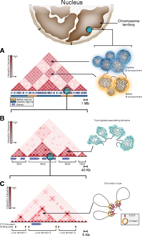
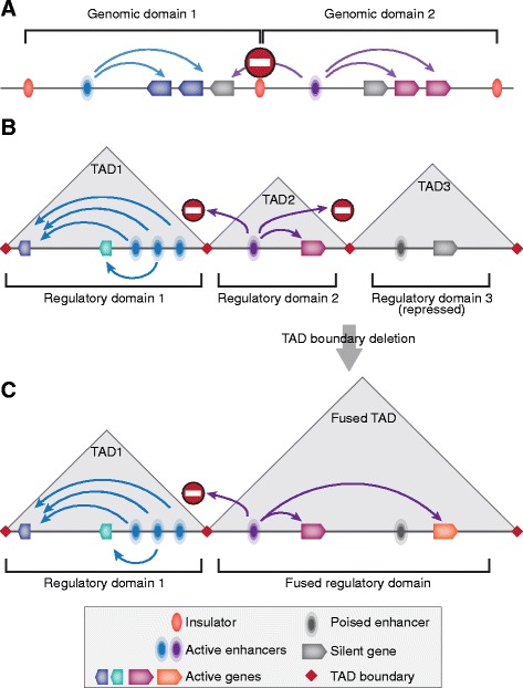
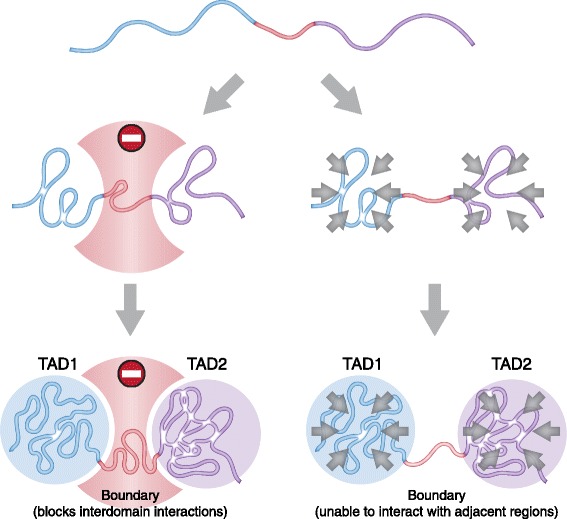
In mammals, genomic DNA that is roughly 2 m long is folded to fit the size of the cell nucleus that has a diameter of about 10 μm. The folding of genomic DNA is mediated via assembly of DNA-protein complex, chromatin. In addition to the reduction of genomic DNA linear dimensions, the assembly of chromatin allows to discriminate and to mark active (transcribed) and repressed (non-transcribed) genes. Consequently, epigenetic regulation of gene expression occurs at the level of DNA packaging in chromatin. Taking into account the increasing attention of scientific community toward epigenetic systems of gene regulation, it is very important to understand how DNA folding in chromatin is related to gene activity. For many years the hierarchical model of DNA folding was the most popular. It was assumed that nucleosome fiber (10-nm fiber) is folded into 30-nm fiber and further on into chromatin loops attached to a nuclear/chromosome scaffold. Recent studies have demonstrated that there is much less regularity in chromatin folding within the cell nucleus. The very existence of 30-nm chromatin fibers in living cells was questioned. On the other hand, it was found that chromosomes are partitioned into self-interacting spatial domains that restrict the area of enhancers action. Thus, TADs can be considered as structural-functional domains of the chromosomes. Here we discuss the modern view of DNA packaging within the cell nucleus in relation to the regulation of gene expression. Special attention is paid to the possible mechanisms of the chromatin fiber self-assembly into TADs. We discuss the model postulating that partitioning of the chromosome into TADs is determined by the distribution of active and inactive chromatin segments along the chromosome. This article was specially invited by the editors and represents work by leading researchers.

Keywords: Active chromatin; Enhancers; Epigenetic regulatory mechanisms; Hi-C; Self-organization; TADs.


Conflict of interest statement

Ethics approval and consent to participate

The authors state that no ethical approval was required for this work.

Consent for publication

Not applicable.

Competing interests

The authors declare that they have no competing interests.

Publisher’s Note

Springer Nature remains neutral with regard to jurisdictional claims in published maps and institutional affiliations.

Figures

Fig. 1
Fig. 1
A scheme illustrating the hierarchical structure of interphase chromatin. Chromosome territories (at the top of the picture) are partitioned into A- and B-compartments (a) formed by long-range spatial interactions between distant genome loci and containing active and repressed genome regions, respectively. At a sub-megabase level, chromatin is folded into topologically-associating domains, TADs (b), commonly interpreted as self-interacting globular structures those positions are largely conserved across cell types. The internal structure of TADs is represented by arrays of so-called loop domains formed by spatial contacts between CTCF/cohesin-binding sites (c). Color intensity on illustrative Hi-C maps (on the left side of each panel) reflects average interaction frequency between corresponding genomic bins
Fig. 2
Fig. 2
Chromosome partitioning into TADs reflects genome partitioning into regulatory domains delimiting zones of enhancer influence. Conventional concept of genomic domain implies that the entire genome is partitioned into non-overlapping parts (domains) containing gene clusters and regulatory regions (a), and demarcated with insulators preventing cross-talk between regulatory systems of the adjacent domains. According to current views, zones of enhancer influence (regulatory domains) largely overlap with TADs (b) that spatially confine communication between genes and enhancers located within adjacent regulatory domains. Deletion of TAD boundary leads to TAD fusion and, consequently, to fusion of corresponding regulatory domains resulting in abnormal enhancer-promoter communication and transcription dysregulation (c)
Fig. 3
Fig. 3
A scheme illustrating two proposal mechanisms of TAD boundary action. Left panel: boundary plays an active role in TAD demarcation preventing interdomain interactions. Right panel: boundary represents a genomic region unable to fold into higher-order structures and/or to interact with adjacent regions. In contrast, TAD is comprised of chromatin regions which tend to interact with each other forming globular structures

Similar articles

Cited by

References

    1. Getzenberg RH, Pienta KJ, Ward WS, Coffey DS. Nuclear structure and the three-dimensional organization of DNA. J Cell Biochem. 1991;47:289–299. doi: 10.1002/jcb.240470402. - DOI - PubMed
    1. Woodcock CL, Dimitrov S. Higher-order structure of chromatin and chromosomes. Curr Opin Genet Dev. 2001;11:130–135. doi: 10.1016/S0959-437X(00)00169-6. - DOI - PubMed
    1. Woodcock CL. Chromatin architecture. Curr Opin Struct Biol. 2006;16:213–220. doi: 10.1016/j.sbi.2006.02.005. - DOI - PubMed
    1. Finch JT, Klug A. Solenoidal model for superstructure in chromatin. Proc Natl Acad Sci U S A. 1976;73:1897–1901. doi: 10.1073/pnas.73.6.1897. - DOI - PMC - PubMed
    1. Richmond TJ, Finch JT, Rushton B, Rhodes D, Klug A. Structure of the nucleosome core particle at 7 a resolution. Nature. 1984;311:532–537. doi: 10.1038/311532a0. - DOI - PubMed

LinkOut - more resources