Review
. 2017 Aug 14:16:355-368.
doi: 10.1016/j.nicl.2017.08.015. eCollection 2017.

Clinical neuroimaging in the preterm infant: Diagnosis and prognosis

Affiliations
Review

Clinical neuroimaging in the preterm infant: Diagnosis and prognosis

Manuel Hinojosa-Rodríguez et al. Neuroimage Clin. .

Abstract

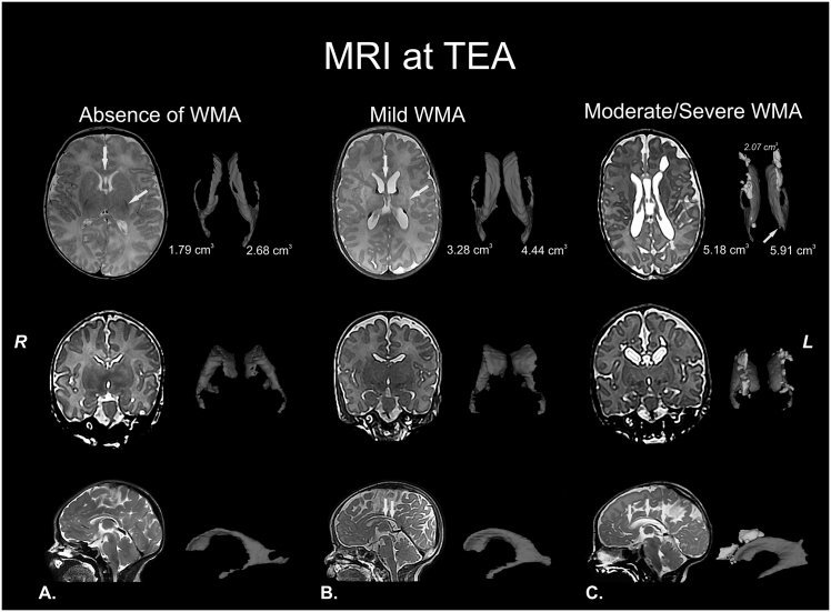
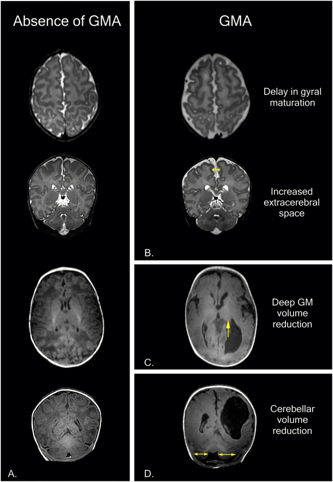
Perinatal care advances emerging over the past twenty years have helped to diminish the mortality and severe neurological morbidity of extremely and very preterm neonates (e.g., cystic Periventricular Leukomalacia [c-PVL] and Germinal Matrix Hemorrhage - Intraventricular Hemorrhage [GMH-IVH grade 3-4/4]; 22 to < 32 weeks of gestational age, GA). However, motor and/or cognitive disabilities associated with mild-to-moderate white and gray matter injury are frequently present in this population (e.g., non-cystic Periventricular Leukomalacia [non-cystic PVL], neuronal-axonal injury and GMH-IVH grade 1-2/4). Brain research studies using magnetic resonance imaging (MRI) report that 50% to 80% of extremely and very preterm neonates have diffuse white matter abnormalities (WMA) which correspond to only the minimum grade of severity. Nevertheless, mild-to-moderate diffuse WMA has also been associated with significant affectations of motor and cognitive activities. Due to increased neonatal survival and the intrinsic characteristics of diffuse WMA, there is a growing need to study the brain of the premature infant using non-invasive neuroimaging techniques sensitive to microscopic and/or diffuse lesions. This emerging need has led the scientific community to try to bridge the gap between concepts or ideas from different methodologies and approaches; for instance, neuropathology, neuroimaging and clinical findings. This is evident from the combination of intense pre-clinical and clinicopathologic research along with neonatal neurology and quantitative neuroimaging research. In the following review, we explore literature relating the most frequently observed neuropathological patterns with the recent neuroimaging findings in preterm newborns and infants with perinatal brain injury. Specifically, we focus our discussions on the use of neuroimaging to aid diagnosis, measure morphometric brain damage, and track long-term neurodevelopmental outcomes.

Keywords: Preterm neonates; infants; magnetic resonance imaging; neonatal neurology; perinatal brain injury; white matter abnormalities.


Figures

Fig. 1
Fig. 1
White matter abnormalities (WMA). T2 structural MRI with corresponding volumetric measurements of the lateral ventricles (in blue) at term-equivalent age (TEA) or nearly TEA. A) Normal MRI at TEA. Healthy neonate at 41 weeks of postmenstrual age with normal MRI: absence of cystic abnormalities, volume and size of corpus callosum (yellow arrow in upper panel) and lateral ventricles are normal, myelination of the corpus callosum, posterior limb of internal capsule (PLIC; yellow arrow in upper panel) and corona radiata corresponding with the age and cerebral white matter signal and volume are normal. B) Mild WMA at nearly TEA. Preterm infant (born at 28 gestational weeks) at 43 weeks of postmenstrual age with mild diffuse WMA: absence of cystic abnormalities, partial thinning of the corpus callosum (yellow arrows in upper and lower panel), mild dilated lateral ventricles, PLIC with myelination delay and focal signal abnormalities in relation to punctate white matter lesions (orange arrows in upper panel), probably associated with hemorrhagic etiology and congestion of medullary veins. C) Moderate-to-severe WMA at TEA. Preterm infant (born at 29 gestational weeks) at 41 weeks of postmenstrual age with cystic WMA: bilateral presence of multiple cysts (3D reconstruction and corresponding volumetric measurement are in yellow in upper panel), global thinning of the corpus callosum (orange arrow in upper panel and yellow arrows in lower panel), dilated lateral ventricles, severe myelination delay, diffuse and extensive signal abnormalities, white matter loss (double direction arrow in upper panel), prominent sulci near the ventricles (orange arrow in upper panel) and communication between small cyst and left lateral ventricle (yellow arrow in upper panel). The features of the last structural MRI are compatible with cystic-PVL. Images in radiological convention. (For interpretation of the references to colour in this figure legend, the reader is referred to the web version of this article.)
Fig. 2
Fig. 2
Examples of gray matter abnormalities (GMA) in MRI at term-equivalent age (TEA) in the first year of life. A) Normal T2 and T1 structural MRI at TEA. Healthy neonate at 41 weeks of postmenstrual age with normal MRI: cortical gray matter (signal, gyral maturation and subarachnoid space), deep/subcortical gray matter and cerebellum (signal and volume) are normal. B) Preterm infant (born at 31 gestational weeks) at 38 weeks of postmenstrual age with mild-to-moderate GMA: delay in gyral maturation and increase in extracerebral space (double direction arrow). C-D) Preterm infant (born at 31 gestational weeks) at 9 months of corrected age with porencephalic cyst and severe GMA. C) Deep GMA with volume reduction and asymmetry in the thalamus (arrow). D) Cerebellar volume reduction and asymmetry in cerebellar hemispheres (double direction arrow). The features of the last structural MRI (C-D) are compatible with a complex spectrum of encephalopathy of prematurity (diagnosis: periventricular hemorrhagic infarction + diffuse WMA compatible with PVL + severe GMA = encephalopathy of prematurity; mixed patterns with two or more lesions in different stages and with distinct evolution in each one). Images in radiological convention.
Fig. 3
Fig. 3
A) Volumetric MRI at term-equivalent age in preterm infant with cystic-PVL. Note the cysts or focal component of cystic-PVL (yellow) and dilated left lateral ventricle (blue). B) Automated segmentation, parcellation and 3D reconstruction of the brain and cerebellum in healthy child at 8 years old. Right cerebral and cerebellar hemispheres: white matter 3D reconstruction. Left cerebral and cerebellar hemispheres: cortical parcellation and 3D reconstruction. Image B in radiological convention. (For interpretation of the references to colour in this figure legend, the reader is referred to the web version of this article.)
Fig. 4
Fig. 4
Structural MRI and Diffusion Tensor Imaging (DTI). Male preterm infant (born at 30 gestational weeks) with cystic-PVL diagnosis and neurological follow-up in the first 2 years. A-B) T2 structural MRI and DTI at nearly term-equivalent age (TEA), around 47 weeks of postmenstrual age. A) T2 structural MRI with bilateral cystic lesions (arrows) and punctate white matter lesions. B) Color-coded fractional anisotropy (FA) map with evident white matter loss in the right posterior limb of the internal capsule (PLIC; right white oval) and bilateral reduction of FA values (right PLIC FA 0.21 vs. left PLIC FA 0.36), mainly in right side. A motor prognosis for this type of cases is possible; the major clinical outcome in these cases is a location-dependent spastic cerebral palsy (hemiplegia, diplegia, quadriplegia, etc.). C) T2 structural MRI at 24 months of corrected age with loss of cysts by cyst walls adjoining or “ventriculomegaly ex-vacuo.” The evidence of cyst in cystic-PVL can be lost at this stage or before (this is one reason why MRI at TEA and follow-up for years is suggested). However, the diffuse component of cystic-PVL (arrows) is still evident at this age. The current diagnosis of this child is triparetic cerebral palsy with left side more affected. Images in radiological convention.
Fig. 5
Fig. 5
Sensorimotor network by resting-state fMRI at term-equivalent age during physiological sleep (without anesthesia or sedation). A-C) Healthy female term infant. D-F) Male preterm infant (born at 35 gestational weeks) with clinical history of right periventricular hemorrhagic infarction. Currently with diagnosis by structural MRI of porencephalic cyst with ipsilateral communication between lateral ventricular system and the subarachnoid space. Note the change in resting-state pattern of sensorimotor network in relation to the healthy term infant. Images A, B, D and E are in radiological convention.

Similar articles

Cited by

References

    1. Abernethy L.J., Palaniappan M., Cooke R.W. Quantitative magnetic resonance imaging of the brain in survivors of very low birth weight. Arch. Dis. Child. 2002;87(4):279–283. - PMC - PubMed
    1. Adhikari M.H., Raja Beharelle A., Griffa A., Hagmann P., Solodkin A., McIntosh A.R., Deco G. Computational modeling of resting-state activity demonstrates markers of normalcy in children with prenatal or perinatal stroke. J. Neurosci. 2015;35(23):8914–8924. - PMC - PubMed
    1. Ajayi-Obe M., Saeed N., Cowan F., Rutherford M., Edwards A. Reduced development of cerebral cortex in extremely preterm infants. Lancet. 2000;356(9236):1162–1163. - PubMed
    1. Ancel P.-Y., Goffinet F., Kuhn P., Langer B., Matis J., Hernandorena X.…Kaminski M. Survival and morbidity of preterm children born at 22 through 34 weeks' gestation in France in 2011: results of the EPIPAGE-2 cohort study. JAMA Pediatr. 2015;169(3):230–238. - PubMed
    1. Anderson P.J., Cheong J.L.Y., Thompson D.K. The predictive validity of neonatal MRI for neurodevelopmental outcome in very preterm children. Semin. Perinatol. 2015;39(2):147–158. - PubMed

MeSH terms