Case Reports
. 2017 Aug;5(16):327.
doi: 10.21037/atm.2017.05.04.

Cystic adventitial disease-case series and review of literature

Affiliations
Case Reports

Cystic adventitial disease-case series and review of literature

Shuo Li et al. Ann Transl Med. 2017 Aug.

Abstract

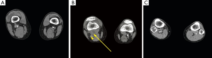
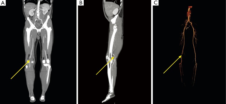
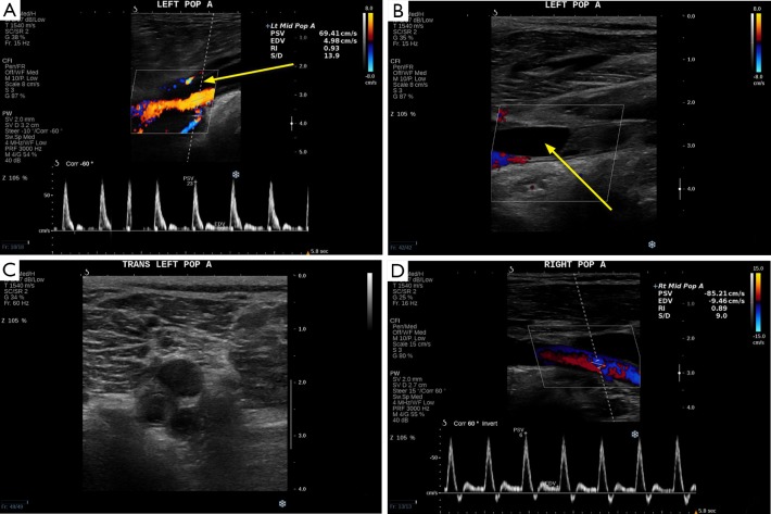
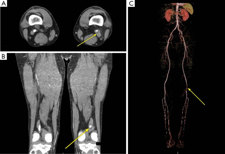
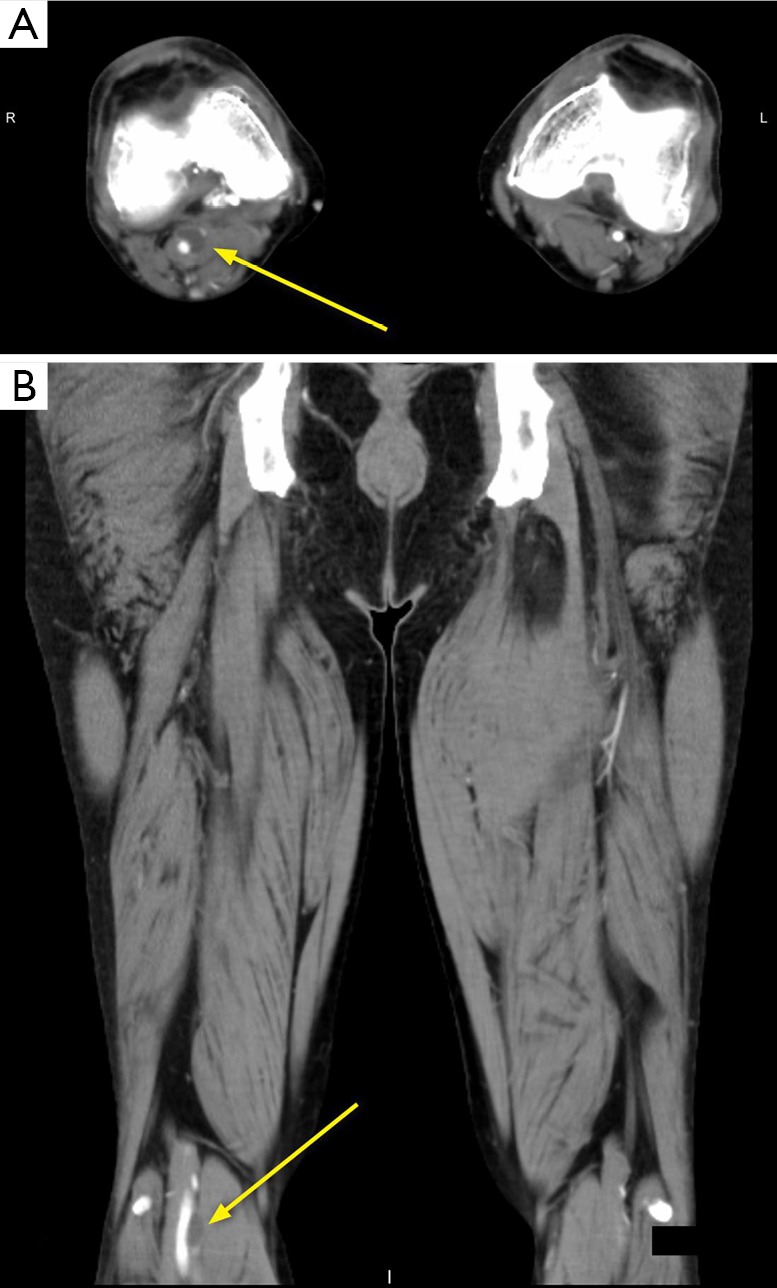
Cystic adventitial disease (CAD) is a rare vascular disorder that involves the arteries and rarely the veins, most commonly found in the popliteal artery of male patients. Etiology of CAD is uncertain and currently without a consensus agreement. Clinically, the most common presenting symptom is claudication. Diagnosis requires a strong clinical suspicion in patients with intermittent claudication, but without other risk factors for atherosclerotic disease. Angiography, ultrasound (US), computed tomography (CT) and magnetic resonance imaging (MRI) can all be used for diagnosis. Treatment of CAD can be done via surgical resection or percutaneous intervention such as aspiration. CAD can rarely recur after treatment.

Keywords: Cystic adventitial disease (CAD); Ishikawa sign; computed tomography angiography (CTA); scimitar sign; ultrasound (US).


Conflict of interest statement

Conflicts of Interest: The authors have no conflicts of interest to declare.

Figures

Figure 1
Figure 1
CTA abdominal and pelvis with runoff. (A–C) Selective axial images through the knees. (A) Normal opacification of bilateral above the knee popliteal arteries; (B) taken at the level of the knee; two adjacent hypodense lesions can be seen in the region of the right popliteal fossa (arrow). The right popliteal artery is near completely occluded, with only a focal area of faint opacification in the posterolateral portion (arrowhead). The left popliteal artery is normal; (C) reconstitution of the distal, below the knee right popliteal artery. Incidentally, there was a right suprapatellar knee effusion and an expansile intramedullary lesion in the head of the proximal right fibula.
Figure 2
Figure 2
Maximal intensive projection (MIP) and 3D volume rendered reconstruction using data from Figure 1. (A,B) Coronal and sagittal MIP images; notice the short segment non-opacification of the right popliteal artery just above the knee (arrow); (C) a 3D AP view volume rendering, with soft tissue and bone segmented out to show only vessels again showing a short segment non-opacified right popliteal artery (arrow). The right popliteal artery lumen before and after the occlusion shows asymmetric shouldering.
Figure 3
Figure 3
Color Doppler and Grayscale ultrasound image of the left popliteal artery with right popliteal artery as comparison. (A) A color Doppler image of the left popliteal artery. There is normal, triphasic flow within the narrowed lumen. An anechoic lesion is seen adjacent to the left popliteal artery lumen (arrow). (B) is taken more distally compared to (A). An anechoic lesion is seen here completely filling the left popliteal artery lumen (arrow), with a thin echogenic line separating this anechoic lesion from the popliteal lumen, representing the vessel intima and media. (C) is transverse grayscale US at the left popliteal fossa, showing a well circumscribed hypoechoic lesion adjacent the popliteal artery and vein. No clear communication is seen between this hypoechoic lesion and the popliteal artery or vein. (D) is a normal comparison US of the right popliteal artery.
Figure 4
Figure 4
CTA abdomen and pelvis runoff, including MIP as well as 3D volume rendered reconstructions. (A) is an axial slice from the patient’s CTA showing a hypodense lesion with a bilobed appearance, intimately associated with the opacified left popliteal artery lumen (arrow). (B) is a coronal MIP image of the legs also showing the hypodense lesion surrounding the opacified lumen (arrow). (C) is a volume rendered image with soft tissue and bone segmented out, showing focal short segment narrowing of the left popliteal artery (arrow). The comparison right popliteal artery is normal in appearance.
Figure 5
Figure 5
CTA abdomen and pelvis with runoff. (A) is an axial slice at the level of the knee from the patient’s CTA abdomen and pelvis with runoff showing a hypodense lesion contacting the opacified right popliteal artery lumen (arrow). (B) is the coronal reformatted image, showing a well circumscribed hypodense lesion adjacent to the right popliteal artery (arrow). The lumen of the right popliteal artery is mildly narrowed but remain patent and opacified.

References

    1. Atkins HJ, Key JA. A case of myxomatous tumour arising in the adventitia of the left external iliac artery. Br J Surg 1947;34:426. 10.1002/bjs.18003413618 - DOI - PubMed
    1. Ksepka M, Li A, Norman S. Cystic Adventitial Disease. Ultrasound Q 2015;31:224-6. 10.1097/RUQ.0000000000000160 - DOI - PubMed
    1. Linquette M, Mesmacque R, Beghin B, et al. Cystic degeneration of the adventitia of the popliteal artery. Apropos of a further case. Sem Hop 1967;43:3005-13. - PubMed
    1. Stallworth JM, Brown AG, Burges GE, et al. Cystic adventitial disease of the popliteal artery. Am Surg 1985;51:455-9. - PubMed
    1. Levien LJ, Benn CA. Adventitial cystic disease: a unifying hypothesis. J Vasc Surg 1998;28:193-205. 10.1016/S0741-5214(98)70155-7 - DOI - PubMed

Publication types

LinkOut - more resources