Multicenter Study
. 2018 Jan;31(1):141-149.
doi: 10.1038/modpathol.2017.103. Epub 2017 Sep 1.

Molecular testing for the clinical diagnosis of fibrolamellar carcinoma

Affiliations
Multicenter Study

Molecular testing for the clinical diagnosis of fibrolamellar carcinoma

Rondell P Graham et al. Mod Pathol. 2018 Jan.

Abstract

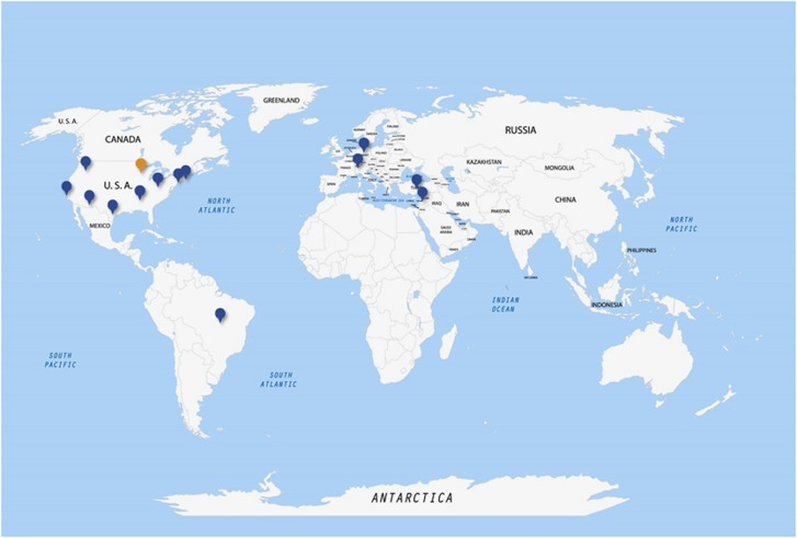
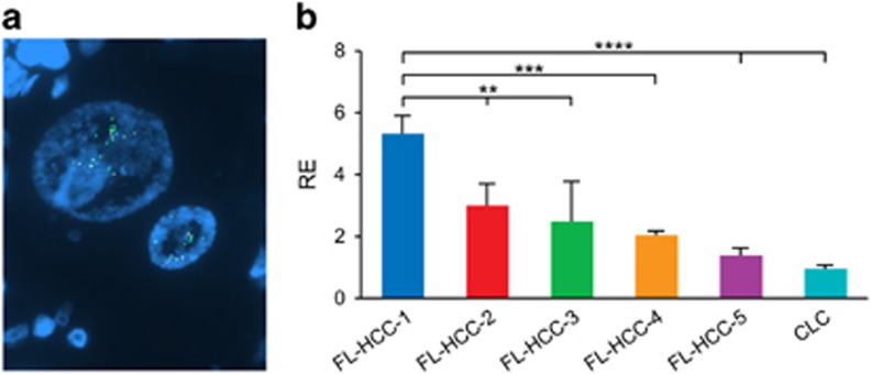
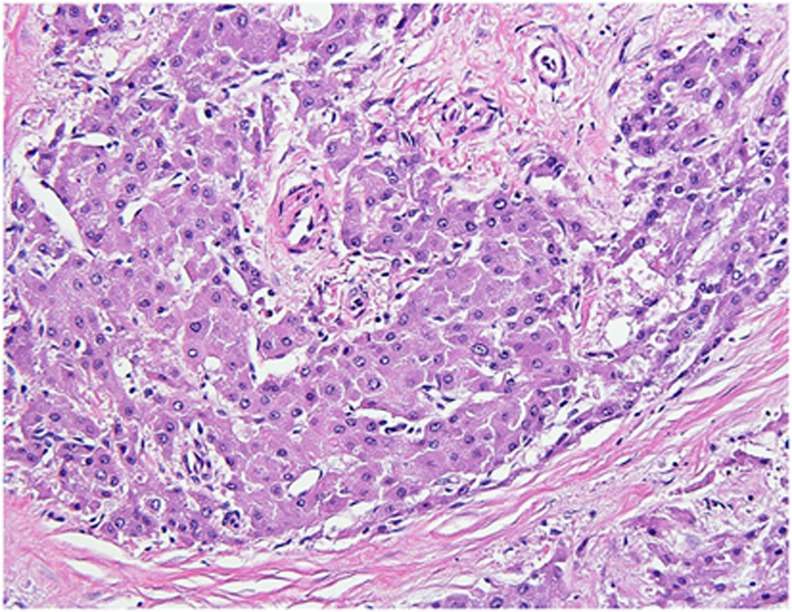
Fibrolamellar carcinoma has a distinctive morphology and immunophenotype, including cytokeratin 7 and CD68 co-expression. Despite the distinct findings, accurate diagnosis of fibrolamellar carcinoma continues to be a challenge. Recently, fibrolamellar carcinomas were found to harbor a characteristic somatic gene fusion, DNAJB1-PRKACA. A break-apart fluorescence in situ hybridization (FISH) assay was designed to detect this fusion event and to examine its diagnostic performance in a large, multicenter, multinational study. Cases initially classified as fibrolamellar carcinoma based on histological features were reviewed from 124 patients. Upon central review, 104 of the 124 cases were classified histologically as typical of fibrolamellar carcinoma, 12 cases as 'possible fibrolamellar carcinoma' and 8 cases as 'unlikely to be fibrolamellar carcinoma'. PRKACA FISH was positive for rearrangement in 102 of 103 (99%) typical fibrolamellar carcinomas, 9 of 12 'possible fibrolamellar carcinomas' and 0 of 8 cases 'unlikely to be fibrolamellar carcinomas'. Within the morphologically typical group of fibrolamellar carcinomas, two tumors with unusual FISH patterns were also identified. Both cases had the fusion gene DNAJB1-PRKACA, but one also had amplification of the fusion gene and one had heterozygous deletion of the normal PRKACA locus. In addition, 88 conventional hepatocellular carcinomas were evaluated with PRKACA FISH and all were negative. These findings demonstrate that FISH for the PRKACA rearrangement is a clinically useful tool to confirm the diagnosis of fibrolamellar carcinoma, with high sensitivity and specificity. A diagnosis of fibrolamellar carcinoma is more accurate when based on morphology plus confirmatory testing than when based on morphology alone.


Conflict of interest statement

The authors declare no conflict of interest.

Figures

Figure 1
Figure 1
Fibrolamellar carcinoma origins—The geographic origin of the cases of fibrolamellar carcinoma included in this study.
Figure 2
Figure 2
Typical fibrolamellar carcinoma results. (a) Typical fibrolamellar carcinoma. The neoplastic cells are characterized by abundant eosinophilic cytoplasm, nuclei with open chromatin, and prominent nucleoli. The tumor cells form trabecula which are separated by bands of fibrosis. (b) Cytokeratin 7 is positive in the tumor cells. (c) A CD68 immunostain shows characteristic cytoplasmic staining. (d) PRKACA break apart FISH in a typical fibrolamellar carcinoma. The FISH result is positive. The tumor cells show separate green signals and intact yellow signals (due to overlapping red and green signals).
Figure 3
Figure 3
Fibrolamellar carcinoma with PRKACA locus amplification. (a) PRKACA break Apart FISH results are consistent with amplification of the PRKACA locus. The fibrolamellar carcinoma showed typical morphology. There are greater than 10 separate green signals per tumor cell nucleus with fewer intact yellow signals. (b) Quantitative reverse transcription-PCR shows a 2–5-fold increase in PRKACA mRNA compared to four randomly selected fibrolamellar carcinomas (FL-HCC-2-5) and one randomly selected cholangiocarcinoma (CLC).
Figure 4
Figure 4
Fibrolamellar carcinoma with uncommon FISH results (a) PRKACA break-apart FISH shows separate red and green signals in a morphologically typical fibrolamellar carcinoma. This FISH signal pattern raised the possibility of either a different fusion partner for PRKACA or heterozygous loss of the PRKACA locus without involvement in a gene fusion event. (b). Array comparative genomic hybridization data at chromosome 19p demonstrated heterozygous loss of the PRKACA locus due to a 10 Mb deletion (orange bidirectional arrow) including the hybridization site of the FISH probe (shown in green).
Figure 5
Figure 5
Possible fibrolamellar carcinoma. An example of a case classified as a ‘possible fibrolamellar carcinoma’ based on morphology, characterized by intratumoral fibrosis and neoplastic cells with granular cytoplasm. The cytoplasm of the tumor cells is amphophilic in nature. This case was positive for PRKACA break-apart by FISH testing.
Figure 6
Figure 6
Unlikely fibrolamellar carcinoma An example of a case classified as ‘unlikely to be fibrolamellar carcinoma’ based on morphology. (a) The tumor shows more nuclear pleomorphism than is seen in typical fibrolamellar carcinomas. (b) The growth patterns shows pseudoacinar areas with cholestasis. This case was negative for PRKACA break-apart by FISH testing.

References

    1. Van Eyken P, Sciot R, Brock P et al. Abundant expression of cytokeratin 7 in fibrolamellar carcinoma of the liver. Histopathology 1990;17:101–107. - PubMed
    1. Ward SC, Huang J, Tickoo SK et al. Fibrolamellar carcinoma of the liver exhibits immunohistochemical evidence of both hepatocyte and bile duct differentiation. Mod Pathol 2010;23:1180–1190. - PubMed
    1. Ross HM, Daniel HD, Vivekanandan P et al. Fibrolamellar carcinomas are positive for CD68. Mod Pathol 2011;24:390–395. - PMC - PubMed
    1. El-Serag HB, Davila JA. Is fibrolamellar carcinoma different from hepatocellular carcinoma? A US population-based study. Hepatology 2004;39:798–803. - PubMed
    1. Torbenson M. Fibrolamellar carcinoma: 2012 update. Scientifica 2012;2012:743790. - PMC - PubMed

Publication types

MeSH terms

Substances

Supplementary concepts