Review
. 2017 Sep 1;127(9):3240-3249.
doi: 10.1172/JCI90606. Epub 2017 Sep 1.

Microglia in Alzheimer's disease

Affiliations
Review

Microglia in Alzheimer's disease

Heela Sarlus et al. J Clin Invest. .

Abstract

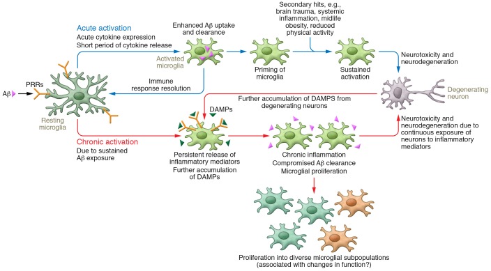
Microglia are brain-resident myeloid cells that mediate key functions to support the CNS. Microglia express a wide range of receptors that act as molecular sensors, which recognize exogenous or endogenous CNS insults and initiate an immune response. In addition to their classical immune cell function, microglia act as guardians of the brain by promoting phagocytic clearance and providing trophic support to ensure tissue repair and maintain cerebral homeostasis. Conditions associated with loss of homeostasis or tissue changes induce several dynamic microglial processes, including changes of cellular morphology, surface phenotype, secretory mediators, and proliferative responses (referred to as an "activated state"). Activated microglia represent a common pathological feature of several neurodegenerative diseases, including Alzheimer's disease (AD). Cumulative evidence suggests that microglial inflammatory activity in AD is increased while microglial-mediated clearance mechanisms are compromised. Microglia are perpetually engaged in a mutual interaction with the surrounding environment in CNS; thus, diverse microglial reactions at different disease stages may open new avenues for therapeutic intervention and modification of inflammatory activities. In this Review, the role of microglia in the pathogenesis of AD and the modulation of microglia activity as a therapeutic modality will be discussed.


Conflict of interest statement

Conflict of interest: The authors have declared that no conflict of interest exists.

Figures

Figure 1
Figure 1. Acute versus chronic activation of microglia in AD.
Aβ binds to PRRs, leading to activation of resting microglia. Acutely activated microglia express cytokines, which drive enhanced phagocytosis, uptake, and clearance of Aβ. Long-term activation of microglia drives proliferation as well as a chronic inflammatory state that causes neurotoxicity and neurodegeneration. Sustained activation of microglia, induced by brain trauma, systemic inflammation, obesity, and reduced physical activity, also drives neurotoxicity and neurodegeneration. DAMPs that arise from these processes further activate microglia, leading to compromised Aβ phagocytosis and propagating chronic inflammation. Chronic inflammation and accumulation of Aβ are well-established clinical features of AD.

References

    1. Beers DR, et al. Wild-type microglia extend survival in PU.1 knockout mice with familial amyotrophic lateral sclerosis. Proc Natl Acad Sci U S A. 2006;103(43):16021–16026. doi: 10.1073/pnas.0607423103. - DOI - PMC - PubMed
    1. Kierdorf K, et al. Microglia emerge from erythromyeloid precursors via Pu.1- and Irf8-dependent pathways. Nat Neurosci. 2013;16(3):273–280. doi: 10.1038/nn.3318. - DOI - PubMed
    1. McKercher SR, et al. Targeted disruption of the PU.1 gene results in multiple hematopoietic abnormalities. EMBO J. 1996;15(20):5647–5658. - PMC - PubMed
    1. Schulz C, et al. A lineage of myeloid cells independent of Myb and hematopoietic stem cells. Science. 2012;336(6077):86–90. doi: 10.1126/science.1219179. - DOI - PubMed
    1. Alliot F, Godin I, Pessac B. Microglia derive from progenitors, originating from the yolk sac, and which proliferate in the brain. Brain Res Dev Brain Res. 1999;117(2):145–152. doi: 10.1016/S0165-3806(99)00113-3. - DOI - PubMed

MeSH terms