Comparative Study
. 2017 Aug 19;6(8):e005838.
doi: 10.1161/JAHA.117.005838.

A Systems Biology Approach to Investigating Sex Differences in Cardiac Hypertrophy

Affiliations
Comparative Study

A Systems Biology Approach to Investigating Sex Differences in Cardiac Hypertrophy

Josephine Harrington et al. J Am Heart Assoc. .

Abstract

Background: Heart failure preceded by hypertrophy is a leading cause of death, and sex differences in hypertrophy are well known, although the basis for these sex differences is poorly understood.

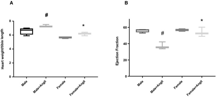
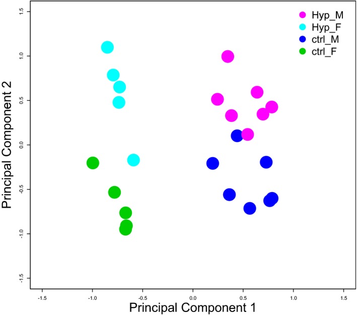
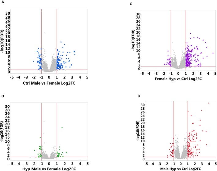
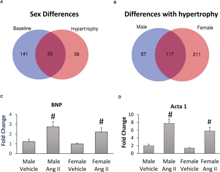
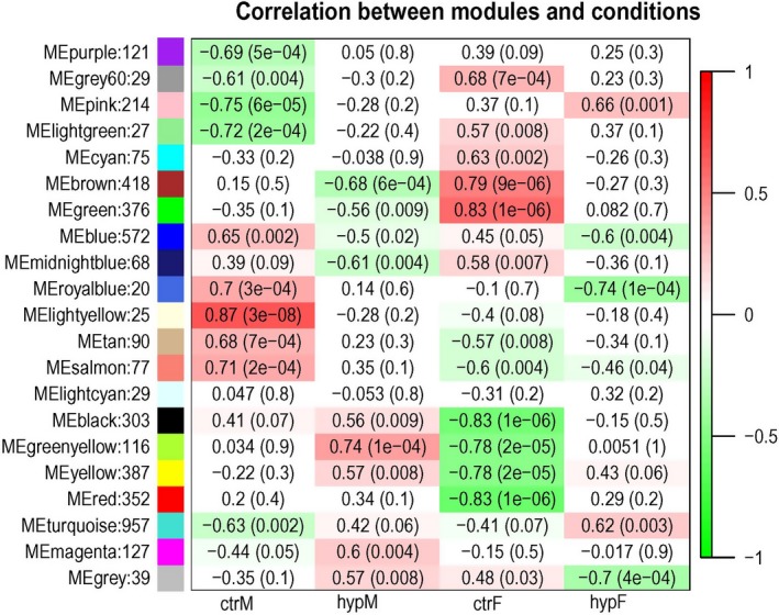
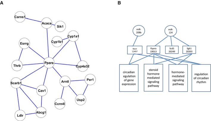
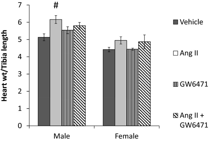
Methods and results: This study used a systems biology approach to investigate mechanisms underlying sex differences in cardiac hypertrophy. Male and female mice were treated for 2 and 3 weeks with angiotensin II to induce hypertrophy. Sex differences in cardiac hypertrophy were apparent after 3 weeks of treatment. RNA sequencing was performed on hearts, and sex differences in mRNA expression at baseline and following hypertrophy were observed, as well as within-sex differences between baseline and hypertrophy. Sex differences in mRNA were substantial at baseline and reduced somewhat with hypertrophy, as the mRNA differences induced by hypertrophy tended to overwhelm the sex differences. We performed an integrative analysis to identify mRNA networks that were differentially regulated in the 2 sexes by hypertrophy and obtained a network centered on PPARα (peroxisome proliferator-activated receptor α). Mouse experiments further showed that acute inhibition of PPARα blocked sex differences in the development of hypertrophy.

Conclusions: The data in this study suggest that PPARα is involved in the sex-dimorphic regulation of cardiac hypertrophy.

Keywords: hypertrophy; sex; systems biology.


Figures

Figure 1
Figure 1
Sex differences in hypertrophy and ejection fraction following 3 weeks treatment with angiotensin II (AngII). Changes in (A) heart weight to tibia length and (B) ejection fraction in males and females following 3 weeks of AngII treatment. Data are mean±SEM, n=3 to 4. We performed a 2‐way ANOVA. The heart weight/tibia length data showed significant differences based on sex and Ang II treatment, but there was not a significant interaction between sex and hypertrophy. The male vehicle‐treated hearts were significantly different than male AngII‐treated hearts, and male and female AngII‐treated hearts were significantly different. The ejection fraction (EF) data showed a significant interaction between sex and hypertrophy. The males were significantly different between vehicle and AngII, and the males and females showed a significant difference with hypertrophy. #Significantly different compared with vehicle treated. *Significantly different from male Ang II treatment. P<0.05 was considered significant.
Figure 2
Figure 2
Principal component analysis for mRNA in male (M) and female (F) mice at baseline and with hypertrophy (Hyp), n=5 to 7 per group. Ctrl indicates control.
Figure 3
Figure 3
Volcano plots of mRNA differences. We used a filter of log2 fold change (FC) ≥1 and a false discovery rate (FDR) of 10% to select the transcripts with significant differences, which are shown in color. A, Data for mRNA in control (Ctrl) male vs female mice, with 174 transcripts (in blue) showing significant differences. B, 71 mRNA transcripts (in green) with significant differences between male and female hearts treated with angiotensin II (AngII). C, 328 transcripts (in purple) show significant differences in females with and without AngII treatment. D, 174 transcripts (in red) exhibit significant differences in males with and without AngII treatment. mRNA was measured in hearts from male and female mice treated with vehicle or AngII for 2 weeks. Hyp indicates hypertrophy.
Figure 4
Figure 4
Sex differences in mRNA with angiotensin II (Ang II) treatment. The Venn diagram in (A) shows transcripts with significant sex differences at baseline and with Ang II treatment. Overall, 141 transcripts show differences only at baseline, 33 show differences at both baseline and hypertrophy, and 38 show a significant sex difference only with hypertrophy. B, Transcripts with a significant difference following Ang II treatment in males vs females. C, Quantitative polymerase chain reaction analysis showing changes in brain natriuretic peptide (BNP; mean and SEM) in fold change normalized to control males (n=5–7; #Significantly different compared with the vehicle control using 2‐way ANOVA [P<0.05]).
Figure 5
Figure 5
The 20 coexpression network modules shared by all samples and their correlation to experimental conditions. Each row corresponds to a module eigengene and each column to a condition. Each cell contains the corresponding Pearson correlation of the eigengene's expression and the condition vector, and in parentheses is the P value of the correlation. The table is color‐coded by correlation value. Ctrl indicates control; F, female; hyp, hypertrophy, M, male.
Figure 6
Figure 6
Pathways exhibiting alterations with sex and hypertrophy. Listed are pathways with enriched presence in genes that showed significant, sex‐dependent differential hypertrophy‐induced changes. This is a visualization of the results to help present the overall theme of the enriched pathways, in which the names in gray are representative and summarize pathway categories, and many redundant pathways are removed. IP‐10 indicates chemokine (c‐c motif) ligand 10.
Figure 7
Figure 7
Network interactions. A, The top protein–protein interaction subnetwork associated with sex‐dependent, hypertrophy‐induced differential changes in gene expression are illustrated. B, Top microRNAmRNA network relevant to sex–hypertrophy interaction.
Figure 8
Figure 8
Inhibition of peroxisome proliferator‐activated receptor α (PPARα) eliminates sex differences in cardiac hypertrophy. Angiotensin II (Ang II) and the PPARα inhibitor GW6471 (4 mg/kg per day) were administered for 3 weeks via osmotic minipumps, n=5 to 6. #Significantly different compared with the vehicle group. Values represented as mean±SEM. Significance was determined by ANOVA followed by a post hoc test. P<0.05 was considered significant.

References

    1. Barrett‐Connor E. Sex differences in coronary heart disease. Why are women so superior? The 1995 Ancel Keys Lecture. Circulation. 1997;95:252–264. - PubMed
    1. Hayward CS, Kelly RP, Collins P. The roles of gender, the menopause and hormone replacement on cardiovascular function. Cardiovasc Res. 2000;46:28–49. - PubMed
    1. Krumholz HM, Larson M, Levy D. Sex differences in cardiac adaptation to isolated systolic hypertension. Am J Cardiol. 1993;72:310–313. - PubMed
    1. Lee DS, Gona P, Vasan RS, Larson MG, Benjamin EJ, Wang TJ, Tu JV, Levy D. Relation of disease pathogenesis and risk factors to heart failure with preserved or reduced ejection fraction: insights from the Framingham Heart Study of the National Heart, Lung, and Blood Institute. Circulation. 2009;119:3070–3077. - PMC - PubMed
    1. Scantlebury DC, Borlaug BA. Why are women more likely than men to develop heart failure with preserved ejection fraction? Curr Opin Cardiol. 2011;26:562–568. - PubMed

Publication types

MeSH terms

LinkOut - more resources