Case Reports
. 2018 Sep;37(5):473-476.
doi: 10.1097/PGP.0000000000000434.

Inflammatory Pseudotumor Presenting as a Mesosalpingeal Mass

Affiliations
Case Reports

Inflammatory Pseudotumor Presenting as a Mesosalpingeal Mass

Simona Stolnicu et al. Int J Gynecol Pathol. 2018 Sep.

Abstract

We describe a case in which the clinical and pathologic features of a mesosalpingeal mass led to a diagnosis of inflammatory pseudotumor, an unusual tumor found in a rare location. The differential diagnosis is rather broad and includes lesions ranging from Castleman disease to an immunoglobulin G4-related fibrosclerosing tumor. The patient is alive and well at last follow-up.


Conflict of interest statement

The authors have no conflicts of interest to declare.

Figures
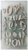
Figure 1
Figure 1
Macroscopy: multiple tissue fragments of white color and soft consistency
Figure 2
Figure 2
(A) Low-power microscopic appearance highlighting the vasculature, diffusely distributed and aggregated inflammatory infiltrates with a lollipop-like lymphoid follicle. (B) High-power microscopic appearance demonstrating bland spindle cells and scattered inflammatory cells
Figure 2
Figure 2
(A) Low-power microscopic appearance highlighting the vasculature, diffusely distributed and aggregated inflammatory infiltrates with a lollipop-like lymphoid follicle. (B) High-power microscopic appearance demonstrating bland spindle cells and scattered inflammatory cells
Figure 3
Figure 3
CD21 (A) and CD35 (B) negative in the spindle cells but highlighting the architecture of the follicles within the lesion. The inflammatory infiltrate showed reactivity for IgG4 (C); however, the number of plasma cells that stained positive for IgG4 was less than 50 per 10 high power fields
Figure 3
Figure 3
CD21 (A) and CD35 (B) negative in the spindle cells but highlighting the architecture of the follicles within the lesion. The inflammatory infiltrate showed reactivity for IgG4 (C); however, the number of plasma cells that stained positive for IgG4 was less than 50 per 10 high power fields

References

    1. Gerald W, Kostianovsky M, Rosai J. Development of vascular neoplasia in Castleman's disease. Am J Surg Pathol. 1990;14:603–14. - PubMed
    1. Zen Y, Nakanuma Y. IgG4-related disease: a cross-sectional study of 114 cases. Am J Surg Pathol. 2010;34:1812–9. - PubMed
    1. Ryu JH, Sekiguchi ES, Yi ES. Pulmonary manifestation of immunoglobulin G4-related sclerosing disease. Eur Respir J. 2012;39:180–6. - PubMed
    1. Rzechorzek NM, Smith C, Schwarz T, et al. Idiopathic sclerosing orbital inflammation mimicking a malignant spindle cell tumor in a dog. Clin Case Rep. 2016;4:935–3. - PMC - PubMed
    1. Kamisawa T, Funata N, Hayashi Y, et al. A new clinicopathological entity of IgG4-related autoimmune disease. J Gastroenterol. 2003;38:982–4. - PubMed

Publication types

LinkOut - more resources