BRCA1 protein expression and subcellular localization in primary breast cancer: Automated digital microscopy analysis of tissue microarrays
- PMID: 28863181
- PMCID: PMC5581176
- DOI: 10.1371/journal.pone.0184385
BRCA1 protein expression and subcellular localization in primary breast cancer: Automated digital microscopy analysis of tissue microarrays
Abstract
Purpose: Mutations in BRCA1 are associated with familial as well as sporadic aggressive subtypes of breast cancer, but less is known about whether BRCA1 expression or subcellular localization contributes to progression in population-based settings.
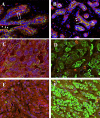
Methods: We examined BRCA1 expression and subcellular localization in invasive breast cancer tissues from an ethnically diverse sample of 286 patients and 36 normal breast tissue controls. Two different methods were used to label breast cancer tissues for BRCA1: (1) Dual immunofluoresent staining with BRCA1 and cytokeratin 8/18 and (2) immunohistochemical staining using the previously validated MS110 mouse monoclonal antibody. Slides were visualized and quantified using the VECTRA Automated Multispectral Image Analysis System and InForm software.
Results: BRCA1 staining was more intense in normal than in invasive breast tissue for both cytoplasmic (p<0.0001) and nuclear (p<0.01) compartments. BRCA1 nuclear to cytoplasmic ratio was higher in breast cancer cells than in normal mammary epithelial cells. Reduced BRCA1 expression was associated with high tumor grade and negative hormone receptors (estrogen receptor, progesterone receptor and Her2). On the other hand, high BRCA1 expression correlated with basal-like tumors (high CK5/6 and EGFR), and high nuclear androgen receptor staining. Lower nuclear to cytoplasmic ratio of BRCA1 correlated significantly with high Ki67 labeling index (p< 0.05) and family history of breast cancer (p = 0.001).
Conclusion: Findings of this study indicate that alterations in BRCA1 protein expression and subcellular localization in breast cancer correlate with poor prognostic markers and aggressive tumor features. Further large-scale studies are required to assess the potential relevance of BRCA1 protein expression and localization in routine classification of breast cancer.
Conflict of interest statement
Figures


Similar articles
-
Expression of BRCA1 protein in breast cancer and its prognostic significance.Hum Pathol. 2008 Jun;39(6):857-65. doi: 10.1016/j.humpath.2007.10.011. Epub 2008 Apr 8. Hum Pathol. 2008. PMID: 18400253
-
BRCA1 expression and molecular alterations in familial breast cancer.Histol Histopathol. 2009 Jan;24(1):69-76. doi: 10.14670/HH-24.69. Histol Histopathol. 2009. PMID: 19012246
-
Loss of nuclear BRCA1 expression in breast cancers is associated with a highly proliferative tumor phenotype.Cancer Genet Cytogenet. 1998 Mar;101(2):109-15. doi: 10.1016/s0165-4608(97)00267-7. Cancer Genet Cytogenet. 1998. PMID: 9494611
-
Promotion of BRCA1-associated triple-negative breast cancer by ovarian hormones.Curr Opin Obstet Gynecol. 2008 Feb;20(1):68-73. doi: 10.1097/GCO.0b013e3282f42237. Curr Opin Obstet Gynecol. 2008. PMID: 18197009 Review.
-
The cell of origin of BRCA1 mutation-associated breast cancer: a cautionary tale of gene expression profiling.J Mammary Gland Biol Neoplasia. 2011 Apr;16(1):51-5. doi: 10.1007/s10911-011-9202-8. Epub 2011 Feb 19. J Mammary Gland Biol Neoplasia. 2011. PMID: 21336547 Review.
Cited by
-
Correlation between BRCA1 expression and the advanced stage of triple‑negative breast cancer.Mol Clin Oncol. 2025 Jan 30;22(4):32. doi: 10.3892/mco.2025.2827. eCollection 2025 Apr. Mol Clin Oncol. 2025. PMID: 39989604 Free PMC article.
-
New, fast and cheap prediction tests for BRCA1 gene mutations identification in clinical samples.Sci Rep. 2023 May 5;13(1):7316. doi: 10.1038/s41598-023-34588-9. Sci Rep. 2023. PMID: 37147448 Free PMC article.
-
Prognostic Value of SGK1 and Bcl-2 in Invasive Breast Cancer.Cancers (Basel). 2023 Jun 11;15(12):3151. doi: 10.3390/cancers15123151. Cancers (Basel). 2023. PMID: 37370761 Free PMC article.
-
High BRCA1 gene expression increases the risk of early distant metastasis in ER+ breast cancers.Sci Rep. 2022 Jan 7;12(1):77. doi: 10.1038/s41598-021-03471-w. Sci Rep. 2022. PMID: 34996912 Free PMC article.
-
Glucose Concentration in Cell Culture Medium Influences the BRCA1-Mediated Regulation of the Lipogenic Action of IGF-I in Breast Cancer Cells.Int J Mol Sci. 2020 Nov 17;21(22):8674. doi: 10.3390/ijms21228674. Int J Mol Sci. 2020. PMID: 33212987 Free PMC article.
References
-
- DeSantis CE, Fedewa SA, Goding Sauer A, Kramer JL, Smith RA, Jemal A. Breast cancer statistics, 2015: Convergence of incidence rates between black and white women. CA Cancer J Clin. 2016;66(1):31–42. doi: 10.3322/caac.21320 . - DOI - PubMed
-
- Stuckey AR, Onstad MA. Hereditary breast cancer: an update on risk assessment and genetic testing in 2015. Am J Obstet Gynecol. 2015;213(2):161–5. doi: 10.1016/j.ajog.2015.03.003 . - DOI - PubMed
-
- Jiang Q, Greenberg RA. Deciphering the BRCA1 Tumor Suppressor Network. J Biol Chem. 2015;290(29):17724–32. doi: 10.1074/jbc.R115.667931 ; PubMed Central PMCID: PMCPMC4505021. - DOI - PMC - PubMed
-
- Chen Y, Chen CF, Riley DJ, Allred DC, Chen PL, Von Hoff D, et al. Aberrant subcellular localization of BRCA1 in breast cancer. Science. 1995;270(5237):789–91. . - PubMed
-
- Goodwin PJ, Phillips KA, West DW, Ennis M, Hopper JL, John EM, et al. Breast cancer prognosis in BRCA1 and BRCA2 mutation carriers: an International Prospective Breast Cancer Family Registry population-based cohort study. J Clin Oncol. 2012;30(1):19–26. doi: 10.1200/JCO.2010.33.0068 . - DOI - PubMed
MeSH terms
Substances
Grants and funding
LinkOut - more resources
Full Text Sources
Other Literature Sources
Medical
Research Materials
Miscellaneous

