. 2017 Jul 1;76(7):595-604.
doi: 10.1093/jnen/nlx040.

Characterization of 2 Novel Ependymoma Cell Lines With Chromosome 1q Gain Derived From Posterior Fossa Tumors of Childhood

Affiliations

Characterization of 2 Novel Ependymoma Cell Lines With Chromosome 1q Gain Derived From Posterior Fossa Tumors of Childhood

Vladimir Amani et al. J Neuropathol Exp Neurol. .

Abstract

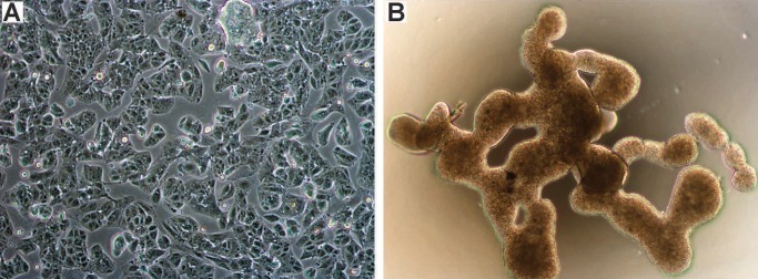
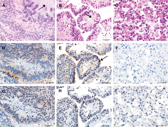
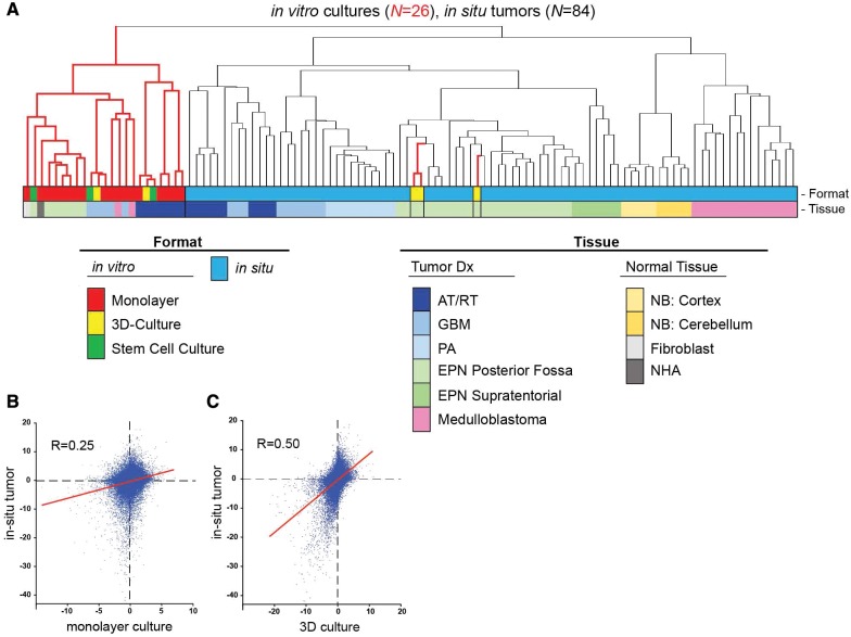
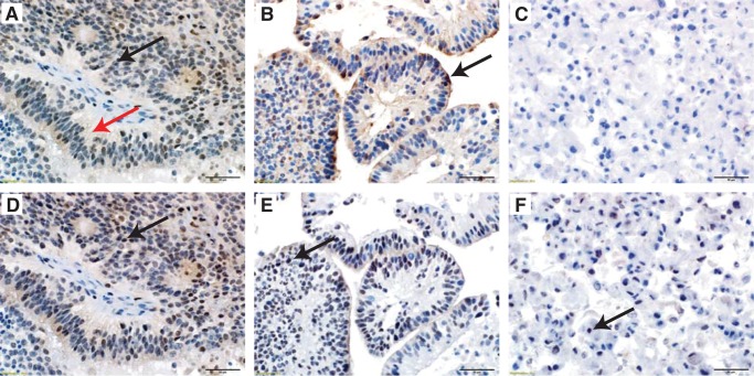
Ependymoma (EPN) is a common brain tumor of childhood that, despite standard surgery and radiation therapy, has a relapse rate of 50%. Clinical trials have been unsuccessful in improving outcome by addition of chemotherapy, and identification of novel therapeutics has been hampered by a lack of in vitro and in vivo models. We describe 2 unique EPN cell lines (811 and 928) derived from recurrent intracranial metastases. Both cell lines harbor the high-risk chromosome 1q gain (1q+) and a derivative chromosome 6, and both are classified as molecular group A according to transcriptomic analysis. Transcriptional enrichment of extracellular matrix-related genes was a common signature of corresponding primary tumors and cell lines in both monolayer and 3D formats. EPN cell lines, when cultured in 3D format, clustered closer to the primary tumors with better fidelity of EPN-specific transcripts than when grown as a monolayer. Additionally, 3D culture revealed ependymal rosette formation and cilia-related ontologies, similar to in situ tumors. Our data confirm the validity of the 811 and 928 cell lines as representative models of intracranial, posterior fossa 1q+ EPN, which holds potential to advance translational science for patients affected by this tumor.

Keywords: Cell line; Chromosome 1q gain; Ependymoma.


Figures

FIGURE 1
FIGURE 1
Morphology of EPN cell lines grown in monolayer and 3D culture formats. Phase contrast microscopy images showing appearance of cell line 811 grown in (A) monolayer (unstained, 100×), and (B) 3D culture (unstained, 40×).
FIGURE 2
FIGURE 2
Histological comparison of EPN in vitro cell lines with in situ tumor of origin. (A) Representative histological images of 811 in situ patient tumor revealing classic features of EPN including syncytial networks of monomorphic cells with round to oval nuclei, speckled chromatin, and perivascular pseudorosettes. The inset magnification highlights fibrillary material (arrow) surrounding the vascular spaces (H&E). (B) 811 3D culture in vitro format derived from the same tumor demonstrates similar appearing cells with regularly arranged monomorphic nuclei with the retention of the syncytial networks but with a pseudopapillary arrangement and centrally placed fibrillary material (arrow) and no vasculature (H&E). (C) 811 cells grown in monolayer format show complete loss of architecture, syncytial networks, and the fibrillary material with discohesive cells and well-defined cell borders (H&E). (D) EMA immunoreactivity in 811 in situ tumor demonstrating a blush of dot like cytoplasmic staining that was conserved in (E) 811 3D culture format. (F) By contrast, 811 grown in monolayer format showed irregularly increased cytoplasmic positivity when compared to the in situ section (EMA). (G) Immunoreactivity for GFAP was observed in fibrillar material and perivascular nuclear-free zones of 811 in situ tumor. (H) 3D format demonstrated accentuated GFAP expression within the fibrillar material (I) that was almost completely lost in the monolayer culture (GFAP). (Panels A–I: 400× magnification, scale bar=50 µm).
FIGURE 3
FIGURE 3
Fidelity of EPN cells cultured in 3D format with EPN in situ tumor. (A) Unsupervised clustering of transcriptomic profiles of 84 in situ tumor and 26 in vitro cultures. Dendrogram branch colors correspond to in vitro (red) and in situ (black) samples. Dot plots portraying transcriptome signature correlation of (B) monolayer, and (C) EPN 3D culture, with EPN in situ tumor. Axes are log10 of p values of expression differences for all transcriptomic probe sets (n=54 675) between EPN and nonEPN samples that have been assigned directionality (negative=overexpressed in EPN; positive=under expressed in EPN). Linear regression lines are shown in red.
FIGURE 4
FIGURE 4
Conservation of EPN-specific cilia-associated proteins, calcyphosine and C9orf24, in EPN in situ tumor and 3D culture format. Representative images of (A) section of 811 in situ tumor with cytoplasmic (black arrow) and fibrillary material (red arrow) immunoreactivity for calcyphosine. (B) 811 3D culture suspension retains the calcyphosine immunoreactivity within the cytoplasm and fibrillary material with an additional accentuation at the outer surface of the pseudopapillary structures (arrow). (C) 811 monolayer culture demonstrates complete loss of calcyphosine expression. (D) Serial step section of the same in situ 811 tumor with nuclear immunoreactivity for C9orf24 (arrow). (E) 811 3D culture suspension retains the C9orf24 nuclear immunoreactivity (arrow). (F) 811 monolayer culture demonstrates partial loss of C9orf24 immunoreactivity (arrow). All images: hematoxylin counterstain, 400× magnification, scale bars=50 μm.

References

    1. Gajjar A, Packer RJ, Foreman NK, et al.Children’s Oncology Group’s 2013 blueprint for research: central nervous system tumors. Pediatr Blood Cancer 2013;60:1022–6 - PMC - PubMed
    1. Korshunov A, Witt H, Hielscher T, et al.Molecular staging of intracranial ependymoma in children and adults. J Clin Oncol 2010;28:3182–90 - PubMed
    1. Kilday JP, Rahman R, Dyer S, et al.Pediatric ependymoma: biological perspectives. Mol Cancer Res 2009;7:765–86 - PubMed
    1. Parker M, Mohankumar KM, Punchihewa C, et al.C11orf95-RELA fusions drive oncogenic NF-kappaB signalling in ependymoma. Nature 2014;506:451–5 - PMC - PubMed
    1. Hoffman LM, Donson AM, Nakachi I, et al.Molecular sub-group-specific immunophenotypic changes are associated with outcome in recurrent posterior fossa ependymoma. Acta Neuropathol 2014;127:731–45 - PMC - PubMed

MeSH terms