

Review
doi: 10.1016/j.chest.2017.08.013.
Epub 2017 Aug 31.
Tracheobronchopathy From Inhaled Corticosteroids
Affiliations
- PMID: 28864055
- PMCID: PMC6026226
- DOI: 10.1016/j.chest.2017.08.013
Item in Clipboard
Review
Tracheobronchopathy From Inhaled Corticosteroids
Chest.
2017 Dec.
Erratum in
-
Errors in Table 1 in: Tracheobronchopathy From Inhaled Corticosteroids.Chest. 2019 Jan;155(1):246. doi: 10.1016/j.chest.2018.11.012. Chest. 2019. PMID: 30616735 Free PMC article. No abstract available.
Abstract
Inhaled corticosteroids (ICSs) have become the mainstay of asthma control. They are also recommended as an add-on therapy to long-acting beta agonists and anticholinergics in moderate to severe COPD with recurrent exacerbations. Ultimately this clinical practice has led to the widespread use of ICSs, which are supported by a more favorable side effect profile than that of systemic steroids.
Keywords: excessive dynamic airway collapse; inhaled corticosteroids; tracheobronchomalacia; tracheomalacia.
Copyright © 2017 American College of Chest Physicians. Published by Elsevier Inc. All rights reserved.
Figures

Effects of inhaled corticosteroids on airway vasculature and supporting structure.
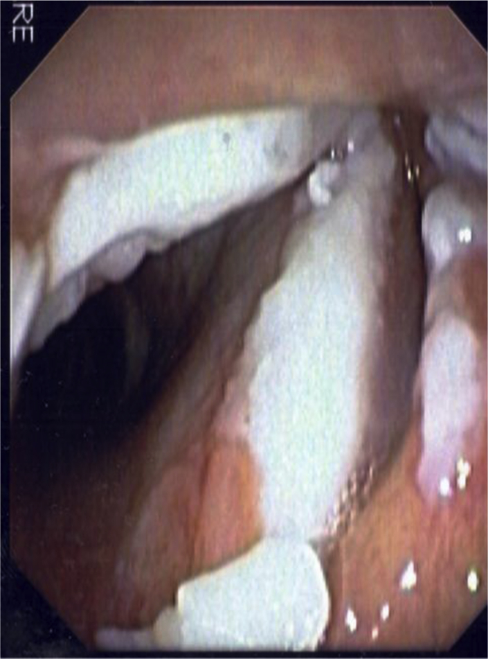
Vocal cord candidiasis in an immunocompromised patient taking inhaled corticosteroids.

Inspiration and expiration CT scans of the chest, revealing excessive dynamic collapse of airways. Note compromised tracheal lumen from the lax posterior wall, whereas the integrity of the cartilage is well maintained.

Flexible bronchoscopy revealing excessive dynamic collapse of airways. Note compromised tracheal lumen from the lax posterior wall, whereas the integrity of the cartilage is well maintained. Courtesy of Dr Tim Murgu.

Tracheomalacia. Note the loss of integrity of the tracheal wall cartilages. Courtesy of Dr Tim Murgu.

CT scan of the chest revealing tracheomalacia. Note loss of integrity of the tracheal wall cartilage. Courtesy of Dr Tim Murgu.

Tracheobronchial smooth muscle atrophy and separation. Note separation of the medial end of the cartilages of the right main bronchus (arrow) from the posterior wall.

Histologic examination of the airway wall from the explanted lung, revealing total absence of airway smooth muscle in a patient with tracheobronchial smooth muscle atrophy and separation (H&E). C = cartilage; CT = connective tissue; G = mucosal glands).
References
-
- Centers for Medicare and Medicaid Services, Medicare Drug Spending Dashboard 2015. Office of Enterprise Data & Analytics (OEDA), 2016. https://www.cms.gov/Research-Statistics-Data-and-Systems/Statistics-Tren.... Accessed July 19, 2017.
-
- National Asthma, Education, and Prevention Program, Expert Panel Report 3 (EPR-3): Guidelines for the diagnosis and management of asthma—summary report 2007. J Allergy Clin Immunol. 2007;120(5 suppl):S94–S138. - PubMed
-
- Sharafkhaneh A., Southard J.G., Goldman M., Uryniak T., Martin U.J. Effect of budesonide/formoterol pMDI on COPD exacerbations: a double-blind, randomized study. Respir Med. 2012;106(2):257–268. - PubMed
Publication types
MeSH terms
Substances
Grants and funding
LinkOut - more resources
Full Text Sources
Other Literature Sources
Medical
Molecular Biology Databases
