Review
. 2017 Sep:60:1-19.
doi: 10.1016/j.preteyeres.2017.08.003. Epub 2017 Aug 31.

Age-related cataracts: Role of unfolded protein response, Ca2+ mobilization, epigenetic DNA modifications, and loss of Nrf2/Keap1 dependent cytoprotection

Affiliations
Review

Age-related cataracts: Role of unfolded protein response, Ca2+ mobilization, epigenetic DNA modifications, and loss of Nrf2/Keap1 dependent cytoprotection

Palsamy Periyasamy et al. Prog Retin Eye Res. 2017 Sep.

Abstract

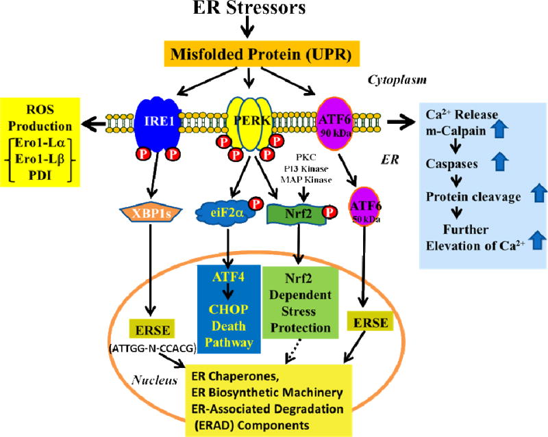
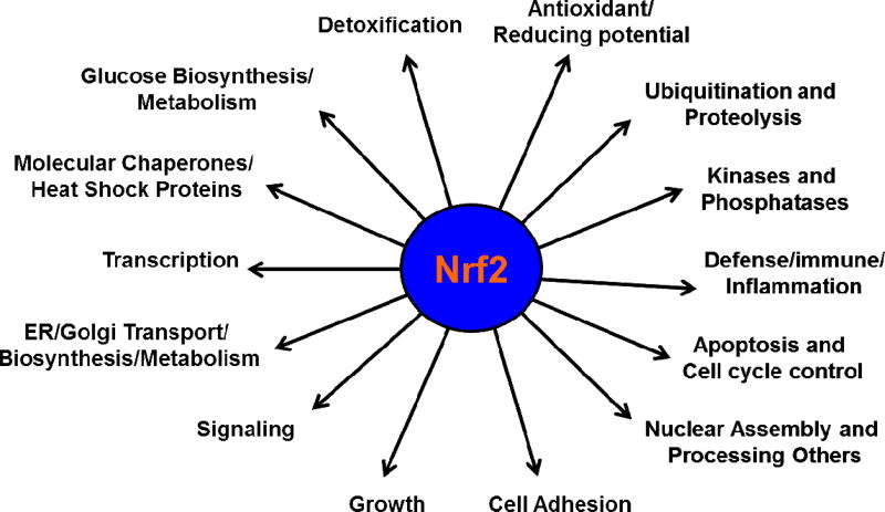
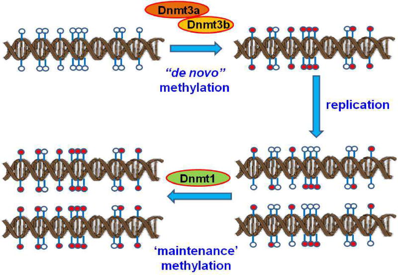
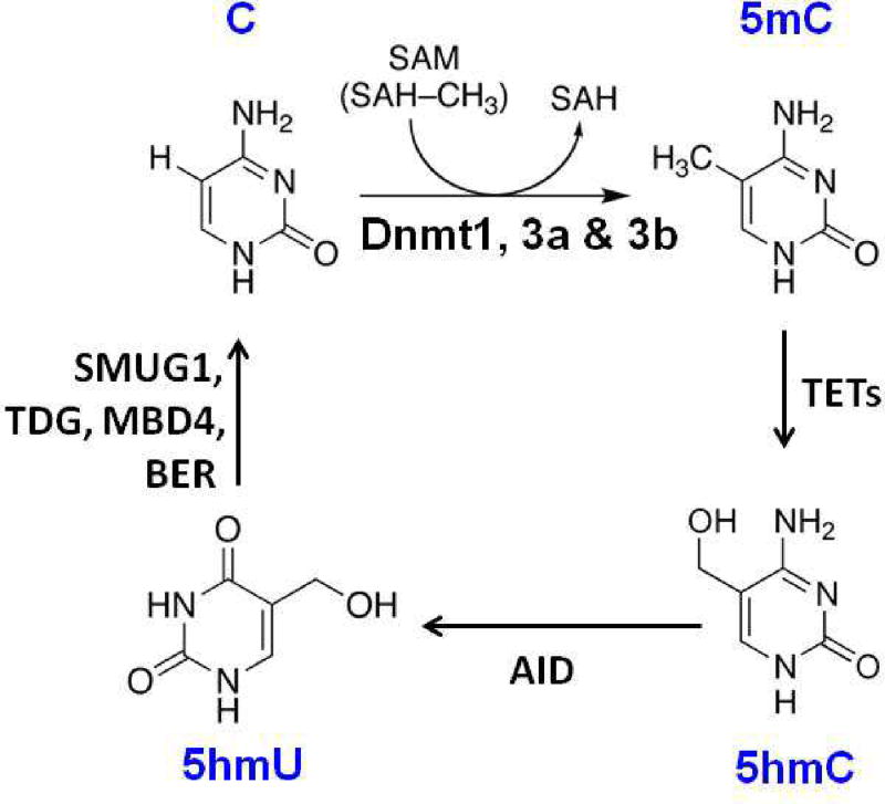
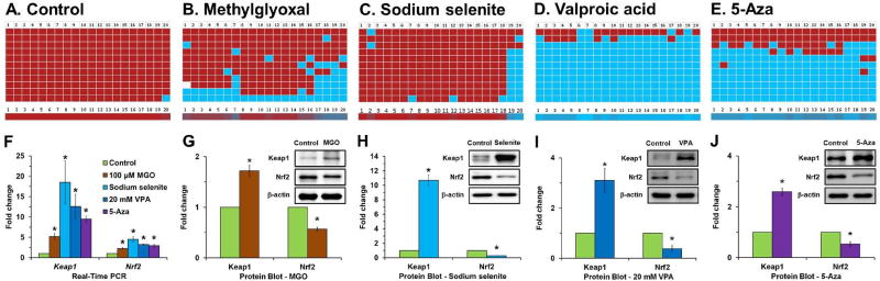
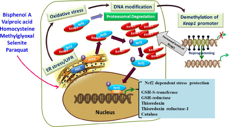
Age-related cataracts are closely associated with lens chronological aging, oxidation, calcium imbalance, hydration and crystallin modifications. Accumulating evidence indicates that misfolded proteins are generated in the endoplasmic reticulum (ER) by most cataractogenic stresses. To eliminate misfolded proteins from cells before they can induce senescence, the cells activate a clean-up machinery called the ER stress/unfolded protein response (UPR). The UPR also activates the nuclear factor-erythroid-2-related factor 2 (Nrf2), a central transcriptional factor for cytoprotection against stress. Nrf2 activates nearly 600 cytoprotective target genes. However, if ER stress reaches critically high levels, the UPR activates destructive outputs to trigger programmed cell death. The UPR activates mobilization of ER-Ca2+ to the cytoplasm and results in activation of Ca2+-dependent proteases to cleave various enzymes and proteins which cause the loss of normal lens function. The UPR also enhances the overproduction of reactive oxygen species (ROS), which damage lens constituents and induce failure of the Nrf2 dependent cytoprotection. Kelch-like ECH-associated protein 1 (Keap1) is an oxygen sensor protein and regulates the levels of Nrf2 by the proteasomal degradation. A significant loss of DNA methylation in diabetic cataracts was found in the Keap1 promoter, which overexpresses the Keap1 protein. Overexpressed Keap1 significantly decreases the levels of Nrf2. Lower levels of Nrf2 induces loss of the redox balance toward to oxidative stress thereby leading to failure of lens cytoprotection. Here, this review summarizes the overall view of ER stress, increases in Ca2+ levels, protein cleavage, and loss of the well-established stress protection in somatic lens cells.

Keywords: 2′,7′-dichlorodihydrofluorescein diacetate; 5-Aza; 5-aza-2′-deoxycytidine; 5-methylcytosine; 5mC; AID; ARCs; ARE; ATF6; Age-related cataracts; BiP; DNA methylation; DNA methyltransferases; Dnmts; ER; ER oxidoreductin 1; ER stress; ER-associated degradation; ERAD; Ero1; GSH; H2DCFDA; HIF-1; IRE1; Ig binding protein; Keap1; Kelch-like ECH-associated protein 1; NF-κB; Nrf2; PDI; PERK; PKC; PMCA; ROS; SERCA; TDG; TET1; UPR; Unfolded protein response; X-box transcription factor-1; XBP-1; activating transcription factor 6; activation-induced cytidine deaminase; age-related cataracts; antioxidant response element; eIF2α; endoplasmic reticulum; eukaryotic translation initiation factor 2α; glutathione; hypoxia-inducible factor-1; inositol-requiring kinase 1; nuclear factor-erythroid-2-related factor 2; nuclear factor-κB; pVHL; plasma membrane Ca(2+)-ATPases; protein disulfide isomerase; protein kinase C; protein kinase R (PKR)-like endoplasmic reticulum kinase; reactive oxygen species; sarco/endoplasmic reticulum Ca(2+)-ATPases; ten-eleven translocation 1; thymine-DNA glycosylase; unfolded protein response; von Hippel-Lindau.


Figures

Fig. 1
Fig. 1
Schematic diagram for molecular mechanisms of activation of ER stress. Accumulation of misfolded protein triggers the phosphorylation of PERK and IRE1 and cleavage of ATF6 to activate the UPR. This rapidly reduces the misfolded protein load through reversible translational attenuation and transcriptional induction of ER resident proteins to further assist protein folding and through induction of chaperones, ER biosynthetic machinery, and ER-associated degradation (ERAD) components. Chronic ER stress induces the apoptotic UPR which generates excessive levels of ROS, through the involvement of Ero1-Lα and-Lβ and PDI. Furthermore, the UPR releases Ca2+ from the ER to activate m-calpain and caspases. P-PERK phosphorylates eIF2α which further activates ATF4 and the CHOP-death pathway. P-PERK, PKC, PI3 kinase and MAP kinase phosphorylate Nrf2; the P-Nrf2 dissociates from Keap1 and translocates into the nucleus to bind to the antioxidant response element (ARE), and this activates the transcription of more than 200 stress/antioxidant enzymatic genes such as glutathione, glutathione reductase, thioredoxin, thioredoxin-S-transferase, and catalase (see Fig. 2). These stress/antioxidant enzymes regenerate reduced glutathione, and the resultant reduced glutathione eliminates ROS so the cell can survive and recover from the stress. Chronic UPR activates the death pathway and results in senescence or death.
Fig. 2
Fig. 2
The Nrf2 target genes. The transcriptional factor, Nrf2 binds to the ARE site in the promoter of cytoprotective genes, and activates (>2.0 fold) or suppresses (<1/2 fold) more than 200–600 target genes in non-lens tissues.
Fig. 3
Fig. 3
Passive DNA demethylation pathway. Passive DNA demethylation is caused by a reduction in activity or absence of Dnmts. Dnmt3a and 3b are responsible for de novo DNA methylation of parent DNA. When methylated DNA is replicated, the daughter strands of DNA are unmethylated. This hemimethylated DNA is recognized by the maintenance DNA methyltransferase1 (Dnmt1) and restores parental DNA methylation patterns through successive rounds of cell division. If Dnmt1 is inhibited or absent when the cell divides, the newly synthesized strand of DNA will not be methylated, and successive rounds of cell division will result in passive demethylation. In this figure, unmethylated CpGs are shown by empty circles, and methylated CpGs are indicated by red circles.
Fig. 4
Fig. 4
Active DNA demethylation pathway. Active DNA demethylation takes place through the enzymatic replacement of 5mC with cytosine via the base-excision DNA repair pathway (BER) pathway. Cytosine is methylated by Dnmt1, 3a, and 3b and the resulting 5mC is oxidized to 5-hydroxymethylcytosine (5hmC) by TET1 proteins. 5hmC is then deaminated by AID into 5-hydroxy uracil (5hmU). Finally, 5hmU can be excised by TDG and repaired by the BER pathway with unmethylated cytosines. Active DNA demethylation pathway is induced in the non-replicating cells.
Fig. 5
Fig. 5
Loss of Keap1 promoter DNA methylation in human lens epithelial cells treated with methylglyoxal, sodium selenite, valproic acid (VPA) and 5-Aza-2'-deoxycytidine (5- Aza). Control human lens epithelial cells do not show any loss of DNA methylation of the Keap1 promoter (A). However, significant loss of DNA methylation of the Keap1 promoter is found in human lens epithelial cells treated with either 100 µM methylglyoxal for 1 day (B), 1 µM sodium selenite for 1 day (C), 20 mM of VPA for 5 days (D) and/or 10 µM 5-Aza for 7 days (E). Ten individual clones of the bisulfite-converted DNA sequences of human lens epithelial cells were analyzed for DNA methylation in fragment-1 (contains 20 CpG dinucleotides) of the Keap1 promoter using BISMA software with default filtering threshold settings (http://biochem.jacobs-university.de/BDPC/BISMA/). Each row of red squares represents methylated CpG dinucleotides. Blue squares represent unmethylated CpG dinucleotides, and white squares represent an undetermined CpG status. The color gradient bar shows that the red region contains more methylated CpG dinucleotides, and the blue region contains more unmethylated CpG dinucleotides. Fold changes in the mRNA expression levels of Keap1 and Nrf2 in control human lens epithelial cells and treated with methylglyoxal, sodium selenite, and 5-Aza (F). The corresponding fold variations in the protein levels of Keap1 and Nrf2 in human lens epithelial cells treated with methylglyoxal (G), sodium selenite (H), VPA (I) and 5-Aza (J).
Fig. 6
Fig. 6
A schematic diagram showing the involvement of various cataractogenic stressors mediating ER stress mediated Nrf2 dependent antioxidant system failure via Keap1 promoter DNA demethylation.

References

    1. Abraham MA, Korula A, Jayakrishnan K, John GT, Thomas PP, Jacob CK. Prognostic factors in diffuse proliferative lupus nephritis. J Assoc Physicians India. 1999;47:862–865. - PubMed
    1. Alam J, Stewart D, Touchard C, Boinapally S, Choi AM, Cook JL. Nrf2, a Cap'n'Collar transcription factor, regulates induction of the heme oxygenase-1 gene. J Biol Chem. 1999;274:26071–26078. - PubMed
    1. Aldana JP, Marcovich R, Singhal P, Reddy K, Morgenstern N, El-Hakim A, Smith AD, Lee BR. Immune response to laparoscopic bowel injury. J Endourol. 2003;17:317–322. - PubMed
    1. Allan GF, Leng X, Tsai SY, Weigel NL, Edwards DP, Tsai MJ, O'Malley BW. Hormone and antihormone induce distinct conformational changes which are central to steroid receptor activation. J Biol Chem. 1992;267:19513–19520. - PubMed
    1. Alves MS, Reis PA, Dadalto SP, Faria JA, Fontes EP, Fietto LG. A novel transcription factor, ERD15 (Early Responsive to Dehydration 15), connects endoplasmic reticulum stress with an osmotic stress-induced cell death signal. J Biol Chem. 2011;286:20020–20030. - PMC - PubMed

Publication types

MeSH terms