Review
. 2017 Sep 1;9(9):a022046.
doi: 10.1101/cshperspect.a022046.

Intermediate Filaments and the Regulation of Cell Motility during Regeneration and Wound Healing

Affiliations
Review

Intermediate Filaments and the Regulation of Cell Motility during Regeneration and Wound Healing

Fang Cheng et al. Cold Spring Harb Perspect Biol. .

Abstract

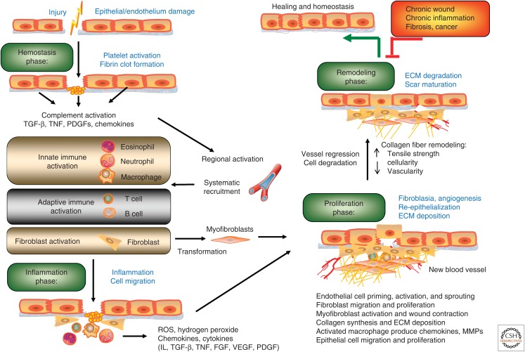
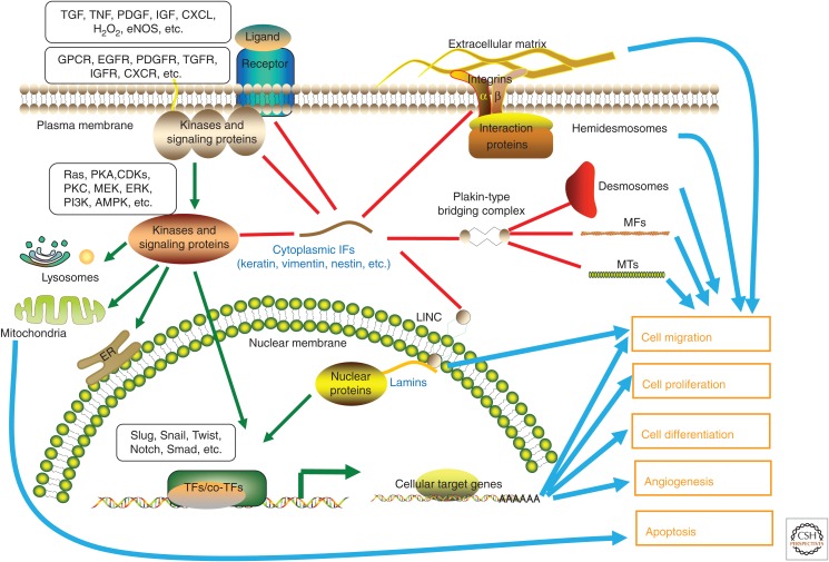
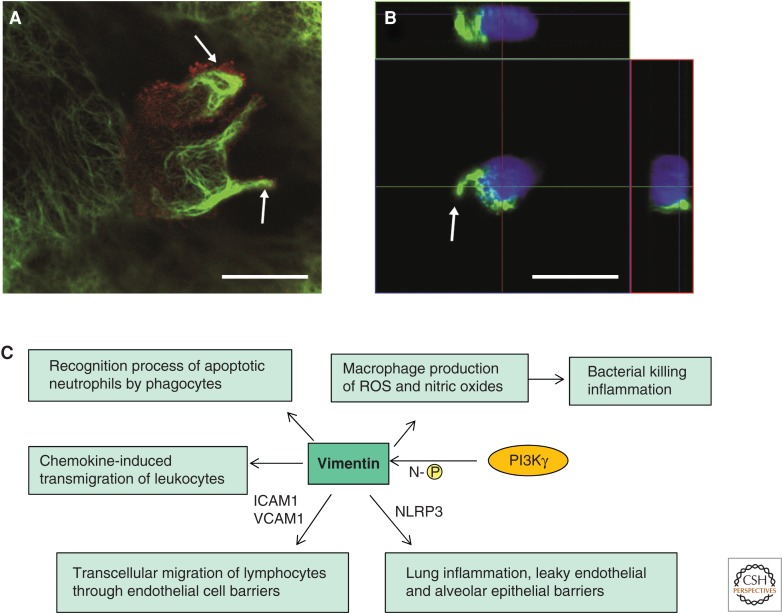
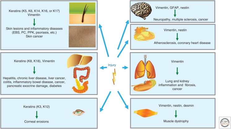
SUMMARYIntermediate filaments (IFs) comprise a diverse group of flexible cytoskeletal structures, the assembly, dynamics, and functions of which are regulated by posttranslational modifications. Characteristically, the expression of IF proteins is specific for tissues, differentiation stages, cell types, and functional contexts. Recent research has rapidly expanded the knowledge of IF protein functions. From being regarded as primarily structural proteins, it is now well established that IFs act as powerful modulators of cell motility and migration, playing crucial roles in wound healing and tissue regeneration, as well as inflammatory and immune responses. Although many of these IF-associated functions are essential for tissue repair, the involvement of IF proteins has been established in many additional facets of tissue healing and regeneration. Here, we review the recent progress in understanding the multiple functions of cytoplasmic IFs that relate to cell motility in the context of wound healing, taking examples from studies on keratin, vimentin, and nestin. Wound healing and regeneration include orchestration of a broad range of cellular processes, including regulation of cell attachment and migration, proliferation, differentiation, immune responses, angiogenesis, and remodeling of the extracellular matrix. In this respect, IF proteins now emerge as multifactorial and tissue-specific integrators of tissue regeneration, thereby acting as essential guardian biopolymers at the interface between health and disease, the failing of which contributes to a diverse range of pathologies.


Figures

Figure 1.
Figure 1.
Characteristic stages of wound healing. Epithelial wound repair starts (1) from clot formation (hemostasis phase), followed by (2) an inflammation phase, (3) a proliferation phase, and (4) a tissue-remodeling phase. Proper wound repair requires a close coordination of different cell components such that they are at the right places at the right times. Any significant delay of this self-limiting process is likely to result in pathogenesis. ECM, extracellular matrix; FGF, fibroblast growth factor; IL, interleukin; MMP, matrix metalloprotease; PDGF, platelet-derived growth factor; ROS, reactive oxygen species; TGF-β, transforming growth factor β; TNF, tumor necrosis factor; VEGF, vascular endothelial growth factor.
Figure 2.
Figure 2.
The roles of intermediate filaments (IFs) in cell motility and cell-fate decision-making during wound repair. This overview shows some of the separate and shared functions of cytoplasmic IF proteins (keratin, vimentin, and nestin) in the organization and regulation of signal transduction in a standardized cell. These IFs are also coupled to microfilaments (MFs), microtubules (MTs), IF-anchoring plaques of cell–cell junctions (desmosomes), and cell–matrix junctions (hemidesmosomes) by the plakin-type protein complexes. Focal adhesions are formed through IF complexes and integrins at the base to the extracellular matrix (ECM). Cytoskeletal filaments are, furthermore, coupled to both inner and outer nuclear membranes and lamins by the linker of nucleoskeleton and cytoskeleton (LINC) complex. During wound repair, the alterations in IF homeostasis and dynamics help to promote the correct cellular response at any given time, determining when cells should change their motility to migrate, divide, differentiate, or die. AMPK, AMP-activated protein kinase; CDK, cyclin-dependent kinase; CXCL, C-X-C chemokine ligand; CXCR, C-X-C chemokine receptor; EGFR, epidermal growth factor receptor; eNOS, endothelial nitric oxide synthase; ERK, extracellular signal-regulated kinase; GPCR, G-protein-coupled receptor; H2O2, hydrogen peroxide; IGF, insulin-like growth factor; IGFR, insulin-like growth factor receptor; MEK, MAP kinase kinase; PDGF, platelet-derived growth factor; PDGFR, platelet-derived growth factor receptor; PI3K, phosphoinositide 3-kinase; PKA, protein kinase A; PKC, protein kinase C; TF, transcription factor; TGF, transforming growth factor; TGFR, transforming growth factor receptor; TNF, tumor necrosis factor.
Figure 3.
Figure 3.
Vimentin intermediate filaments (IFs) function in leukocyte transcellular migration. (A) Reorganization of vimentin IFs (green) of a migrating peripheral blood mononuclear cell (PBMC) toward the uropods (white arrows) of two migrating PBMCs. The staining by antibody against CD44 (red), a membrane marker for the PBMC, outlines the borders of the PBMC. The focal plane is at the upper level of the endothelial cell (EC), so only parts of the EC vimentin IFs can be seen around the migrating cell. Scale bar, 10 μm. (B) A similar polarization and reorganization of vimentin IFs (green) also occur when the cells are incubated for 30 min on gelatin-coated coverslips. z-axis side views are shown from two different planes (colored lines on the top and on the right-hand side of the large image show the maximum projection). Scale bar, 10 μm. (C) Vimentin IFs have the ability to bind and modulate the activity of signaling proteins, thereby influencing inflammatory signals in response to injury. The figure summarizes the key vimentin-controlled interactions and signaling pathways that influence innate immunity and inflammation. Vimentin itself can be phosphorylated by phosphoinositide 3-kinase γ (PI3Kγ) at its amino terminus (N-[P]). ICAM1, intercellular adhesion molecule 1; NLRP3, NLRP3 inflammasome; ROS, reactive oxygen species; VCAM1, vascular cell-adhesion molecule 1. (A,B, Reprinted, with permission, from Nieminen et al. 2006.)
Figure 4.
Figure 4.
Involvement of intermediate filament (IF) expression in different tissues on injury and relevant associated disease. IF proteins can be expressed in different compositions and combinations depending on the cell type, differentiation state, and functional settings. The broad and complex distribution of IFs regulates cellular activities in different organs on specific physiological stimuli, especially during tissue repair. The mutation of genes encoding IFs, or disturbance of IF functions, can ultimately lead to tissue-wide pathologic alterations, ranging from skin lesions to cancer of various kinds. EBS, epidermolysis bullosa simplex; GFAP, glial fibrillary acidic protein; K, keratin; PC, pachyonychia congenital; PPK, palmoplantar keratoderma.

References

    1. Alam H, Kundu ST, Dalal SN, Vaidya MM. 2011a. Loss of keratins 8 and 18 leads to alterations in α6β4-integrin-mediated signalling and decreased neoplastic progression in an oral-tumour-derived cell line. J Cell Sci 124: 2096–2106. - PubMed
    1. Alam H, Sehgal L, Kundu ST, Dalal SN, Vaidya MM. 2011b. Novel function of keratins 5 and 14 in proliferation and differentiation of stratified epithelial cells. Mol Biol Cell 22: 4068–4078. - PMC - PubMed
    1. Arwert EN, Hoste E, Watt FM. 2012. Epithelial stem cells, wound healing and cancer. Nat Rev Cancer 12: 170–180. - PubMed
    1. Barberis L, Pasquali C, Bertschy-Meier D, Cuccurullo A, Costa C, Ambrogi C, Vilbois F, Chiarle R, Wymann M, Altruda F, et al. 2009. Leukocyte transmigration is modulated by chemokine-mediated PI3K γ-dependent phosphorylation of vimentin. Eur J Immunol 39: 1136–1146. - PubMed
    1. Baribault H, Wilson-Heiner M, Muller W, Penner J, Bakhiet N. 1997. Functional analysis of mouse keratin 8 in polyoma middle T-induced mammary gland tumours. Transgenic Res 6: 359–367. - PubMed

LinkOut - more resources