. 2018 Jan;32(1):130-142.
doi: 10.1096/fj.201601142R. Epub 2017 Sep 1.

A novel role for ceramide synthase 6 in mouse and human alcoholic steatosis

Affiliations

A novel role for ceramide synthase 6 in mouse and human alcoholic steatosis

Bianca Williams et al. FASEB J. 2018 Jan.

Abstract

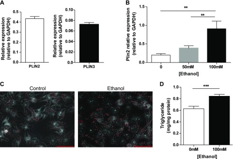
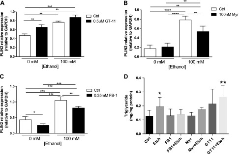
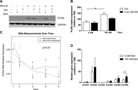
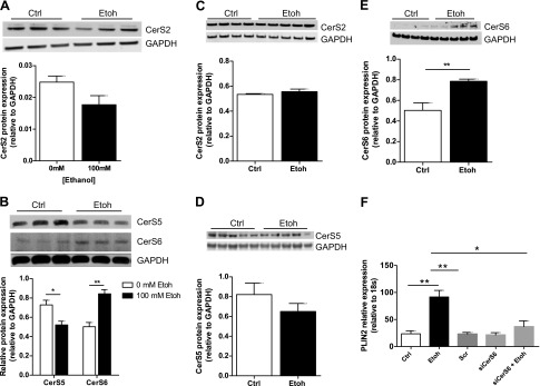
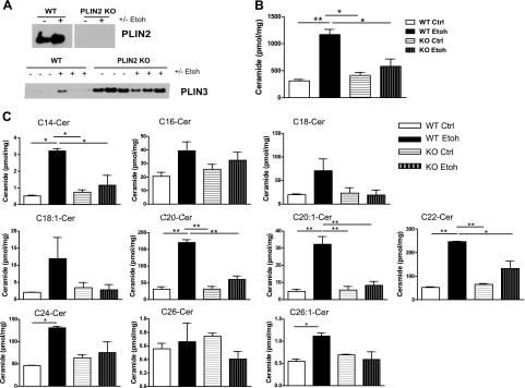
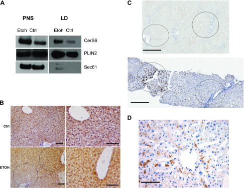
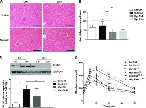
Perilipin 2 (PLIN2) is a lipid-droplet protein that is up-regulated in alcoholic steatosis and associated with hepatic accumulation of ceramides, bioactive lipids implicated in alcoholic liver disease pathogenesis. The specific role of ceramide synthetic enzymes in the regulation of PLIN2 and promotion of hepatocellular lipid accumulation is not well understood. We examined the effects of pharmacologic ceramide synthesis inhibition on hepatic PLIN2 expression, steatosis, and glucose and lipid homeostasis in mice with alcoholic steatosis and in ethanol-incubated human hepatoma VL17A cells. In cells, pharmacologic inhibition of ceramide synthase reduced lipid accumulation by reducing PLIN2 RNA stability. The subtype ceramide synthase (CerS)6 was specifically up-regulated in experimental alcoholic steatosis in vivo and in vitro and was up-regulated in zone 3 hepatocytes in human alcoholic steatosis. In vivo ceramide reduction by inhibition of de novo ceramide synthesis reduced PLIN2 and hepatic steatosis in alcohol-fed mice, but only de novo synthesis inhibition, not sphingomyelin hydrolysis, improved glucose tolerance and dyslipidemia. These findings implicate CerS6 as a novel regulator of PLIN2 and suggest that ceramide synthetic enzymes may promote the earliest stage of alcoholic liver disease, alcoholic steatosis.-Williams, B., Correnti, J., Oranu, A., Lin, A., Scott, V., Annoh, M., Beck, J., Furth, E., Mitchell, V., Senkal, C. E., Obeid, L., Carr, R. M. A novel role for ceramide synthase 6 in mouse and human alcoholic steatosis.

Keywords: CerS6; PLIN2; alcohol; glucose tolerance; lipid droplet; liver.


Figures

Figure 1.
Figure 1.
VL17A cells, an in vitro model of alcoholic steatosis. A) VL17A cells PLIN2 and PLIN3 gene expression at baseline (24 h of plating). B) PLIN2 gene expression in response to 0, 50, or 100 mM Etoh incubation. C) Photomicrograph of Oil-Red-O staining of VL17A cells incubated in Ctrl or Etoh medium (40 times). Scale bars, 100 µm. D) Cellular triglyceride content in response to Ctrl or Etoh medium. **P < 0.01, ***P < 0.0001.
Figure 2.
Figure 2.
FB1 ceramide synthase inhibition reduces PLIN2 in Ctrl and Etoh cells. A–C) Densitometry quantification of PLIN2 protein expression relative to GAPDH in Ctrl and Etoh cells incubated with GT-11 (A), Myr (B), or FB1 (C). D) Triglyceride content after treatment with ceramide synthetic inhibitors. *P ≤ 0.05, **P ≤ 0.01, ***P ≤ 0.001, ****P ≤ 0.0001.
Figure 3.
Figure 3.
Transcriptional and posttranslational regulation of PLIN2 by FB1. A) Immunoblot of PLIN2 protein isolated from VL17A cells incubated with Ctrl or Etoh medium, with or without FB1, and with or without the proteasomal inhibitor MG132. B) PLIN2 mRNA level relative to 18S in Ctrl and Etoh cells incubated with FB1. C) PLIN2 degradation. Data are presented as PLIN2 mRNA level relative to 18S in Etoh cells incubated with or without FB1 and actinomycin over time. D) CerS1–6 gene expression relative to 18S in VL17A cells incubated 48 h in Ctrl or Etoh medium. *P ≤ 0.05, **P ≤ 0.01.
Figure 4.
Figure 4.
CerS6 reduction ameliorates Etoh-induced PLIN2 up-regulation in VL17A cells. A, B) Immunoblot and densitometry quantification of CerS2 (A) and CerS5 and CerS6 (B) protein in VL17A cells. CE) Immunoblot and densitometry quantification of CerS2 (C), CerS5 (D), and CerS6 (E) protein in mouse liver. F) PLIN2 gene expression relative to 18S after siRNA knockdown of CerS6 in VL17A cells incubated in Ctrl or Etoh medium. *P ≤ 0.05, **P ≤ 0.01.
Figure 5.
Figure 5.
Absence of PLIN2 prevents Etoh-induced, long-chain ceramide up-regulation in LDs. A) PLIN2 and PLIN3 immunoblots of LDs isolated from wild-type (WT) or PLIN2-null mice fed Ctrl or Etoh diets for 4 wk. B, C) Mass spectrometric analysis of total long-chain ceramides (B) and individual ceramide species (C) in LDs isolated from WT and PLIN2-null mice fed Ctrl or Etoh diets; n = 5 mice/group. KO, knockout. *P ≤ 0.05, **P ≤ 0.01.
Figure 6.
Figure 6.
CerS6 localizes to the LD fraction of VL17A cells and is up-regulated in zone 3 hepatocytes in murine and human alcoholic steatosis. A) Sucrose gradient fractionation immunoblots of VL17A LD and PNS fractions incubated with Ctrl or Etoh medium. B) CerS6 IHC of Ctrl and Etoh-fed wild-type mouse livers. Original magnification, ×20. Scale bars, 100 μm. C) CerS6 IHC in nonsteatotic human liver (upper) and human alcoholic steatosis (lower) showing hepatic zones 1–3. Scale bars, 500 μm. D) CerS6 IHC of zone 3 in a patient with mild alcoholic steatosis. Original magnification, ×40. Scale bar, 100 μm. Dashed circle, zone 1; solid circle, zone 3. Sec61, endoplasmic reticulum membrane protein.
Figure 7.
Figure 7.
Myr reduces hepatic steatosis and PLIN2 and improves glucose tolerance in alcohol-fed mice. A) Hematoxylin and eosin stain of liver sections from Ctrl or alcohol-fed wild-type mice injected with Sal or Myr. Original magnification, ×40. Scale bars, 100 μm. B) Liver triglyceride content. *P ≤ 0.05, **P ≤ 0.01, ***P ≤ 0.001. C) Immunoblot and densitometry quantification of mouse liver PLIN2 protein. *P ≤ 0.05. D) Glucose tolerance test, n = 3–5 mice/group. **P ≤ 0.01, ***P ≤ 0.0001 vs. Sal:Ctrl; §§P ≤ 0.01 vs. Sal:Etoh; ##P < 0.05 vs. Myr:Ctrl; AP ≤ 0.05 vs. Myr:Etoh; BP ≤ 0.001 vs. Myr:Ctrl; CP ≤ 0.001 vs. Myr:Etoh (D).

References

    1. Rehm J.,, Samokhvalov A. V.,, Shield K. D. (2013) Global burden of alcoholic liver diseases. J. Hepatol. 59, 160–168 - PubMed
    1. Carr R. M.,, Dhir R.,, Yin X.,, Agarwal B.,, Ahima R. S. (2013) Temporal effects of ethanol consumption on energy homeostasis, hepatic steatosis, and insulin sensitivity in mice. Alcohol. Clin. Exp. Res. 37, 1091–1099 - PMC - PubMed
    1. Raynard B.,, Balian A.,, Fallik D.,, Capron F.,, Bedossa P.,, Chaput J. C.,, Naveau S. (2002) Risk factors of fibrosis in alcohol-induced liver disease. Hepatology 35, 635–638 - PubMed
    1. Carr R. M.,, Ahima R. S. (2016) Pathophysiology of lipid droplet proteins in liver diseases. Exp. Cell Res. 340, 187–192 - PMC - PubMed
    1. Carr R. M.,, Dhir R.,, Mahadev K.,, Comerford M.,, Chalasani N. P.,, Ahima R. S. (2017) Perilipin staining distinguishes between steatosis and nonalcoholic steatohepatitis in adults and children. Clin. Gastroenterol. Hepatol. 15, 145–147 - PMC - PubMed

Publication types

MeSH terms