. 2017 Oct 20;292(42):17236-17249.
doi: 10.1074/jbc.M117.806893. Epub 2017 Sep 1.

Negatively charged residues in the first extracellular loop of the L-type CaV1.2 channel anchor the interaction with the CaVα2δ1 auxiliary subunit

Affiliations

Negatively charged residues in the first extracellular loop of the L-type CaV1.2 channel anchor the interaction with the CaVα2δ1 auxiliary subunit

Benoîte Bourdin et al. J Biol Chem. .

Abstract

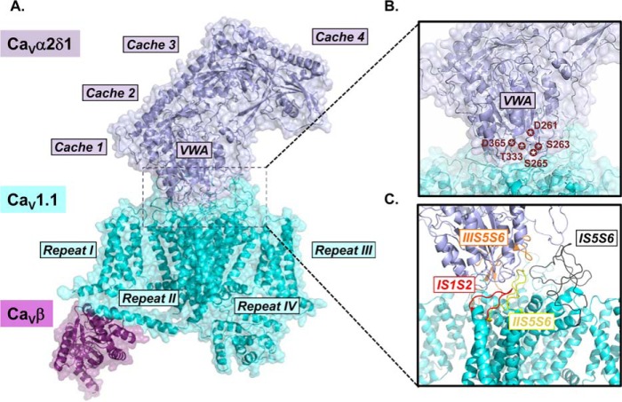
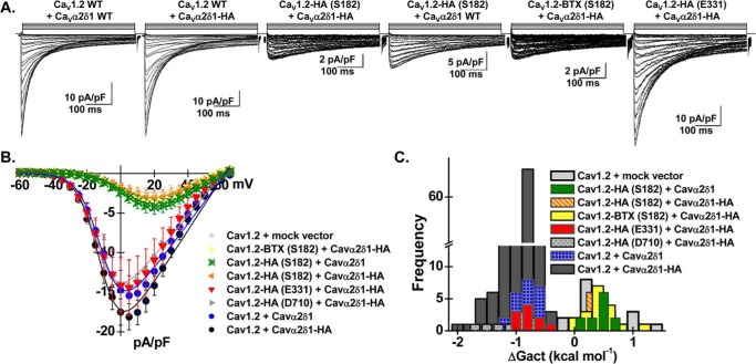
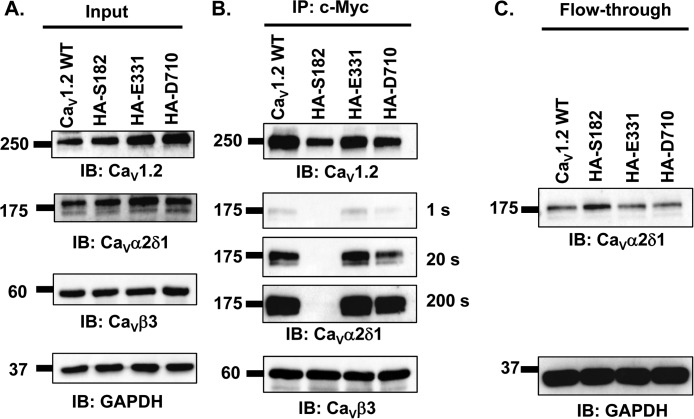
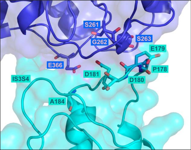
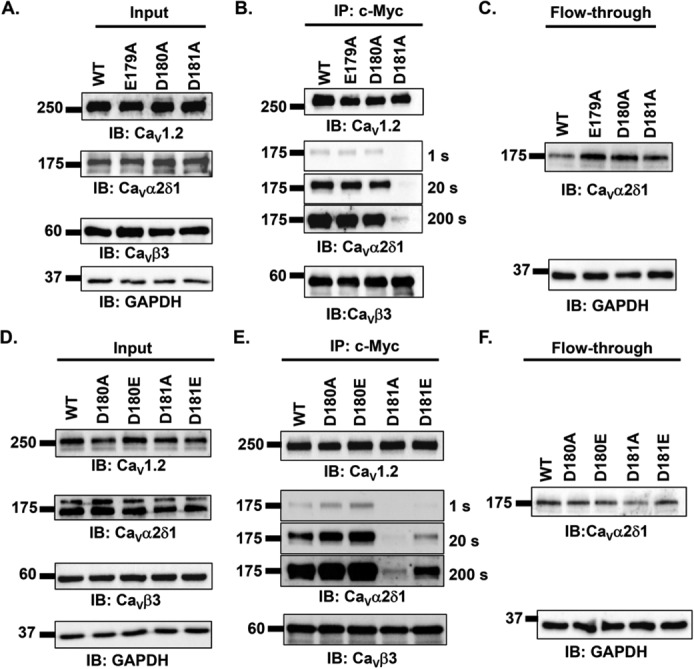
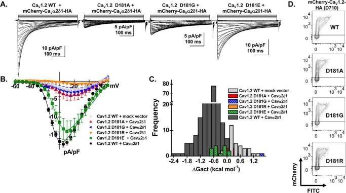
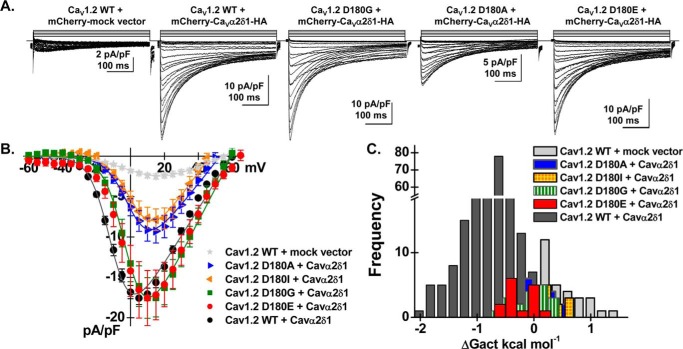
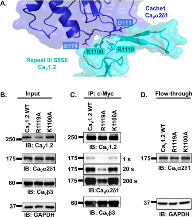
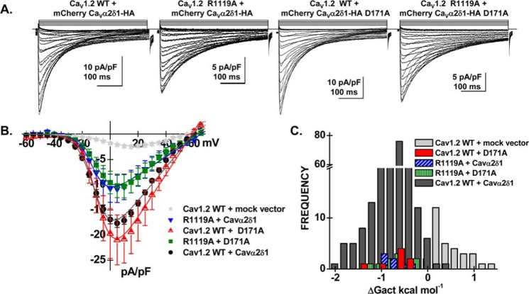
Voltage-gated L-type CaV1.2 channels in cardiomyocytes exist as heteromeric complexes. Co-expression of CaVα2δ1 with CaVβ/CaVα1 proteins reconstitutes the functional properties of native L-type currents, but the interacting domains at the CaV1.2/CaVα2δ1 interface are unknown. Here, a homology-based model of CaV1.2 identified protein interfaces between the extracellular domain of CaVα2δ1 and the extracellular loops of the CaVα1 protein in repeats I (IS1S2 and IS5S6), II (IIS5S6), and III (IIIS5S6). Insertion of a 9-residue hemagglutinin epitope in IS1S2, but not in IS5S6 or in IIS5S6, prevented the co-immunoprecipitation of CaV1.2 with CaVα2δ1. IS1S2 contains a cluster of three conserved negatively charged residues Glu-179, Asp-180, and Asp-181 that could contribute to non-bonded interactions with CaVα2δ1. Substitutions of CaV1.2 Asp-181 impaired the co-immunoprecipitation of CaVβ/CaV1.2 with CaVα2δ1 and the CaVα2δ1-dependent shift in voltage-dependent activation gating. In contrast, single substitutions in CaV1.2 in neighboring positions in the same loop (179, 180, and 182-184) did not significantly alter the functional up-regulation of CaV1.2 whole-cell currents. However, a negatively charged residue at position 180 was necessary to convey the CaVα2δ1-mediated shift in the activation gating. We also found a more modest contribution from the positively charged Arg-1119 in the extracellular pore region in repeat III of CaV1.2. We conclude that CaV1.2 Asp-181 anchors the physical interaction that facilitates the CaVα2δ1-mediated functional modulation of CaV1.2 currents. By stabilizing the first extracellular loop of CaV1.2, CaVα2δ1 may up-regulate currents by promoting conformations of the voltage sensor that are associated with the channel's open state.

Keywords: calcium channel; electrophysiology; gating; molecular modeling; protein-protein interaction.


Conflict of interest statement

The authors declare that they have no conflicts of interest with the contents of this article

Figures

Figure 1.
Figure 1.
Three-dimensional cryo-electron microscopy structure of the CaV1.1 channel. A, surface representation of the rabbit CaV1.1 channel (cyan) in complex with CaVα2δ1 (deep blue) and CaVβ2 (violet) (PDB: 5GJV). The structural domains of CaVα2δ1 and the relative orientation of repeats I–IV in the pore-forming CaVα1 subunit of CaV1.1 are identified. For the correlation between the primary sequence and the structural domains of CaVα2δ1, see Fig. 2 in Ref. . The extensive interface between the two proteins is surrounded by a dashed black square, and this region is shown enlarged in B and C. B, structural details of the VWA domain of CaVα2δ1 are emphasized, and the residues forming the MIDAS are identified by stars. C, extracellular loops of CaV1.1 forming the interface with CaVα2δ1 are shown. Only the main chains are shown. The S1S2 loop in repeat I (IS1S2, residues 71–82) is in red; the turret and external pore region S5S6 in repeat I (IS5S6, residues 219–278) is in black; the turret and external pore region S5S6 in repeat II (IIS5S6, residues 581–600) is in yellow, and the turret and external pore region S5S6 in repeat III (IIIS5S6, residues 950–998) is in orange. Images were produced with PyMOL (Molecular Graphics System, Version 1.8 Schrödinger, LLC).
Figure 2.
Figure 2.
Inserting epitopes within the first extracellular loop of CaV1.2 prevents functional modulation of whole-cell current by CaVα2δ1. Stable recombinant HEKT cells expressing CaVβ3 were transiently transfected with pmCherry–CaVα2δ1–HA (or pmCherry–CaVα2δ1 WT) and pCMV–CaV1.2 WT or mutants as indicated over each current trace. In all cases, CaVα2δ1 was tagged in the C terminus by an mCherry epitope. The epitopes HA or BTX were inserted after the identified residues without altering the primary sequence on each side of the insertion site. A, representative whole-cell Ca2+ current traces obtained after recombinant expression of CaV1.2 WT + CaVα2δ1 WT, CaV1.2 WT + CaVα2δ1–HA, CaV1.2-HA (Ser-182) + CaVα2δ1–HA, CaV1.2–HA (Ser-182) + CaVα2δ1 WT, CaV1.2–BTX (Ser-182) + CaVα2δ1–HA, and CaV1.2–HA (Glu-331) + CaVα2δ1–HA (from left to right). CaV1.2 currents were modulated to the same extent in the presence of mCherry–CaVα2δ1–HA or mCherry–CaVα2δ1. Furthermore, similar results were obtained with CaV1.2–HA (Glu-331). Currents were recorded in the presence of 2 mm Ca2+ from a holding potential of −100 mV. Time scale is 100 ms throughout. The current density scale ranged from 2 to 10 pA/pF, as indicated. B, averaged current-voltage relationships. The absolute peak current densities measured with mCherry–CaVα2δ1–HA varied from −4 to −46 pA/pF over the 12-month recording period with a mean of −18 ± 1 pA/pF (n = 243). Averaged peak current densities obtained with the mock mCherry vector are shown. Co-expression with CaVα2δ1 left-shifted the voltage dependence of activation of CaV1.2 WT/CaVβ3 from E0.5, act = +8 ± 2 mV (n = 25) to E0.5, act = −8.3 ± 0.2 mV (n = 243) for CaV1.2 WT/CaVβ3 with mCherry–CaVα2δ1–HA. Statistical analyses were performed with a one-way ANOVA test: *, p < 0.01, and **, p < 0.001, against the mock vector. See Table 1 for details. C, distribution of the free energies of activation. The free energies of activation (ΔGact) measured in the presence of the mock vector and in the presence of mCherry–CaVα2δ1–HA are centered at 0.5 ± 0.1 and −0.78 ± 0.03 kcal mol−1, respectively. The distribution of the ΔGact values for the following combinations CaV1.2–HA (Ser-182) with CaVα2δ1–HA, CaV1.2-HA (Ser-182) with CaVα2δ1 WT, and CaV1.2–BTX (Ser-182) with CaVα2δ1–HA overlapped with the ΔGact values measured in the presence of the mock vector (no CaVα2δ1).
Figure 3.
Figure 3.
Epitope insertion in the first extracellular loop of CaV1.2 impairs the co-immunoprecipitation of CaVα2δ1 with CaV1.2/CaVβ3 proteins. HEKT cells were transiently transfected with pmCherry–CaVα2δ1–HA and pCMV–CaVβ3–c-Myc and either pCMV–CaV1.2 WT, pCMV–CaV1.2–HA (Ser-182), pCMV–CaV1.2–HA (Glu-331), or pCMV–CaV1.2–HA (Asp-710). Cell lysates were immunoprecipitated (IP) overnight with anti-c-Myc magnetic beads to capture CaVβ3, eluted in a Laemmli buffer, and fractionated by SDS-PAGE using 8% gels. A, immunoblotting was carried out on total proteins (20 μg) collected from the cell lysates for each of the three proteins (CaV1.2, CaVα2δ1, and CaVβ3) before the immunoprecipitation assay (Input) to confirm that each protein was translated at the expected molecular mass. Each experimental condition is identified by the specific CaV1.2 construct. The signal for the housekeeping protein GAPDH is shown below each blot. B, immunoblotting (IB) was carried out after eluting the protein complexes from the anti-c-Myc beads with anti-CaV1.2, anti-CaVα2δ1, and anti-CaVβ3 antibodies (from top to bottom, as indicated). Images for CaVα2δ1 were captured after short (1 s) or long exposure times (20 and 200 s). CaVβ3 and CaV1.2 proteins migrated at 60 and 250 kDa, respectively. All CaVα2δ1 proteins migrated at ≈175 kDa, which is consistent with the molecular mass of the mCherry–CaVα2δ1–HA in previous studies (30). All immunoblots were carried out in parallel under the same transfection and extraction conditions. C, proteins that did not bind to the antibody-bead complex (referred to as the flow-through fraction) were collected, diluted in a Laemmli buffer, and fractionated by SDS-PAGE using an 8% gel and revealed with the anti-CaVα2δ1. As seen, mCherry–CaVα2δ1–HA is present in the flow-through fraction at the expected molecular mass (175 kDa) confirming that the proteins were appropriately translated and were present in the preparation in detectable quantities throughout. These experiments were carried out three times with the mutants and 10 times for the WT construct over a period of 5 months and yielded qualitatively similar results.
Figure 4.
Figure 4.
Three-dimensional model of the extracellular loops of the rabbit CaV1.2 in complex with the VWA domain of the rat CaVα2δ1 protein. The 3D model of the region spanning the first transmembrane helix S1 to the fourth transmembrane helix S4 in the first repeat in CaV1.2 (IS1S4) is shown in cyan, and the VWA domain of CaVα2δ1 is shown in deep blue. Residues Pro-178, Glu-179, Asp-180, Asp-181, and Ala-184 of CaV1.2 and residues Ser-261, Gly-262, Ser-263, and Glu-366 in CaVα2δ1 (three of the five residues in the MIDAS) are shown in stick representation with oxygen and nitrogen atoms colored in red and in blue, respectively. The model does not predict strong electrostatic interactions between CaV1.2 Asp-180 and residues in CaVα2δ1. Intramolecular interactions with residues in the extracellular IS3S4 loop are not ruled out. CaV1.2 Asp-181 appears to be appropriately oriented to form electrostatic interactions with Gly-262 and Ser-263 in CaVα2δ1. Modeling was achieved with Modeler 9.17. The figure was produced using PyMOL.
Figure 5.
Figure 5.
Mutations of the aspartate residue at position 181 impair the co-immunoprecipitation of CaVα2δ1 with CaV1.2/CaVβ3 proteins. A–C, HEKT cells were transiently transfected with pmCherry–CaVα2δ1–HA and pCMV–CaVβ3–c-Myc and either pCMV–CaV1.2 WT, pCMV–CaV1.2 E179A, pCMV–CaV1.2 D180A, or pCMV–CaV1.2 D181A. Cell lysates were immunoprecipitated (IP) overnight with anti-c-Myc magnetic beads to capture CaVβ3, eluted in a Laemmli buffer, and fractionated by SDS-PAGE using 8% gels. A, immunoblotting was carried out on total proteins (20 μg) collected from the cell lysates for each of the three proteins (CaV1.2, CaVα2δ1, and CaVβ3) before the immunoprecipitation assay (Input) to confirm that each protein was translated at the expected molecular mass. Each experimental condition is identified by the specific CaV1.2 construct. The signal for the housekeeping protein GAPDH is shown below each blot. B, immunoblotting was carried out after eluting the protein complexes from the beads with anti-CaV1.2, anti-CaVα2δ1, and anti-CaVβ3 antibodies (from top to bottom, as indicated). Images for CaVα2δ1 were captured after short (1 s) or longer exposure times (20 and 200 s). All immunoblots were carried out in parallel under the same transfection and extraction conditions. C, proteins that did not bind to the antibody-bead complex (flow-through fraction) were collected, diluted in a Laemmli buffer, and fractionated by SDS-PAGE using an 8% gel and revealed with the anti-CaVα2δ1. D–F, HEKT cells were transiently transfected with pmCherry–CaVα2δ1–HA and pCMV–CaVβ3–c-Myc and either pCMV–CaV1.2 WT, pCMV–CaV1.2 D180A, pCMV–CaV1.2 D180E, pCMV–CaV1.2 D181A, or pCMV–CaV1.2 D181E. Cell lysates were immunoprecipitated overnight with anti-c-Myc magnetic beads to capture CaVβ3, eluted in a Laemmli buffer, and fractionated by SDS-PAGE using 8% gels. D, immunoblotting was carried out on total proteins (20 μg) collected from the cell lysates for each of the three proteins (CaV1.2, CaVα2δ1, and CaVβ3) before the immunoprecipitation assay (Input) to confirm that each protein was translated at the expected molecular mass. Each experimental condition is identified by the specific CaV1.2 construct. The signal for the housekeeping protein GAPDH is shown below each blot. E, immunoblotting (IB) was carried out with anti-CaV1.2, anti-CaVα2δ1, and anti-CaVβ3 antibodies (from top to bottom, as indicated) after eluting the protein complexes from the beads. Images for CaVα2δ1 were captured after short (1 s) or longer exposure times (20 and 200 s). All immunoblots were carried out in parallel under the same transfection and extraction conditions. F, proteins that did not bind to the antibody-bead complex (flow-through fraction) were collected, diluted in a Laemmli buffer, and fractionated by SDS-PAGE using an 8% gel and revealed with the anti-CaVα2δ1. As seen, mCherry–CaVα2δ1–HA is present in the flow-through fraction at the expected molecular mass (175 kDa) confirming that the proteins were appropriately translated and were present in the preparation in detectable quantities throughout. These experiments were carried out four times with the mutants and 10 times for the WT construct over a period of 5 months and yielded reproducible results.
Figure 6.
Figure 6.
Mutations at Asp-181 prevent up-regulation of CaV1.2 currents. HEKT cells were transiently transfected with pCMV–CaVβ3 and pmCherry–CaVα2δ1–HA with the CaV1.2 constructs (D181A, D181G, D181R, and D181E). A, whole-cell Ca2+ current traces were recorded in the presence of 2 mm Ca2+ from a holding potential of −100 mV for the constructs as identified. The current traces with the largest currents are shown for CaV1.2 constructs D181G and D181E. Time scale is 100 ms throughout. The current density scale is either 5 or 10 pA/pF as indicated. B, averaged current-voltage relationships. Peak current densities versus voltage relationships were measured for CaV1.2 WT and CaV1.2 mutants (as shown). Currents traces obtained with the empty mCherry (mock) vector are also shown. CaV1.2 constructs D181A, D181G, and D181R generated currents that were not significantly up-regulated by mCherry–CaVα2δ1–HA WT. Statistical analyses were performed with a one-way ANOVA test: *, p < 0.01, and **, p < 0.001 against the mock vector. See Table 1 for details. C, distribution of the free energies of activation. The values for the free energy of activation (ΔGact) measured for CaV1.2 constructs (D181A, D181G, D181E, and D181R) overlapped with the values measured for the mock vector. D, representative two-dimensional plots of mCherry versus FITC fluorescence. The cell-surface expression of the CaV1.2 mutants was evaluated by introducing the mutation in the mCherry–CaV1.2–HA construct. The surface fluorescence was estimated from the relative intensity of the fluorescence emitted by the fluorescein isothiocyanate (FITC)-conjugated anti-HA as measured using a flow cytometry assay (10,000 intact cells). The construct allows for detection of intracellular and extracellular fluorescence using FITC-conjugated anti-HA (“x axis”) and an anti-mCherry (“y axis”), respectively. The robust mCherry signal (y axis) confirms that the proteins were translated up to the end of the coding sequence. The cell-surface fluorescence for FITC, calculated as ΔMedFI as explained under “Experimental procedures,” was slightly lower for the CaV1.2 mutants (D181A, D181G, and D181R) than for the WT construct. Nonetheless, all constructs significantly fluoresced at the cell surface supporting the view that the absence of function did not result from a complete absence of trafficking to the cell membrane. Furthermore, the ΔMedFI signal for the total protein was similar for all WT and mutant constructs demonstrating that proteins were appropriately translated, an observation also obtained from carrying out routine Western blotting.
Figure 7.
Figure 7.
Asp-180 in CaV1.2 controls the CaVα2δ1-induced shift in voltage-dependent gating of CaV1.2 currents. A, representative whole-cell Ca2+ current traces obtained after the transient expression of pCMV–CaVβ3 and pmCherry–CaVα2δ1–HA with the CaV1.2 constructs (D180I, D180A, D180G, and D180E) in HEKT cells. Note that the leftmost current trace was obtained in the presence of CaV1.2 WT and the mock mCherry vector. Recordings were made in the presence of 2 mm Ca2+ from a holding potential of −100 mV. Time scale is 100 ms throughout. The current density scale ranged from 2 to 10 pA/pF as indicated. B, averaged current-voltage relationships. Peak current densities versus voltage relationships were measured for CaV1.2 WT and CaV1.2 mutants (as shown). Averaged peak current densities obtained with the mock mCherry vector are shown in light gray stars. All CaV1.2 constructs (D180I, D180A, D180G, and D180E) were up-regulated by mCherry–CaVα2δ–HA albeit to variable extent. Statistical analyses were performed with a one-way ANOVA test: *, p < 0.01, and **, p < 0.001, against the mock mCherry vector. Nonetheless, the current-voltage relationships measured with the CaV1.2 mutants were clearly shifted to the right when compared with CaV1.2 WT. See Table 1 for details. C, distribution of the free energies of activation. The free energies of activation (ΔGact) for CaV1.2 D180E, D180I, D180A, and D180G did not overlap with the values measured with CaV1.2 WT. ΔGact values for CaV1.2 D180A and D180I were not significantly different from ΔGact values measured for the mock vector, whereas ΔGact values for CaV1.2 D180G and D180E were significantly different at p < 0.01.
Figure 8.
Figure 8.
Extracellular loop S5S6 in repeat III has a modest impact on the interaction between CaVα2δ1 and CaV1. 2/CaVβ3 proteins. A, 3D model of IIIS5S6 in CaV1.2 (residues 1059–1204) is shown in cyan, and the cache1 domain (residues 104–223) in CaVα2δ1 is shown in deep blue. The carboxyl group on the side chain of Arg-1119 in CaV1.2 is located close enough (< 3 Å) to the main-chain atoms of Glu-171 in CaVα2δ1 to potentially contribute to the formation of hydrogen bonds. In contrast, the 3D model does not predict a favorable interaction between Lys-1100 in CaV1.2 and Glu-174 in CaVα2δ1 with a minimum distance estimated to be at 4.6 Å. Modeling was achieved with Modeler 9.17. The figure was produced using PyMOL. B–D, HEKT cells were transiently transfected with pmCherry–CaVα2δ1–HA WT and pCMV–CaVβ3–c-Myc and either pCMV–CaV1.2 WT, pCMV–CaV1.2 R1119A, or pCMV–CaV1.2 K1100A. Cell lysates were immunoprecipitated overnight with anti-c-Myc magnetic beads to capture CaVβ3, eluted in a Laemmli buffer, and fractionated by SDS-PAGE using 8% gels. B, immunoblotting (IB) was carried out on total proteins (20 μg) collected from the cell lysates for each of the three proteins (CaV1.2, CaVα2δ1, and CaVβ3) before the immunoprecipitation assay (Input) to confirm that each protein was translated at the expected molecular mass. Each experimental condition is identified by the specific CaV1.2 construct. C, immunoblotting was carried out as detailed earlier. Images for CaVα2δ1 were captured after 1, 20, and 200 s of exposure. The signal for the anti-CaVα2δ1 was detected only after a 200-s exposure when probed in the presence of CaV1.2 R1119A/CaVβ3. D, protein lysates that ran through without binding to the antibody-bead complex (flow-through fraction) were collected, diluted in a Laemmli buffer, and fractionated by SDS-PAGE using an 8% gel and revealed with the anti-CaVα2δ1. As seen, mCherry–CaVα2δ1–HA is present in all flow-through fractions at the expected molecular mass (175 kDa) confirming that the proteins were appropriately translated and were present in the preparation in detectable quantities throughout.
Figure 9.
Figure 9.
Double mutant analysis of charged residues in repeat III in CaV1.2. HEKT cells were transiently transfected with CaVβ3, pCMV–CaV1.2 WT or mutant and pmCherry–CaVα2δ1–HA WT or mutant. A, representative whole-cell Ca2+ current traces were recorded in the presence of 2 mm Ca2+ from a holding potential of −100 mV. From left to right: CaV1.2 WT with mCherry–CaVα2δ1–HA WT; CaV1.2 R1119A with mCherry–CaVα2δ1–HA WT; CaV1.2 WT with mCherry–CaVα2δ1–HA D171A, and CaV1.2 R1119A with mCherry–CaVα2δ1–HA D171A. Time scale is 100 ms throughout. The current density scale is either 5 or 10 pA/pF as indicated. B, averaged current-voltage relationships. Peak current densities versus voltage relationships were measured for the WT construct and the mutants (as shown). Current traces obtained with the empty mCherry vector (mock vector) are also shown. Statistical analyses were performed with a one-way ANOVA test: *, p < 0.01, and **, p < 0.001, against the mock vector. See Table 1 for details. C, distribution of the free energies of activation. The values of the free energy of activation (ΔGact) for all conditions were significantly different from the values measured with the mock vector.

References

    1. Bers D. M. (2000) Calcium fluxes involved in control of cardiac myocyte contraction. Circ. Res. 87, 275–281 - PubMed
    1. Jaleel N., Nakayama H., Chen X., Kubo H., MacDonnell S., Zhang H., Berretta R., Robbins J., Cribbs L., Molkentin J. D., and Houser S. R. (2008) Calcium influx through T- and L-type calcium channels have different effects on myocyte contractility and induce unique cardiac phenotypes. Circ. Res. 103, 1109–1119 - PMC - PubMed
    1. Gao H., Wang F., Wang W., Makarewich C. A., Zhang H., Kubo H., Berretta R. M., Barr L. A., Molkentin J. D., and Houser S. R. (2012) Calcium influx through L-type calcium channels and transient receptor potential channels activates pathological hypertrophy signaling. J. Mol. Cell. Cardiol. 53, 657–667 - PMC - PubMed
    1. Bannister J. P., Bulley S., Narayanan D., Thomas-Gatewood C., Luzny P., Pachuau J., and Jaggar J. H. (2012) Transcriptional upregulation of α2δ1 elevates arterial smooth muscle cell voltage-dependent Ca2+ channel surface expression and cerebrovascular constriction in genetic hypertension. Hypertension 60, 1006–1015 - PMC - PubMed
    1. Yue L., Feng J., Gaspo R., Li G. R., Wang Z., and Nattel S. (1997) Ionic remodeling underlying action potential changes in a canine model of atrial fibrillation. Circ. Res. 81, 512–525 - PubMed

Publication types

Substances

Grants and funding