Review
. 2018:35:31-53.
doi: 10.1007/7854_2017_7.

Oxytocin Modulation of Neural Circuits

Affiliations
Review

Oxytocin Modulation of Neural Circuits

Mariela Mitre et al. Curr Top Behav Neurosci. 2018.

Abstract

Oxytocin is a hypothalamic neuropeptide first recognized as a regulator of parturition and lactation which has recently gained attention for its ability to modulate social behaviors. In this chapter, we review several aspects of the oxytocinergic system, focusing on evidence for release of oxytocin and its receptor distribution in the cortex as the foundation for important networks that control social behavior. We examine the developmental timeline of the cortical oxytocin system as demonstrated by RNA, autoradiographic binding, and protein immunohistochemical studies, and describe how that might shape brain development and behavior. Many recent studies have implicated oxytocin in cognitive processes such as processing of sensory stimuli, social recognition, social memory, and fear. We review these studies and discuss the function of oxytocin in the young and adult cortex as a neuromodulator of central synaptic transmission and mediator of plasticity.

Keywords: Cortex; Inhibition; Neuromodulation; Oxytocin; Synaptic plasticity.


Figures

Fig. 1
Fig. 1
OXTR-2 expression profile in the brain. (a) Schematics summarizing OXTR-2 expression in mothers, virgin females, and males using immunohistochemistry. Shown are four anterior–posterior coronal sections. Color indicates percentage of DAPI-positive cells that were OXTR-2 per region. Brain regions identified and quantified: auditory cortex (ACtx), anterior hypothalamus (AHP), basolateral amygdaloid nucleus (BL), central amygdaloid nucleus (Ce), anterior olfactory nucleus (AO), bed nucleus of stria terminalis (BST), hippocampal areas CA1-CA3, dentate gyrus (DG), frontal association cortex (FrA), globus pallidus (LGP), granular cell layer of the olfactory bulb (GrO), lateral hypothalamic area (LH), right lateral septum (LS), motor cortex (M1), nucleus accumbens core (NaC), piriform cortex (PCtx), prelimbic cortex (PrL), paraventricular nucleus of hypothalamus (PVN), median raphe (RN), somatosensory cortex (S1), suprachiasmatic nucleus (SCN), supraoptic nucleus of hypothalamus (SON), visual cortex (V1), and ventromedial hypothalamic nucleus (VMH). Gray areas may have expressed oxytocin receptors but were not quantified here. (b) OXTR-2 immunostaining in piriform cortex of female (left) and male (right) imaged at 10×. Note more OXTR-2 cells in females. Scale bar, 100 μm. (c) OXTR-2 immunostaining of virgin female hippocampus imaged at 20×. Scale, 200 μm. (d) OXTR-2 immunostaining in left auditory cortex (left) and right auditory cortex (right) of virgin female imaged at 20×. Note more staining in left auditory cortex. Scale, 100 μm. Adapted from Mitre et al. (2016)
Fig. 2
Fig. 2
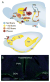
Projections of oxytocin neurons. (a) An rAAV-expressing Venus under the control of the mouse oxytocin promoter was injected into paraventricular and supraoptic nucleus of adult female rats. Viral infection resulted in Venus expression in cell bodies and fibers from oxytocinergic neurons to subcortical (A) and cortical (B) regions. The infected paraventricular nucleus of the hypothalamus in one hemisphere is colored in green. The density of fibers is depicted in the following colors: yellow, orange, red, and violet. The abbreviations of structures are as follows: accumbens nucleus core (AcbC), accumbens nucleus shell (AcbSH), anterior olfactory nucleus (AON), basolateral amygdaloid nucleus (BLA), bed nucleus of the stria terminalis (BNST), field CA1 of hippocampus (CA1), field CA3 of hippocampus (CA3), central amygdaloid nucleus (CeA), cingulate cortex (Cg), caudate putamen (Cg), dentate gyrus (DG), dorsal peduncular cortex (DP), subiculum-dorsal (DS), dorsal taenia tecta (DTT), entorhinal cortex lateral (Entl), frontal association cortex (FrA), nucleus of the horizontal limb of the diagonal band (HDB), insular corticies (I), island of Calleja (ICj), globus pallidus lateral (LGP), lateral septal nucleus (LS), medial amygdaloid nucleus (MeA), medial orbital cortex (MeA), prelimbic cortex (PrL), paraventricular thalamic nuclei (PV), paraventricular nucleus of the hypothalamus (PVN); temporal association cortex (TeA), olfactory tubercle (Tu), ventral orbital cortex (VO), subiculum-ventral (VS), ventral taenia tecta (VTT). Adapted from Knobloch et al. (2012). (b) Section of virgin female hypothalamus from Oxt-IRES-Cre animal expressing YFP via AAV (pAAV-5Ef1a-DIO ChETA-EYFP) stereotaxically injected into left PVN. Immunostained with antibodies to YFP and imaged at 10×. Green, YFP+ axons. Blue, DAPI. Scale: 400 μm. Adapted from Mitre et al. (2016)
Fig. 3
Fig. 3
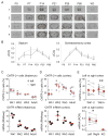
Development of OXTR expression. (a) Receptor autoradiography in C57BL/6J mice at several ages and coronal levels, and lack of specific OXTR ligand binding in OXTR KO brain assessed at P60. (1) accessory (a) and main (b) olfactory bulbs; (2) neocortex (c), septum (d), claustrum (e), endopiriform cortex (f), piriform cortex (g), diagonal band of Broca (h); (3) bed nucleus of the stria terminalis (i), ventral caudatoputamen (j); (4) periventricular thalamus (k), CA3 hippocampus (l), central amygdala (m), medial amygdala (n), hypothalamus (o). Scale bar = 1 cm. (b) Quantification of receptor autoradiography for OXTR in C57BL/6J mice demonstrates transient developmental profiles. OXTR binding with highly selective OXTR ligand is evident in the septum and the somatosensory neocortex. (c) Summary of OXTR-2 labeled cells (top) and OXTR mRNA measured with RNAseq (bottom) at different postnatal weeks (Wk) in auditory thalamus. The first postnatal week had the highest thalamic OXTR-2 expression and mRNA level. Filled symbols, tissue from left hemisphere; open symbols, right hemisphere. (d) Summary of OXTR-2 labeled cells (top) and OXTR mRNA (bottom) at different ages in auditory cortex. The second and third postnatal weeks had highest amount of expression. (e) Summary of oxytocin receptor lateralization in left vs. right virgin female auditory cortex. Top, OXTR-2 expression is higher in left auditory cortex than in right auditory cortex from the same animals during and after postnatal week 3, but not earlier during postnatal weeks 1–2. Bottom, oxytocin receptor mRNA (measured with RT-PCR relative to ribophorin mRNA expression) is higher in left auditory cortex than in right auditory cortex from the same adult virgin females. Oxytocin receptor mRNA was not detected in oxytocin receptor KO mice. *p < 0.05. Adapted from Mitre et al. (2016)
Fig. 4
Fig. 4
Oxytocin and olfactory recognition. (a) Retrograde viral labeling of AON neurons following injection of CAV2-Cre into the MOB of Ai9 reporter mice for dTomato (red), and immunoreactivity for the OXTR (green) in the AON (scale bar, 150 μm). (b) Simultaneous increases in inward sEPSC and outward sIPSC rate following laser stimulation. Top, example traces. Bottom, PSTH of the time course of the simultaneous rate increases for a single stimulation in a regular-firing neuron. (c) Impaired same-sex social recognition in mice following OXTR deletion specifically in the AON. Male mice were placed with an unknown juvenile for 5 min. After 30 min in the home cage, they were placed with the same juvenile and a second unknown juvenile for 3 min. To generate OXTRΔAON mice, rAAV1/2-CBA-Cre was injected in the AON of mice in which the OXTR gene was flanked by loxP sites. Control mice received the same virus injection but had two wild-type OXTR alleles. Total exploration time of social partners during the initial sample phase was longer in OXTRΔAON vs control mice. (d) Social recognition memory was expressed as percentage of exploration time of the new juvenile mouse over the total time exploring both interaction partners for OXTRΔAON vs control mice. (e) Recognition memory for nonsocial odors was determined as the percent of exploration time of a new odorant over the total time exploring both odorants for OXTRΔAON vs control mice. Adapted from Oettl et al. (2016)
Fig. 5
Fig. 5
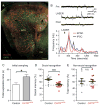
Oxytocin and maternal responses to infant distress vocalizations. (a) Maternal retrieval behavior: isolated pups make ultrasonic distress calls, alerting care-taking mice to find and retrieve lost pups back to the nest; naïve virgins disregard calls. (b) Percentages of animals that retrieved 1+ times within 12 h of being co-housed. Virgin females received either oxytocin injections (red, “OT”), optogenetic PVN stimulation (blue, “Opto”), or saline injections (black). All co-housed dams retrieved pups. (c) Cumulative percentage of initially naïve virgin females retrieving after co-housing. Wild-type animals received saline injections or oxytocin; Oxt-IRES-Cre animals received optical stimulation in PVN. Oxytocin-injected and Oxt-IRES-Cre animals began to retrieve in greater numbers and at a faster rate than saline-injected mice 12 h after co-housing. (d) Example voltage-clamp recording of inhibitory postsynaptic currents (IPSCs) evoked by extracellular stimulation. Top, oxytocin was washed into the bath for 5 min. Red bar, duration of oxytocin washin. Dashed line, baseline IPSC amplitude. Bottom, brain slice from Oxt-IRES-Cre mouse expressing ChETA in oxytocin neurons. Oxytocin release was evoked by blue light (hυ) for 3 min. (e) Pup calls evoke stronger and more temporally precise responses in mother mice compared to naïve virgin females. Top, spectrogram of pup vocalizations; bottom, three representative trials from auditory cortical neurons recorded in vivo. (f) Optogenetic release of oxytocin transforms responses in virgin auditory cortex; before pairing oxytocin with pup calls (Pre), responses were weak and temporally unreliable. After pairing, responses rapidly became stronger, and over 3 h responses also become temporally precise. Adapted from Marlin et al. (2015)

References

    1. Bales KL, Carter CS. Developmental exposure to oxytocin facilitates partner preferences in male prairie voles (Microtus ochrogaster) Behav Neurosci. 2003a;117(4):854–859. - PubMed
    1. Bales KL, Carter CS. Sex differences and developmental effects of oxytocin on aggression and social behavior in prairie voles (Microtus ochrogaster) Horm Behav. 2003b;44(3):178–184. - PubMed
    1. Bales KL, Pfeifer LA, Carter CS. Sex differences and developmental effects of manipulations of oxytocin on alloparenting and anxiety in prairie voles. Dev Psychobiol. 2004;44(2):123–131. doi: 10.1002/dev.10165. - DOI - PubMed
    1. Bartz JA, Zaki J, Bolger N, Ochsner KN. Social effects of oxytocin in humans: context and person matter. Trends Cogn Sci. 2011;15(7):301–309. doi: 10.1016/j.tics.2011.05.002. - DOI - PubMed
    1. Braida D, Donzelli A, Martucci R, Capurro V, Busnelli M, Chini B, Sala M. Neurohypophyseal hormones manipulation modulate social and anxiety-related behavior in zebrafish. Psychopharmacology. 2012;220(2):319–330. doi: 10.1007/s00213-011-2482-2. - DOI - PubMed

LinkOut - more resources