Observational Study
. 2017 Sep 2;18(1):165.
doi: 10.1186/s12931-017-0651-5.

Extracellular histones are clinically relevant mediators in the pathogenesis of acute respiratory distress syndrome

Affiliations
Observational Study

Extracellular histones are clinically relevant mediators in the pathogenesis of acute respiratory distress syndrome

Xin Lv et al. Respir Res. .

Abstract

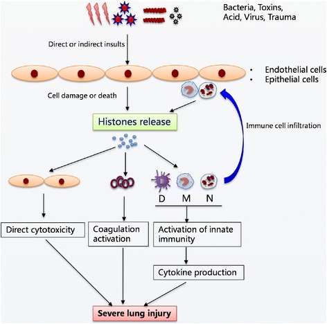
Background: Extracellular histones were recently identified as an inflammatory mediator involved in the pathogenesis of various organ injuries. This study aimed to examine extracellular histone levels and their clinical implications in acute respiratory distress syndrome (ARDS) patients and to explore histone-mediated effects through ex-vivo investigations.

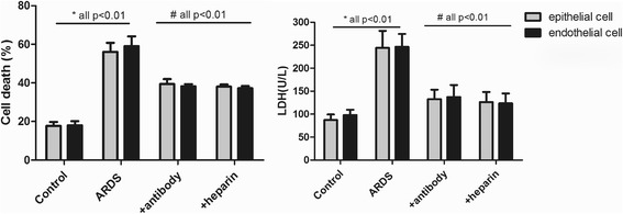
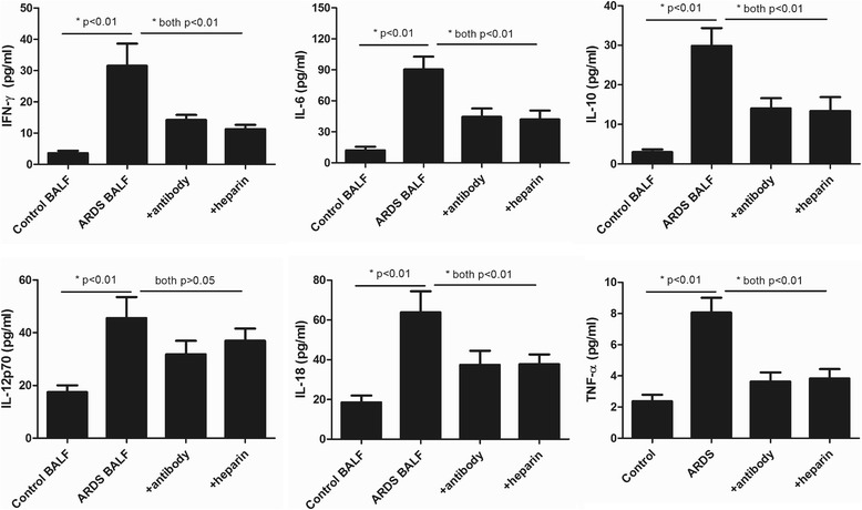
Methods: Extracellular histones, cytokine profiles and clinical data from 96 ARDS patients and 30 healthy volunteers were obtained. Human bronchial epithelial cells (BEAS-2B), human pulmonary artery endothelial cells (HPAEC), and human monocytic U937 cells were exposed to bronchoalveolar lavage fluid (BALF) collected from ARDS patients, and cellular damage and cytokine production were assessed. Furthermore, the effect of histone-targeted interventions by heparin or anti-histone antibody was evaluated.

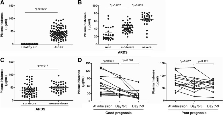
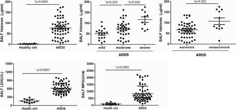
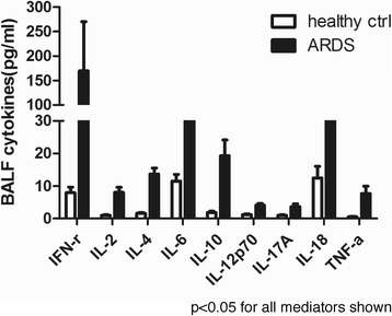
Results: Plasma and BALF extracellular histone levels were much higher in ARDS patients than in healthy controls. There was a significant association between extracellular histones and ARDS severity and mortality. In addition, extracellular histones correlated with an evident systemic inflammation detected in ARDS patients. Ex-vivo analysis further showed that ARDS patient's BALF remarkably induced epithelial and endothelial cell damage and stimulated cytokine production in the supernatant of U937 cells. The adverse effects on these cells could be abrogated by heparin or anti-histone antibody.

Conclusions: Extracellular histones in ARDS patients are excessively increased and may contribute to disease aggravation by inducing cellular damage and promoting systemic inflammation. Targeting extracellular histones may provide a promising approach for treating ARDS.

Keywords: Acute respiratory distress syndrome (ARDS); Cell damage; Extracellular histones; Systemic inflammation.


Conflict of interest statement

Ethics approval and consent to participate

The study protocol was approved by the Ethics Committee of Shanghai Pulmonary Hospital, Tongji University School of Medicine (Shanghai, P.R.China).

Consent for publication

Not applicable

Competing interests

The authors declare that they have no competing interests.

Publisher’s Note

Springer Nature remains neutral with regard to jurisdictional claims in published maps and institutional affiliations.

Figures

Fig. 1
Fig. 1
Elevated extracellular histones in the plasma of ARDS patients. aMedian plasma histones were significantly increased in ARDS patients than in healthy controls (both p < 0.0001). b Plasma histone levels correlated with disease severity. Severe ARDS patients had higher extracellular histone levels than moderate or mild patients (all p < 0.05). c Median plasma histones were higher in nonsurvivor ARDS patients than those in survivors (p = 0.017). d Sequential plasma histone levels in ARDS patients at admission and days 3–5 and days 7–9. According to 28-day mortality, patients surviving the episode of ARDS (good prognosis) had a significant reduction in histone levels whereas nonsurvivors (poor prognosis) did not. Variables were expressed as median (interquartile range)
Fig. 2
Fig. 2
Extracellular histones were detected in the BALF of ARDS patients and their possible sources. Extracellular histones were also significantly higher in the BALF of ARDS patients than in healthy controls. Likewise, severe ARDS patients had higher BALF extracellular histone levels than moderate or mild patients, whereas non-survivor ARDS patients had higher BALF extracellular histone levels than survivors. Quantification of BALF LDH activity, a marker reflecting tissue damage, and BALF MPO activity, a marker of inflammatory cell activation, showed that both LDH and MPO levels were increased remarkably in ARDS patients compared to healthy controls, thus indicating a possible cellular sources for extracellular histones. Variables were expressed as median (interquartile range)
Fig. 3
Fig. 3
Detection of systemic inflammation in the BALF of ARDS patients at admission. Multiplex immunoassay for a panel of multiple cytokines was performed. Only 9 cytokines with significant differences (p < 0.05) between groups were shown. Variables were expressed as median (interquartile range)
Fig. 4
Fig. 4
Induction of lung epithelial and endothelial cell damage by ARDS patient’s BALF. It showed that ARDS patients’ BALF could drastically induce human lung epithelial and endothelial cell damage after overnight incubation, whereas addition of anti-histone H4 antibody or heparin remarkably inhibited cell death caused by ARDS patient’s BALF. Likewise, LDH levels were significantly increased in supernatant of these cells after overnight incubation of ARDS patient’s BALF, whereas addition of anti-histone H4 antibody or heparin could reduce LDH levels. Variables were expressed as mean ± standard deviation (SD)
Fig. 5
Fig. 5
Stimulatory effects of ARDS patient’s BALF on human monocytic cells. It showed that 6 histone-related cytokines were all notably increased in the supernatant of ARDS BALF-treated human monocytic U937 cells, whereas addition of anti-histone H4 antibody or heparin could decrease these cytokine levels (all p < 0.05). Variables were expressed as mean ± standard deviation (SD)
Fig. 6
Fig. 6
The hypothesized mechanistic model of extracellular histones in mediating cellular injury and systemic inflammation associated with ARDS. It is proposed that various insults (lung specific or extrapulmonary) cause extensive death of lung endothelial and/or epithelial cells or induce inflammatory cell infiltration, which subsequently leads to massive release of histones into extracellular spaces. The presence of high levels of extracellular histones may exert multiple biological effects including direct cytotoxicity, platelet aggregation, and activation of systemic inflammation by promoting cytokine production, which in turn attracts more inflammatory cells and enhance inflammation that eventually contribute to the pathogenesis of ARDS

References

    1. Umbrello M, Formenti P, Bolgiaghi L, Chiumello D. Current concepts of ARDS: a narrative review. Int J Mol Sci. 2016;18:64. doi: 10.3390/ijms18010064. - DOI - PMC - PubMed
    1. Matthay MA. The acute respiratory distress syndrome: pathogenesis and treatment. Annu Rev Pathol. 2011;6:147–163. doi: 10.1146/annurev-pathol-011110-130158. - DOI - PMC - PubMed
    1. Gattinoni L, Van HF, Larsson A, Mcauley DF, Ranieri M. Epidemiology, patterns of care, and mortality for patients with acute respiratory distress syndrome in intensive care units in 50 countries. JAMA. 2016;315:788–800. doi: 10.1001/jama.2016.0291. - DOI - PubMed
    1. Standiford TJ, Ward PA. Therapeutic targeting of acute lung injury and acute respiratory distress syndrome. Transl Res. 2016;167:183–191. doi: 10.1016/j.trsl.2015.04.015. - DOI - PMC - PubMed
    1. Matthay MA, Zimmerman GA. Acute lung injury and the acute respiratory distress syndrome: four decades of inquiry into pathogenesis and rational management. Am J Respir Cell Mol Biol. 2005;33:319–27. - PMC - PubMed

Publication types