

Review
doi: 10.1016/j.semradonc.2017.04.001.
Mechanisms of Normal Tissue Injury From Irradiation
Affiliations
- PMID: 28865514
- PMCID: PMC5653270
- DOI: 10.1016/j.semradonc.2017.04.001
Item in Clipboard
Review
Mechanisms of Normal Tissue Injury From Irradiation
Semin Radiat Oncol.
2017 Oct.
Abstract
Normal tissue injury from irradiation is an unfortunate consequence of radiotherapy. Technologic improvements have reduced the risk of normal tissue injury; however, toxicity causing treatment breaks or long-term side effects continues to occur in a subset of patients. The molecular events that lead to normal tissue injury are complex and span a variety of biologic processes, including oxidative stress, inflammation, depletion of injured cells, senescence, and elaboration of proinflammatory and profibrogenic cytokines. This article describes selected recent advances in normal tissue radiobiology.
Published by Elsevier Inc.
Figures
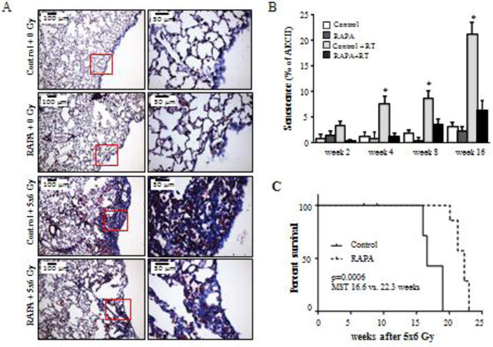
C57BL/6NCr Mice were exposed to 5×6 Gy of thoracic IR and treated with rapamycin or control diet. A) Masson trichrome staining of lung tissue at 16 weeks after IR. Collagen: blue, nuclei: purple, cytoplasm/epithelia: pink. B) The percentage of type II pneumocyte (AECII) cells staining for β-Gal activity at 2, 4, 8, and 16 weeks after IR was scored. A) Representative images at week 16 after IR. Percentage of AECII co-stained for Pro-surfactant-C (Pro-SP-C) at 16 weeks after IR. C) Kaplan-Meier survival analysis demonstrated that administration of rapamycin extended survival compared to mice receiving control diet after IR. Columns: mean, error bars: SD, brackets: p<0.05 by ANOVA. Reproduced with permission from.
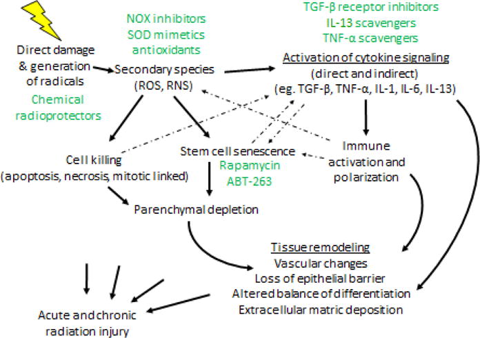
Radiation induces direct damage in normal tissues but also can generate free radicals, which can themselves cause injury or give rise to secondary species that may also cause injury. As a consequence, normal tissue cells are killed through a variety of mechanisms. Normal tissue stem cells may enter a state of senescence, which when combined with the initial cell killing, may result in parenchymal depletion and resulting tissue dysfunction. Simultaneously, ROS can initiate cytokine signaling, as can cell death and senescence. Collectively, these effects lead to immune activation and alterations in immune cell polarization, that can lead to chronic inflammation and further oxidative stress. Potential prevention, mitigation, and treatment approaches are highlighted in green.
References
-
- Martin M, Lefaix J, Delanian S. TGF-beta1 and radiation fibrosis: a master switch and a specific therapeutic target? Int J Radiat Oncol Biol Phys. 2000;47:277–90. - PubMed
-
- von Sontag C. The Chemical Basis of Radiation Biology. Francis Ta; London: 1987.
-
- Khan MA, Van Dyk J, Yeung IW. Partial volume rat lung irradiation; assessment of early DNA damage in different lung regions and effect of radical scavengers. Radiother Oncol. 2003;66:95–102. - PubMed
Publication types
MeSH terms
Substances
Grants and funding
LinkOut - more resources
Full Text Sources
Other Literature Sources
Medical
