

Biophysics of Biochemical Signaling in Dendritic Spines: Implications in Synaptic Plasticity
- PMID: 28866426
- PMCID: PMC5700242
- DOI: 10.1016/j.bpj.2017.07.029
Biophysics of Biochemical Signaling in Dendritic Spines: Implications in Synaptic Plasticity
Abstract
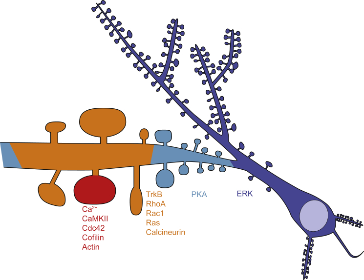
Dendritic spines are mushroom-shaped postsynaptic compartments that host biochemical signal cascades important for synaptic plasticity and, ultimately, learning and memory. Signaling events in spines involve a signaling network composed of hundreds of signaling proteins interacting with each other extensively. Synaptic plasticity is typically induced by Ca2+ elevation in spines, which activates a variety of signaling pathways. This leads to changes in the actin cytoskeleton and membrane dynamics, which in turn causes structural and functional changes of the spine. Recent studies have demonstrated that the activities of these proteins have a variety of spatiotemporal patterns, which orchestrate signaling activity in different subcellular compartments at different timescales. The diffusion and the decay kinetics of signaling molecules play important roles in determining the degree of their spatial spreading, and thereby the degree of the spine specificity of the signaling pathway.
Copyright © 2017 Biophysical Society. Published by Elsevier Inc. All rights reserved.
Figures
References
Publication types
MeSH terms
Substances
LinkOut - more resources
Full Text Sources
Other Literature Sources
Miscellaneous
