. 2017 Dec;25(12):3969-3977.
doi: 10.1007/s00167-017-4688-2. Epub 2017 Sep 2.

The pyramidalis-anterior pubic ligament-adductor longus complex (PLAC) and its role with adductor injuries: a new anatomical concept

Affiliations

The pyramidalis-anterior pubic ligament-adductor longus complex (PLAC) and its role with adductor injuries: a new anatomical concept

Ernest Schilders et al. Knee Surg Sports Traumatol Arthrosc. 2017 Dec.

Abstract

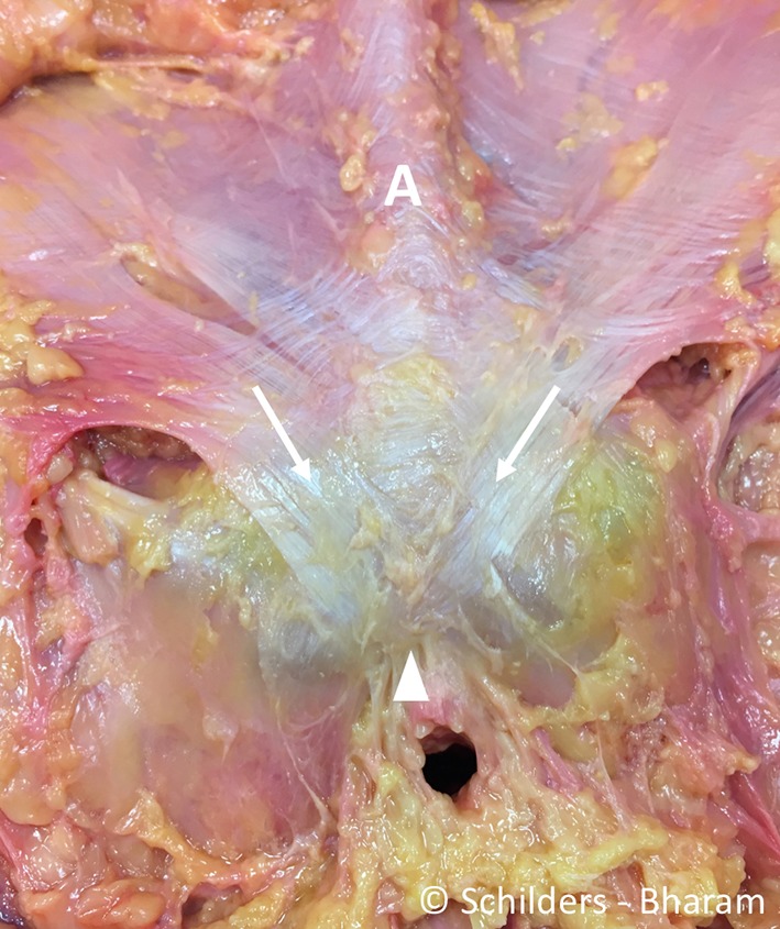
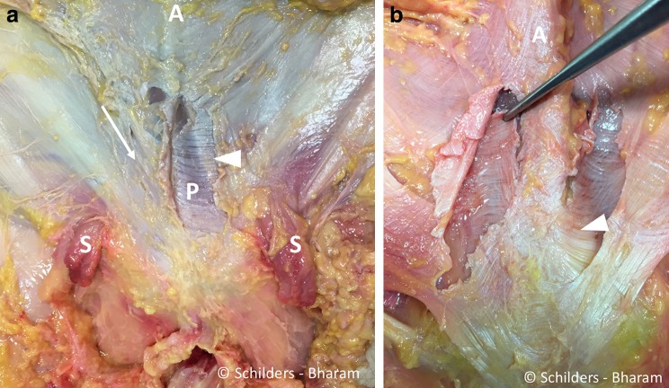
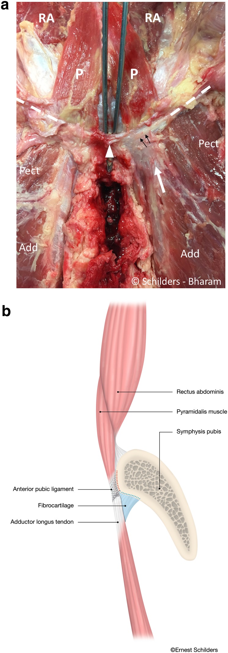
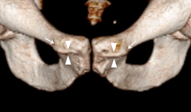
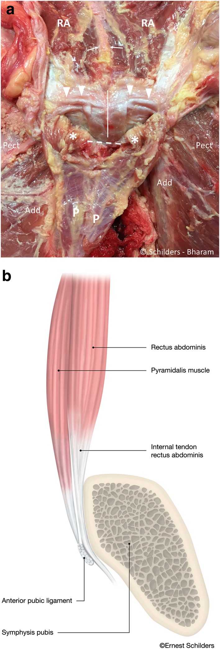
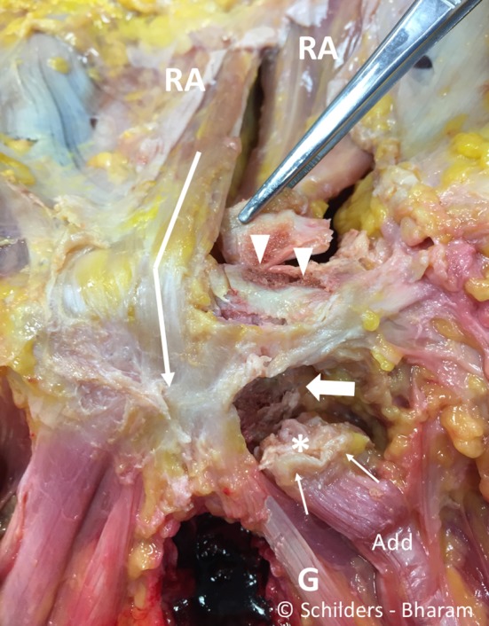
Purpose: Adductor longus injuries are complex. The conflict between views in the recent literature and various nineteenth-century anatomy books regarding symphyseal and perisymphyseal anatomy can lead to difficulties in MRI interpretation and treatment decisions. The aim of the study is to systematically investigate the pyramidalis muscle and its anatomical connections with adductor longus and rectus abdominis, to elucidate injury patterns occurring with adductor avulsions.

Methods: A layered dissection of the soft tissues of the anterior symphyseal area was performed on seven fresh-frozen male cadavers. The dimensions of the pyramidalis muscle were measured and anatomical connections with adductor longus, rectus abdominis and aponeuroses examined.

Results: The pyramidalis is the only abdominal muscle anterior to the pubic bone and was found bilaterally in all specimens. It arises from the pubic crest and anterior pubic ligament and attaches to the linea alba on the medial border. The proximal adductor longus attaches to the pubic crest and anterior pubic ligament. The anterior pubic ligament is also a fascial anchor point connecting the lower anterior abdominal aponeurosis and fascia lata. The rectus abdominis, however, is not attached to the adductor longus; its lateral tendon attaches to the cranial border of the pubis; and its slender internal tendon attaches inferiorly to the symphysis with fascia lata and gracilis.

Conclusion: The study demonstrates a strong direct connection between the pyramidalis muscle and adductor longus tendon via the anterior pubic ligament, and it introduces the new anatomical concept of the pyramidalis-anterior pubic ligament-adductor longus complex (PLAC). Knowledge of these anatomical relationships should be employed to aid in image interpretation and treatment planning with proximal adductor avulsions. In particular, MRI imaging should be employed for all proximal adductor longus avulsions to assess the integrity of the PLAC.

Keywords: Adductor injuries; Adductor longus avulsions; Anterior pubic ligament; Groin anatomy; Groin pain in athletes; Pyramidalis; Pyramidalis–anterior pubic ligament–adductor longus complex (PLAC); Rectus abdominis.


Conflict of interest statement

Conflict of interest

The authors do not report any conflict of interest for this study.

Funding

The authors want to acknowledge Maimonides Medical Center, Brooklyn, NY, USA, for the grant and use of the facilities and Mrs Maya D. Culbertson for the support with the dissection.

Ethical approval

The study did not represent research with living individuals or the use of protected health information and was declared exempt by our institutional review board. The study was undertaken at Maimonides, Medical Center, Brooklyn, NY, USA.

Figures

Fig. 1
Fig. 1
Male cadaver. Removal of the skin and the subcutaneous fat from the level of the anterior superior iliac spines to mid-thigh exposing the aponeurosis of the (asterisk) of the abdominal external oblique muscle, the rectus abdominis, the pyramidalis, the superficial part of the anterior pubic ligament (arrowhead) and the fascia lata (F). S spermatic cords. Arrow external inguinal ring
Fig. 2
Fig. 2
Male cadaver, after removal of the spermatic cords and the penis. There is a cord-like linea alba (A). The fibres of the external oblique aponeurosis that forms the medial part of the external inguinal ring (arrow) interlace with the superficial portion of the anterior pubic ligament (arrowhead)
Fig. 3
Fig. 3
a Male cadaver, after a window was made in the anterior rectus fascia adjacent to the linea alba on the left side. The aponeurosis anterior to the pyramidalis muscle and the rectus abdominis comprises two layers. The superficial layer has oblique oriented fibres (arrow right side), and the fibres of the deeper layer have a more horizontal orientation. The window in the superficial fascia of the left abdominal external oblique muscle demonstrates the transverse orientation of the fibres of the deep layer (arrowhead), anterior to the pyramidalis muscle (P). A linea alba. S spermatic cord. b Forceps holding the deeper layer of the fascia anterior to the right pyramidalis muscle. Note the transversely oriented fibres. These fibres interlace with the fibres from the rectus abdominis aponeurosis and the linea alba (arrowhead). A cord-like linea alba
Fig. 4
Fig. 4
a Male cadaver. Anterior symphyseal area after removal of the entire aponeurosis demonstrates the remnants of the superficial portion of the anterior pubic ligament (arrowhead). The forceps is placed between under the anterior pubic ligament that spans over the symphyseal joint. The pyramidalis muscle (P) arises from the pubic crest and the anterior pubic ligament. The proximal adductor longus tendon (white arrow) interlaces with the deep portion of the anterior pubic ligament (black arrows). The anterior pubic ligament has transversely orientated fibres (black arrows) and extends into the inguinal ligament (dashed line) bilaterally. Pect pectineus muscle. Add adductor longus muscle. RA rectus abdominis muscle. b Sagittal section through the pubic bone medial to the pubic tubercles and lateral to the symphyseal joint. Drawing demonstrating the pyramidalis–anterior pubic ligament–adductor longus complex comprising the pyramidalis muscle (P), anterior pubic ligament (arrow) and adductor longus tendon (arrow head) and fibrocartilage (FC). The rectus abdominis (RA) is posterior to the pyramidalis muscle, and its musculotendinous junction cranial to the superior edge of the symphysis. With traumatic avulsion of the adductor longus, the fibrocartilage (red dots) of the adductor is avulsed in partial tears. Both the fibrocartilage (red dots) and anterior pubic ligament (blue dots) avulse in complete tears
Fig. 5
Fig. 5
3D CT image of a male person showing the osteology of the anterior symphysis with the pubic crest (arrowheads) in between the pubic tubercles (arrows)
Fig. 6
Fig. 6
a Male cadaver, same specimen as Fig. 4. The pyramidalis muscle is sharply detached from the linea alba (white arrows) and folded distally. The internal tendons of the rectus abdominis run anterior to the symphyseal joint (white line), and the external tendon inserts on the superior lateral edge of the pubis (arrowheads). There is a thin aponeurosis on the posterior side of the pyramidalis muscle (P) with transverse fibre orientation. This aponeurosis does not cover the pyramidalis muscle anterior to the pubic bone (asterisk). Pect pectineus muscle. Add adductor longus muscle. A linea alba. Dashed line region of the anterior pubic ligament. RA rectus abdominis muscle. b Sagittal midline drawing demonstrating the anatomical relationship of the anterior pubic ligament (arrowhead), the pyramidalis muscle (P) and the internal tendon of the rectus abdominis (RA) at the level of the symphysis pubis. The internal tendon of the rectus abdominis receives contributions from left and right sides giving the tendon a Y-shape (arrow). The pyramidalis muscle inserts on the anterior pubic ligament, which spans the symphyseal joint and acts like a pulley through its position anterior to the internal tendon of the rectus abdominis muscle
Fig. 7
Fig. 7
Male cadaver. It shows the anterior symphyseal and perisymphyseal area after resection of the pyramidalis muscle and the anterior pubic ligament. It demonstrates that the internal tendon of the rectus abdominis interlaces with the gracilis (G) and fascia lata (arrow). It shows the footprint of the external tendon of the rectus abdominis (arrowheads) and the footprint of the adductor longus fibrocartilage (large arrow). The adductor longus (Add) tendon (thin arrows) and the fibrocartilage (asterisk) are detached. RA rectus abdominis muscle

References

    1. Anson BJ, Beaton LE, McVay CB. The pyramidalis muscle. Anat Rec. 1938;72:7.
    1. Beaton L, Anson B. The pyramidalis muscle: its occurrence and size in American whites and negroes. Am J Phys Anthropol. 1939;25:261–269. doi: 10.1002/ajpa.1330250236. - DOI
    1. Becker I, Woodley SJ, Stringer MD. The adult human pubic symphysis: a systematic review. J Anat. 2010;217:475–487. doi: 10.1111/j.1469-7580.2010.01300.x. - DOI - PMC - PubMed
    1. Bell J, Bell C (1816) The anatomy of the human body. Vol I containing the anatomy of the bones, muscles, and joints, the heart and circulation, and lungs. Vol I. 4th edn. Longman, Hurst, Rees, Orme, and Brown, Paternoster-Row, London. T. Cadell and W. Davies, Strand
    1. Boutin RD, Newman JS. MR imaging of sports-related hip disorders. Magn Reson Imaging Clin N Am. 2003;11:255–281. doi: 10.1016/S1064-9689(03)00029-1. - DOI - PubMed

LinkOut - more resources