Review
. 2017 Oct;33(10):677-702.
doi: 10.1016/j.tig.2017.08.001. Epub 2017 Aug 31.

Signaling and Gene Regulatory Networks in Mammalian Lens Development

Affiliations
Review

Signaling and Gene Regulatory Networks in Mammalian Lens Development

Ales Cvekl et al. Trends Genet. 2017 Oct.

Abstract

Ocular lens development represents an advantageous system in which to study regulatory mechanisms governing cell fate decisions, extracellular signaling, cell and tissue organization, and the underlying gene regulatory networks. Spatiotemporally regulated domains of BMP, FGF, and other signaling molecules in late gastrula-early neurula stage embryos generate the border region between the neural plate and non-neural ectoderm from which multiple cell types, including lens progenitor cells, emerge and undergo initial tissue formation. Extracellular signaling and DNA-binding transcription factors govern lens and optic cup morphogenesis. Pax6, c-Maf, Hsf4, Prox1, Sox1, and a few additional factors regulate the expression of the lens structural proteins, the crystallins. Extensive crosstalk between a diverse array of signaling pathways controls the complexity and order of lens morphogenetic processes and lens transparency.

Keywords: BMP; FGF; Pax6; Wnt signaling; cell determination; crystallins; differentiation; ectoderm; lens; pre-placodal region; retinoic acid.


Figures

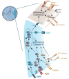
Figure 1
Figure 1. The earliest stages of vertebrate lens formation from the pre-placodal ectoderm
(A) Model of chicken lens cell specification based on the inductive signaling of BMP, the necessity of FGF signaling, and the inhibitory role of Wnt signaling. The precursors for lens and olfactory placodes are intermingled and surround the anterior neural plate domain. Additional cell types, including cells moving into the neural plate/tube and outside towards the ectoderm, are not shown for simplicity. (B) The choice between lens/olfactory and adenohypophyseal cell fates is regulated by Shh signaling promoting adenohypophyseal placode formation, is temporaly regulated the subsequent activation and inhibition of BMP signaling needed for olfactory placodal progenitors. Sustained BMP signaling is required for lens cell formation. (C) Schematic of chicken embryo at the neural fold stage (4–5 somites), Figures A–C adapted and modified from [8].
Figure 2
Figure 2. Stages of lens formation in mouse embryos from the prospective lens ectoderm, and eye/lens structure in the parietal “third” eye in lizard
(A) Mouse E8.5 (surface ectoderm). (B) Mouse E9.0 (prospective lens ectoderm). (C) Mouse E10.5 (invaginating lens placode - lens pit). (D) Mouse E11 (open lens vesicle). (E) Mouse E12.5 (primary lens fiber cell differentiation). (F) Mouse E13.5–E14.5 lens (completion of primary lens fiber cell elongation – initiation of secondary lens fiber cell formation). (G) Mature mouse lens. Note that the initial compartment formed by the primary lens fibers (E12.5–E14.5, panel F) now forms the central lens nucleus devoid of intracellular organelles. H) The parietal “third” eye of lizards. The lens is of neuroectodermal origin and is comprised from elongated nucleated cells immediatelly bellow a transparent layer resembling the cornea. The retina is comprised of photoreceptors and ganglion cells. Anterior lens epithelium, ALE; corneal epithelium, CE; invaginating lens placode, iLP; lens capsule, LC; lens epithelium, Epi; lens placode, LP; neuroretina, NR; optic vesicle, OV; periocular mesenchyme, POM; primary lens fibers, 10 LFs; prospective lens ectoderm, PLE; retinal pigmented epithelium, RPE; secondary lens fibers, 20 LFs; surface ectoderm, SE. Panels (A–E) are from [75], (F–G) [72, 322] and (H) is based on a scheme [323].
Figure 3
Figure 3. Transcriptional regulation of Pax6 in the lens
(A) Regulatory regions of the Pax6 locus. The mouse Pax6 locus resides within a 420 kb region of chromosome 2. The promoters P0 and P1, and EE and SIMO enhancers are indicated. (B) Transcription factors bound to EE include Meis1/2 [100, 284], Pou2f1(Oct1), Sox2 [287], Pax6 [324], Pknox1 [286], and Six3 [44]. Candidate binding sites for AP-1, Ets, and Smads are indicated. (C) Transcription factors bound to SIMO include Meis1/2 [100, 325], Pax6, and Six3 [44]. Candidate binding sites for AP-1, Ets, and Smads are indicated (see also Tables 2 and 3).
Figure 4
Figure 4. GRNs of αA-, βB1-, and γA-crystallin gene expression
Lens cell differentiation (light-moderate-dark blue contours) is illustrated at mouse embryonic stages E10.5, E11.5, E12.5, and E14.5. The individual GRNs are formed from the “core” GRN comprised of a feed-forward loop consisting of Pax6, c-Maf, and αA-crystallin [189] from E10.5. From E11.5 in posterior cells of the lens vesicle, FGF signaling activates the αA-crystallin indirectly via a regulatory region in the c-Maf promoter and through both the αA-crystallin upstream enhancer DCR1 [158] and promoter [189]. Transcriptional regulation of βB1-crystallin is comprised from the common “core” module, expanded by a Pax6 → Prox1 module [139] active from E12.5. Expression of γA-crystallin requires Sox1 [171, 175] and from E14.5 is further augmented by Hsf4 [170] and the FGF/MAPK pathway [187]. Other regulatory layers are involved as Hsf4 inhibits the expression of Fgf1, Fgf4, Fgf7, and Fgfr1 [170], and Prox1 regulates the expression of Fgfr3, Fgfrl1, and Lctl and its expression is downstream of FGF/MAPK cascade [186].
Figure 5
Figure 5. Lens development and extracellular signaling
(A) Schematic diagram of signaling pathways in the epithelial cells at the transitional zone of the lens. FGF and integrin signaling promote cell proliferation and survival, whereas Notch signaling induced by adjacent lens fiber cells suppresses cell differentiation. In addition, Wnt signaling transmitted by Lrp5/6 and Fzd receptors converges with cadherin and Yap to regulate cell differentiation, polarity and adhesion. (B) In differentiating fiber cells, FGF signaling through the Ras-MAPK pathway activates ETS and AP1 family transcription factors to induce expression of lens differentiation factors such as Prox1 and c-Maf. FGF also synergizes with Smad-mediated BMP signaling in cell differentiation and cooperate with Cx50 in p27-mediated cell cycle exit. Cell adhesion molecules integrin and N-cadherin act with Eph/Ephrin receptors to induce small GTPases and Src family kinases to promote actin assembly and cell elongation.

References

    1. Grainger RM. Embryonic lens induction: shedding light on vertebrate tissue determination. Trends Genet. 1992;8:349–55. - PubMed
    1. Piatigorsky J. Lens differentiation in vertebrates. A review of cellular and molecular features. Differentiation. 1981;19:134–53. - PubMed
    1. Ashery-Padan R, et al. Pax6 activity in the lens primordium is required for lens formation and for correct placement of a single retina in the eye. Genes Dev. 2000;14:2701–11. - PMC - PubMed
    1. Beebe DC, Coats JM. The lens organizes the anterior segment: specification of neural crest cell differentiation in the avian eye. Dev. Biol. 2000;220:424–31. - PubMed
    1. Silla ZT, et al. Signals from the lens and Foxc1 regulate the expression of key genes during the onset of corneal endothelial development. Exp. Cell Res. 2014;322:381–8. - PubMed

Publication types

LinkOut - more resources