. 2017 Sep 7;170(6):1175-1183.e11.
doi: 10.1016/j.cell.2017.08.005. Epub 2017 Aug 31.

Mating in the Closest Living Relatives of Animals Is Induced by a Bacterial Chondroitinase

Affiliations

Mating in the Closest Living Relatives of Animals Is Induced by a Bacterial Chondroitinase

Arielle Woznica et al. Cell. .

Abstract

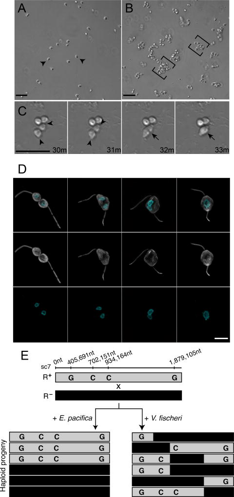
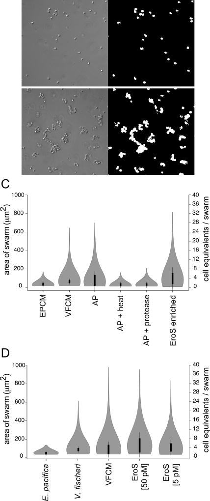
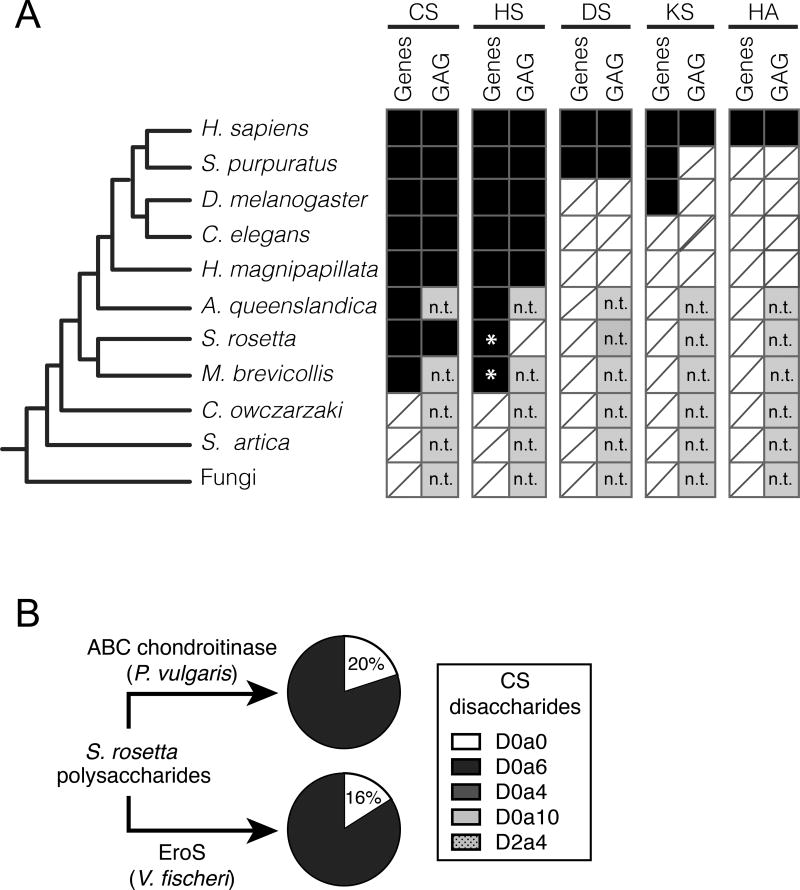
We serendipitously discovered that the marine bacterium Vibrio fischeri induces sexual reproduction in one of the closest living relatives of animals, the choanoflagellate Salpingoeca rosetta. Although bacteria influence everything from nutrition and metabolism to cell biology and development in eukaryotes, bacterial regulation of eukaryotic mating was unexpected. Here, we show that a single V. fischeri protein, the previously uncharacterized EroS, fully recapitulates the aphrodisiac-like activity of live V. fischeri. EroS is a chondroitin lyase; although its substrate, chondroitin sulfate, was previously thought to be an animal synapomorphy, we demonstrate that S. rosetta produces chondroitin sulfate and thus extend the ancestry of this important glycosaminoglycan to the premetazoan era. Finally, we show that V. fischeri, purified EroS, and other bacterial chondroitin lyases induce S. rosetta mating at environmentally relevant concentrations, suggesting that bacteria likely regulate choanoflagellate mating in nature.

Keywords: Vibrio; choanoflagellate; chondroitin lyase; chondroitin sulfate; chondroitinase; host-microbe; mating; swarming; symbiosis.


Figures

Figure 1
Figure 1. V. fischeri bacteria induce swarming and mating in the choanoflagellate, S. rosetta
(A) In the absence of V. fischeri, motile S. rosetta cells (arrowheads) are evenly dispersed. (B) Within 30 minutes of exposure to V. fischeri, S. rosetta motile cells aggregate into large swarms (brackets). Scale bar = 20µm. (C) S. rosetta cells within a swarm pair and fuse. Prior to fusion, cells reposition themselves such that their basal membranes are adjacent and their apical flagella point away (31’; arrowheads mark apical pole of unfused cells). Cell fusion takes only minutes, and occurs along the basal membrane (32’; indicated by arrow), resulting in a single, elongated cell (33’; indicated by arrow). Scale bar = 20µm. (D) Stages of cell and nuclear fusion in S. rosetta mating pairs. Haploid mating pairs are oriented with their basal poles (opposite the flagellum) touching (D1), and cell fusion proceeds along the basal membrane, resulting in a binucleated cell with two flagella (D2). Nuclei then congress towards the midline (D3), where the nuclei undergo nuclear fusion, resulting in a diploid cell (D4). Anti-tubulin antibody (D1’-4’; white) highlights the cell body and flagellum, and Hoechst (D1’’-4’’; cyan) highlights the nucleus. Scale bar = 5µm (E) Evidence for meiotic recombination in S. rosetta following exposure to V. fischeri. Two haploid, genotypically distinct S. rosetta strains [R+(grey shading) and R- (black shading)] were mixed in the presence of either E. pacifica conditioned media (EPCM) or V. fischeri conditioned media (VFCM) for 16 hours. Haploid progeny were clonally isolated and genotyped at polymorphic markers across the genome (Supplemental Data). We show here genotyping results for four representative loci along supercontig 7 (sc7). All clones isolated from EPCM-treated cultures contained unrecombined parental genotypes, while haploid clones isolated from VFCM-treated cultures showed clear evidence of recombination. Top numbers show marker genomic positions along sc7.
Figure 2
Figure 2. Bioactivity-guided isolation of the V. fischeri aphrodisiac
(A, B) Automated image analysis allowed quantification of S. rosetta swarming in response to V. fischeri-derived activity. Pictured are S. rosetta cells 30 minutes after treatment with E. pacifica conditioned media (A) or V. fischeri conditioned media (B). By generating a binary mask (A’, B’) we could measure the area of each swarm and estimate the number of cells (“cell equivalents”) per swarm. (C) The V. fischeri aphrodisiac is a secreted protein. Swarming in S. rosetta is induced by V. fischeri culture supernatant (VFCM), as well as compounds in the ammonium sulfate precipitation of V. fischeri culture supernatant (AP), but not by AP exposed to heat (80°C for 10 minutes; AP + Heat) or proteases (AP + Protease), or by culture supernatant from the prey bacterium E. pacifica (EPCM). Proteins in the ammonium sulfate precipitation of V. fischeri culture supernatant were separated by size exclusion and anion exchange chromatography, and the aphrodisiac activity tracked with a ~90kD protein band (EroS enriched) that was revealed by mass spectrometry to be the V. fischeri EroS protein (also see Figure S2 and STAR Methods. Data are presented as violin box plots, showing the median cell number (white circle), interquartile range (thick line), and range excluding outliers (thin line). Surrounding the box plot is a kernel density trace, plotted symmetrically to show the swarm area frequency distribution. A minimum of 650 swarm areas were plotted for each condition. (D) EroS triggers mating at plausible environmental concentrations. Purified EroS induces swarming in S. rosetta at concentrations as low as 5 pM, and is sufficient to fully recapitulate the aphrodisiac activity of live V. fischeri bacteria and VFCM.
Figure 3
Figure 3. The V. fischeri aphrodisiac is a chondroitinase
(A) Alignment of the V. fischeri EroS amino acid sequence to diverse bacterial GAG lyases reveals that V. fischeri harbors conserved His and Tyr residues (indicated by *) at sites required for catalytic activity in characterized GAG lyases (Han et al., 2014; Linhardt et al., 2006). Amino acids with >50% conservation between sequences are shaded (black shading for identical amino acids and grey shading for similar amino acids. (B) Purified EroS degrades chondroitin sulfate and hyaluronan. EroS was incubated with purified chondroitin sulfate (open circle), hyaluronan (grey hexagon), dermatan sulfate (open square), and heparan sulfate (grey triangle), and GAG lyase activity of EroS was measured by monitoring the abundance of unsaturated oligosaccharide reaction products with an absorbance at 232nm. Chondroitin sulfate and hyaluronan oligosaccharides accumulated rapidly in the presence of EroS, indicating depolymerization, whereas heparan sulfate and dermatan sulfate were not depolymerized by EroS. (C) Alanine substitutions at two predicted catalytic residues in EroS (H278 and Y287) eliminated the protein’s ability to degrade chondroitin sulfate. The chondroitinase activity of either wild type EroS (open circle) or EroS-H278A,Y287A (filled circle) against purified chondroitin sulfate was measured by monitoring the abundance of unsaturated oligosaccharide products with an absorbance at 232nm. (D) The chondroitinase activity of EroS is necessary and sufficient for its function as an aphrodisiac. EroS-H278A,Y287A failed to induce swarming in S. rosetta. P. vulgaris ABC chondroitinase and F. heparinum AC chondroitinase were sufficient to induce swarming at levels similar to EroS, whereas S. hyalurolyticus hyaluronidase failed to induce swarming, indicating that chondroitinase activity is necessary and sufficient for aphrodisiac activity.
Figure 4
Figure 4. S. rosetta produces chondroitin sulfate that can be degraded by EroS
(A) Phylogenetic distribution of diverse GAGs [CS= chondroitin sulfate; HS= heparan sulfate; DS= dermatan sulfate; KS= keratan sulfate; HA= hyaluronan], and their biosynthetic genes. The presence (black box) and absence (white box with slash) of genes required for the biosynthesis of GAGs (“Gene”) and biochemical evidence for GAGs (“GAG”) in S. rosetta and select opisthokonts. *; Ori et al. (2011) identified putative homologs of a subset of HS biosynthetic enzymes in the M. brevicollis genome, and we detect homologs of the same limited set of HS biosynthetic enzymes in S. rosetta. Importantly, these enzymes are shared components of the chondroitin biosynthetic pathway, and digestion of S. rosetta polysaccharides with heparinases failed to liberate heparan sulfate disaccharides, suggesting S. rosetta does not produce heparan sulfate (also refer to Figure S3). n.t.; not tested (experiments have not been performed to biochemically profile GAGs). (B) S. rosetta produces chondroitin that can be degraded by EroS. Polysaccharides isolated from S. rosetta were treated with either P. vulgaris ABC chondroitinase, an enzyme that can degrade many modifications of chondroitin into its disaccharide units (CS disaccharides), or purified EroS. Both ABC chondroitinase and EroS yielded similar amounts of unsulfated chondroitin disaccharide (D0a0) and chondroitin-6-sulfate disaccharide (D0a6) degradation products, indicating that unsulfated chondroitin and chondroitin-6-sulfate are produced by S. rosetta. In contrast, we were unable to detect chondroitin-4-sulfate (D0a4), chondroitin-4,6-sulfate (D0a10), or chondroitin-2,4-sulfate (D2a4) following degradation of S. rosetta polysaccharides with either EroS or ABC chondroitinase.

Comment in

References

    1. Ahn MY, Shin KH, Kim DH, Jung E-A, Toida T, Linhardt RJ, Kim YS. Characterization of a Bacteroides species from human intestine that degrades glycosaminoglycans. Can. J. Microbiol. 2011;44:423–429. - PubMed
    1. Alegado RA, Brown LW, Cao S, Dermenjian RK, Zuzow R, Fairclough SR, Clardy J, King N. A bacterial sulfonolipid triggers multicellular development in the closest living relatives of animals. Elife. 2012;1:e00013. - PMC - PubMed
    1. Avery MI. Lekking in birds: choice, competition and reproductive constraints. Ibis. 1984;126:177–187.
    1. Babcock R, Mundy C, Keesing J, Oliver J. Predictable and unpredictable spawning events: in situ behavioural data from free-spawning coral reef invertebrates. Invertebrate Reproduction & Development. 2011;22:213–227.
    1. Bartels-Hardege HD, Hardege JD, Zeeck E, Müller C, Wu BL, Zhu MY. Sex pheromones in marine polychaetes: a biologically active volatile compound from the coelomic fluid of female Nereis (Neanthes) japonica. Journal of Experimental Marine Biology and Ecology. 1996;201:275–284.

MeSH terms

Substances