. 2017 Oct 28:264:203-210.
doi: 10.1016/j.jconrel.2017.08.037. Epub 2017 Sep 1.

Calcified plaque modification alters local drug delivery in the treatment of peripheral atherosclerosis

Affiliations

Calcified plaque modification alters local drug delivery in the treatment of peripheral atherosclerosis

Abraham R Tzafriri et al. J Control Release. .

Abstract

Background: Calcific atherosclerosis is a major challenge to intraluminal drug delivery in peripheral artery disease (PAD).

Objectives: We evaluated the effects of orbital atherectomy on intraluminal paclitaxel delivery to human peripheral arteries with substantial calcified plaque.

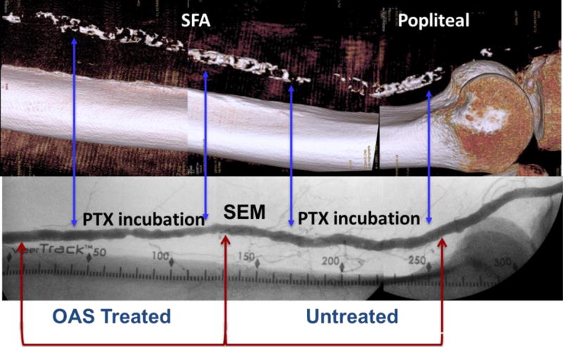
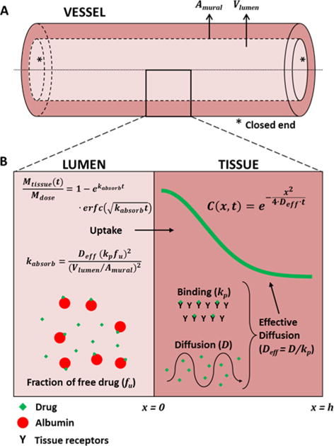
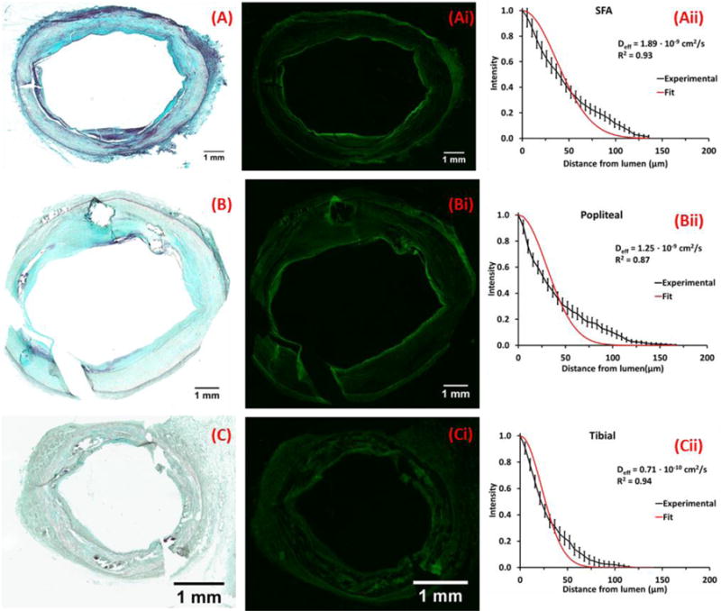
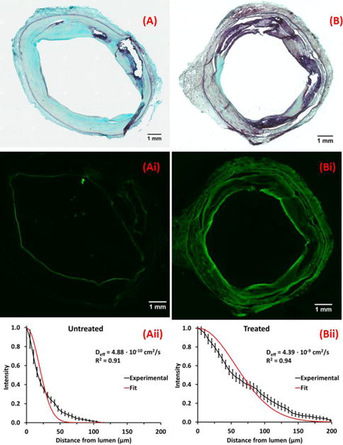
Methods: Diagnostic angiography and 3-D rotational imaging of five fresh human lower limbs revealed calcification in all main arteries. The proximal or distal segment of each artery was treated using an orbital atherectomy system (OAS) under simulated blood flow and fluoroscopy. Explanted arterial segments underwent either histomorphometric assessment of effect or tracking of 14C-labeled or fluorescent-labeled paclitaxel. Radiolabeled drug quantified bulk delivery and fluorescent label established penetration of drug over finer spatial domain in serial microscopic sections. Results were interpreted using a mathematical model of binding-diffusion mediated arterial drug distribution.

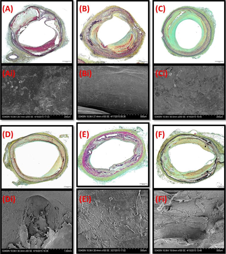
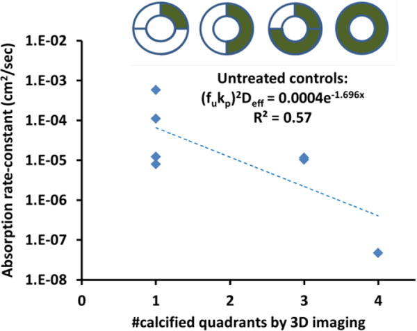
Results: Lesion composition affected paclitaxel absorption and distribution in cadaveric human peripheral arteries. Pretreatment imaging calcium scores in control femoropopliteal arterial segments correlated with a log-linear decline in the bulk absorption rate-constant of 14C-labeled, declining 5.5-fold per calcified quadrant (p=0.05, n=7). Compared to controls, OAS-treated femoropopliteal segments exhibited 180μm thinner intima (p<0.001), 45% less plaque calcification, and 2 log orders higher paclitaxel bulk absorption rate-constants. Correspondingly, fluorescent paclitaxel penetrated deeper in OAS-treated femoropopliteal segments compared to controls, due to a 70% increase in diffusivity (p<0.001).

Conclusions: These data illustrate that calcified plaque limited intravascular drug delivery, and controlled OAS treatment of calcific plaques resulted in greater drug permeability and improved adjunct drug delivery to diseased arteries.

Keywords: Atherectomy; Drug coated balloons; Drug eluting stents; Orbital atherectomy, calcified plaque; Paclitaxel; Peripheral artery disease.


Figures

Figure 1
Figure 1. Experimental paradigm
Representative angiographic image (bottom) of the femoropopliteal artery illustrating treatment location and vessel segmentation. Segments assigned for fluorescent or radiolabeled drug infusion are demarcated by arrows (blue) and their corresponding calcium burden depicted in reconstructed 3-D images (top). Inserts depict cross section views along green demarcation arrows.
Figure 2
Figure 2. Schematic of diffusion-partitioning controlled arterial drug uptake and distribution
(A) Schematic of an excised arterial segment infused with drug solution and both ends were closed. Vlumen is the lumen volume and Amural is the surface area of the infusate/tissue interface (B) High magnification cross sectional view of the lumen and arterial media, illustrating the key processes and equations governing drug distribution from a well-mixed infusate volume (Vlumen). In the lumen, a fraction fu, if the drug is free and the remainder is bound to albumin). Only free drug is absorbed by the artery wall. Once in the tissue, free drug diffuses with diffusion coefficient D or binds nonspecifically to tissue proteins with partition coefficient kp. Drug distribution is dominated by the effective diffusion coefficient Deff, (Eq. 3) whereas total drug uptake is also governed by the partition coefficient, lumen volume, mural area and the fraction of free drug (Eqs. 1–2).
Figure 3
Figure 3. OAS treatment affects neointimal morphology
Representative MP stained cross sections (A-F) and matching enface SEM (Ai-Fi) of untreated (A-C) and OAS-treated (D-Fi) artery segments. (A,D) the SFA, (B,E) popliteal arteries, (C,F) tibial arteries.
Figure 4
Figure 4. Paclitaxel absorption kinetics scale inversely with calcification scores
Bulk absorption rate-constants of paclitaxel (diamonds) decreased exponentially with increasing calcium scores in control femoropopliteal arteries.
Figure 5
Figure 5. Morphology and drug distribution of representative untreated arterial segments
Representative MP stained frozen sections of the SFA (A) popliteal (B) and tibial (C) arteries from a single leg; neointima thicknesses are respectively 372±166μm (A), 737±704μm (B) and 111±44μm (C). Corresponding fluorescent micrographs (Ai, Bi, Ci) and normalized circumferentially averaged intensity plots (Aii, Bii, Cii) are depicted (black lines) to the right of each micrograph along with best fit diffusion-binding predictions (red line, Eq. 3). Best-fit effective diffusivities are respectively 1.90×10⁻⁹ cm2/sec (A), 1.25×10⁻⁹ cm2/sec (B) and 0.71×10⁻⁹ cm2/sec (C).
Figure 6
Figure 6. Morphology and drug distribution in control and OAS treated sections of a representative SFA
Representative MP stained frozen sections of untreated (A) and OAS-treated (B) SFA segments, and corresponding fluorescent micrographs (Ai, Bi) and normalized circumferentially averaged intensity plots (Aii, Bii, Cii) are depicted (black lines) to the right of each micrograph along with best fit diffusion-binding predictions (red line, Eq. 3). Intima thicknesses are 677±461μm (A) and 717±324μm (B). Best-fit effective diffusivities are 0.49×10⁻⁹ cm2/sec (A), 4.39×10⁻⁹ cm2/sec (B).

References

    1. Rocha-Singh KJ, Zeller T, Jaff MR. Peripheral arterial calcification: prevalence, mechanism, detection, and clinical implications. Catheter Cardiovasc Interv. 2014;83:E212–220. - PMC - PubMed
    1. Kashyap VS, Pavkov ML, Bishop PD, Nassoiy SP, Eagleton MJ, Clair DG, Ouriel K. Angiography underestimates peripheral atherosclerosis: lumenography revisited. J Endovasc Ther. 2008;15:117–125. - PMC - PubMed
    1. Fitzgerald PJ, Ports TA, Yock PG. Contribution of localized calcium deposits to dissection after angioplasty. An observational study using intravascular ultrasound. Circulation. 1992;86:64–70. - PubMed
    1. Adlakha S, Sheikh M, Wu J, Burket MW, Pandya U, Colyer W, Eltahawy E, Cooper CJ. Stent fracture in the coronary and peripheral arteries. J Interv Cardiol. 2010;23:411–419. - PubMed
    1. Micari A, Cioppa A, Vadala G, Castriota F, Liso A, Marchese A, Grattoni C, Pantaleo P, Cremonesi A, Rubino P, Biamino G. Clinical evaluation of a paclitaxel-eluting balloon for treatment of femoropopliteal arterial disease: 12-month results from a multicenter Italian registry. JACC Cardiovasc Interv. 2012;5:331–338. - PubMed

MeSH terms

Substances