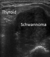
A Cervical Schwannoma Masquerading as a Thyroid Nodule
- PMID: 28868262
- PMCID: PMC5566684
- DOI: 10.1159/000454877
A Cervical Schwannoma Masquerading as a Thyroid Nodule
Abstract
Background: We present a case of a cervical schwannoma, likely originating from the pharyngeal plexus of the vagal nerve. The lesion masqueraded as a thyroid nodule and magnetic resonance imaging (MRI) assisted in preoperative diagnosis. We review the radiographic characteristics of nerve sheath tumors on MRI as well as the diagnostic cytologic stains which can enhance the possibility of a correct preoperative diagnosis.
Case: We describe a 60-year-old female with dysphagia and a neck mass consistent with a nodular goiter. The patient's history, diagnostic images, cytology, pathology, and surgical management are presented and analyzed. The preoperative diagnosis of a cervical schwannoma was suspected by the use of MRI which led to additional specialized cytologic stains.
Conclusion: Pharyngeal wall schwannomas are important to consider in the differential diagnosis of thyroid nodules when fine needle aspiration cytology indicates cells of neural origin. Imaging by MRI can assist in identifying lesions of neural origin masquerading as thyroid nodules.
Keywords: Magnetic resonance imaging; S-100; Schwannoma; Thyroid nodule.
Figures


References
-
- Kandil E, Abdel Khalek M, Abdullah O, Dali D, Faruqui S, Khan A, Friedlander P, Jaffe BM, Crawford B. Primary peripheral nerve sheath tumors of the thyroid gland. Thyroid. 2010;20:583–586. - PubMed
-
- Subramaniam V, Adarsha T. Schwannoma of the thyroid gland - a case report. J Chir. 2010;6:536–538.
-
- Gustafson LM, Liu JH, Rutter MJ, Stern Y, Cotton RT. Primary neurilemoma of the thyroid gland: a case report. Am J Otolaryngol. 2001;22:84–86. - PubMed
LinkOut - more resources
Full Text Sources
Other Literature Sources

