

Evaluation of Renal Blood Flow in Chronic Kidney Disease Using Arterial Spin Labeling Perfusion Magnetic Resonance Imaging
- PMID: 28868513
- PMCID: PMC5575771
- DOI: 10.1016/j.ekir.2016.09.003
Evaluation of Renal Blood Flow in Chronic Kidney Disease Using Arterial Spin Labeling Perfusion Magnetic Resonance Imaging
Abstract
Introduction: Chronic kidney disease (CKD) is known to be associated with reduced renal blood flow. However, data to-date in humans is limited.
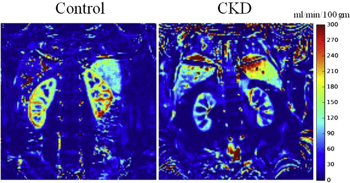
Methods: In this study, non-invasive arterial spin labeling (ASL) MRI data was acquired in 33 patients with diabetes and stage-3 CKD, and 30 healthy controls.
Results: A significantly lower renal blood flow both in cortex (108.4±36.4 vs. 207.3±41.8; p<0.001, d=2.52) and medulla (23.2±8.9 vs. 42.6±15.8; p<0.001, d=1.5) was observed. Both cortical (ρ=0.67, p<0.001) and medullary (ρ=0.62, p<0.001) blood flow were correlated with eGFR, and cortical blood flow was found to be confounded by age and BMI. However, in a subset of subjects that were matched for age and BMI (n=6), the differences between CKD and control subjects remained significant both in cortex (107.4±42.8 vs. 187.51±20.44; p=0.002) and medulla (15.43±8.43 vs. 39.18±11.13; p=0.002). A threshold value to separate healthy and CKD was estimated to be Cor_BF=142.9 and Med_BF=24.1.
Conclusion: These results support the use of ASL in the evaluation of renal blood flow in patients with moderate level of CKD. Whether these measurements can identify subjects at risk of progressive CKD requires further longitudinal follow-up.
Keywords: MRI; arterial spin labeling; chronic kidney disease; eGFR; perfusion; renal blood flow.
Figures
References
-
- Fine L.G., Norman J.T. Chronic hypoxia as a mechanism of progression of chronic kidney diseases: from hypothesis to novel therapeutics. Kidney Int. 2008;74:867–872. - PubMed
-
- Fine L.G., Orphanides C., Norman J.T. Progressive renal disease: the chronic hypoxia hypothesis. Kidney Int Suppl. 1998;65:S74–S78. - PubMed
Grants and funding
LinkOut - more resources
Full Text Sources
Other Literature Sources
Research Materials
Miscellaneous