Observational Study
. 2017 Nov;90(1079):20170082.
doi: 10.1259/bjr.20170082. Epub 2017 Oct 9.

Approach to interpret images produced by new generations of multidetector CT scanners in post-operative spine

Affiliations
Observational Study

Approach to interpret images produced by new generations of multidetector CT scanners in post-operative spine

Rania Zeitoun et al. Br J Radiol. 2017 Nov.

Abstract

Objective: To reach a practical approach to interpret MDCT findings in post-operative spine cases and to change the false belief of CT failure in the setting of instruments secondary to related artefacts.

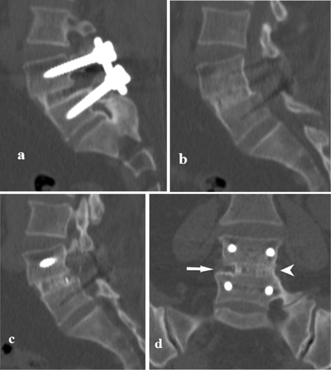
Methods: We performed observational retrospective analysis of premier, early and late MDCT scans in 68 post-operative spine patients, with emphasis on instruments related complications and osseous fusion status. We used a grading system for assessment of osseous fusion in 35 patients and we further analysed the findings in failure of fusion, grade (D).

Results: We observed a variety of instruments related complications (mostly screws medially penetrating the pedicle) and osseous fusion status in late scans. We graded 11 interbody and 14 posterolateral levels as osseous fusion failure, showing additional instruments related complications, end plates erosive changes, adjacent segments spondylosis and malalignment.

Conclusion: Modern MDCT scanners provide high quality images and are strongly recommended in assessment of the instruments and status of osseous fusion. In post-operative imaging of the spine, it is essential to be aware for what you are looking for, in relevance to the date of surgery. Advances in knowledge: Modern MDCT scanners allow assessment of instruments position and integrity and osseous fusion status in post-operative spine. We propose a helpful algorithm to simplify interpreting post-operative spine imaging.


Figures

Figure 1.
Figure 1.
Different grades of malpositioned lumbar pedicular screws on axial images. (a) Left screw: severe medial penetration of the pedicle intruding into the spinal canal, Right screw: adequately positioned, completely contained within the pedicle and surrounded by cortex (b) Right screw: anterior cortex penetration (c) Left screw: severe medial violation of the lateral recess, the screw is in contact with the nerve root (arrow) (d) Left screw: minor medial penetration of the pedicle (e) Right screw: moderate medial penetration touching the nerve root, Left screw: completely contained within the pedicle.
Figure 2.
Figure 2.
Malpositioned screws on sagittal images: screws are penetrating the upper end plate (a) and lower end plate (b) and intruding into the intervertebral disc spaces.
Figure 3.
Figure 3.
Complete osseous fusion at L4/5 interbody space, grade (A): decompression surgery at L4-5 level and internal fixation; 18 months following surgery. Sagittal (a, b) and coronal (c) images: bone bridges are fully covering the interbody space.
Figure 4.
Figure 4.
Incomplete osseous fusion at L4/5 interbody space, grade (B): decompression surgery at L4-5 level and internal fixation. Sagittal (a, b, c) and coronal (d) images: bone bridges are covering middle and left two-thirds of the interbody space around adequately positioned disc cage (arrow head), while the right third does not yet show bone bridges (arrow).
Figure 5.
Figure 5.
Failure of osseous fusion at L4/5 interbody space, grade (D): decompression surgery at L4-5 level and internal fixation. Sagittal images (a, b): lack of bone bridges all through the interbody space in addition to erosive changes of the vertebral end plates (short arrow) and anterior displacement of L4. Axial images (c, d): malpositioned right screw, severely medially penetrating the pedicle and intruding into the spinal canal.
Figure 6.
Figure 6.
Failure of osseous fusion at L4/5 interbody space, grade (D) and chronic spondylodiscitis: decompression surgery at L4-5 level and internal fixation. Sagittal (a) and coronal (b) images: lack of bone bridges all through the interbody space in addition to mixed erosive and sclerotic changes of the vertebral bodies representing sequel of chronic spondylodiscitis. Axial CT images (c, d, e): evident erosions of the vertebral end plates mixed with sclerosis, osteolysis around the pedicular screw (arrow) with subsequent loosening and penetration of the lateral cortex.
Figure 7.
Figure 7.
Failure of osseous fusion at L5/S1 interbody space with pseudoarthrosis, grade (D): decompression surgery at L5-S1 level, disc cage placement and internal fixation using pedicular screws and rods. Sagittal (a, b, c) and coronal (d, e) images: lack of bone bridges all through the intervertebral body space, apart from bone within the disc cage, representing mature pseudoarthrosis (arrow). Anterior displacement of L5, erosive and sclerotic changes of the opposing end plates, vacuum phenomenon and migration of cage material (double arrow).
Figure 8.
Figure 8.
Fusion at L3/4 posterobilateral spaces: solid bone mass completely covering the right posterolateral space (long arrow), while on the left incomplete bone bridges with linear defects and sclerosed bone margins are noted (short arrow) representing pseudoarthrosis.
Figure 9.
Figure 9.
Algorithm for possible complications on MDCT images of post-operative spine.

References

    1. Douglas-Akinwande AC, Buckwalter KA, Rydberg J, Rankin JL, Choplin RH. Multichannel CT: evaluating the spine in postoperative patients with orthopedic hardware. Radiographics 2006; 26 Suppl 1: S97–S110. DOI: https://doi.org/10.1148/rg.26si065512 - DOI - PubMed
    1. Thakkar RS, Malloy JP, Thakkar SC, Carrino JA, Khanna AJ. Imaging the postoperative spine. Radiol Clin North Am 2012; 50: 731–47. DOI: https://doi.org/10.1016/j.rcl.2012.04.006 - DOI - PubMed
    1. Hayashi D, Roemer FW, Mian A, Gharaibeh M, Müller B, Guermazi A. Imaging features of postoperative complications after spinal surgery and instrumentation. AJR Am J Roentgenol 2012; 199: W123–W129. DOI: https://doi.org/10.2214/AJR.11.6497 - DOI - PubMed
    1. Lotfinia I, Sayahmelli S, Gavami M. Postoperative computed tomography assessment of pedicle screw placement accuracy. Turk Neurosurg 2010; 20: 500–7. DOI: https://doi.org/10.5137/1019-5149.JTN.3215-10.1 - DOI - PubMed
    1. Carter JD, Swearingen AB, Chaput CD, Rahm MD. Clinical and radiographic assessment of transforaminal lumbar interbody fusion using HEALOS collagen-hydroxyapatite sponge with autologous bone marrow aspirate. Spine J 2009; 9: 434–8. DOI: https://doi.org/10.1016/j.spinee.2008.11.004 - DOI - PubMed

Publication types

MeSH terms