

doi: 10.1038/nmeth.4400.
Epub 2017 Sep 4.
Red-shifted luciferase-luciferin pairs for enhanced bioluminescence imaging
Affiliations
- PMID: 28869756
- PMCID: PMC5678970
- DOI: 10.1038/nmeth.4400
Item in Clipboard
Red-shifted luciferase-luciferin pairs for enhanced bioluminescence imaging
Nat Methods.
2017 Oct.
Abstract
Red-shifted bioluminescence reporters are desirable for biological imaging. We describe the development of red-shifted luciferins based on synthetic coelenterazine analogs and corresponding mutants of NanoLuc that enable bright bioluminescence. One pair in particular showed superior in vitro and in vivo sensitivity over commonly used bioluminescence reporters. We adapted this pair to develop a bioluminescence resonance-energy-based Antares reporter called Antares2, which offers improved signal from deep tissues.
Figures
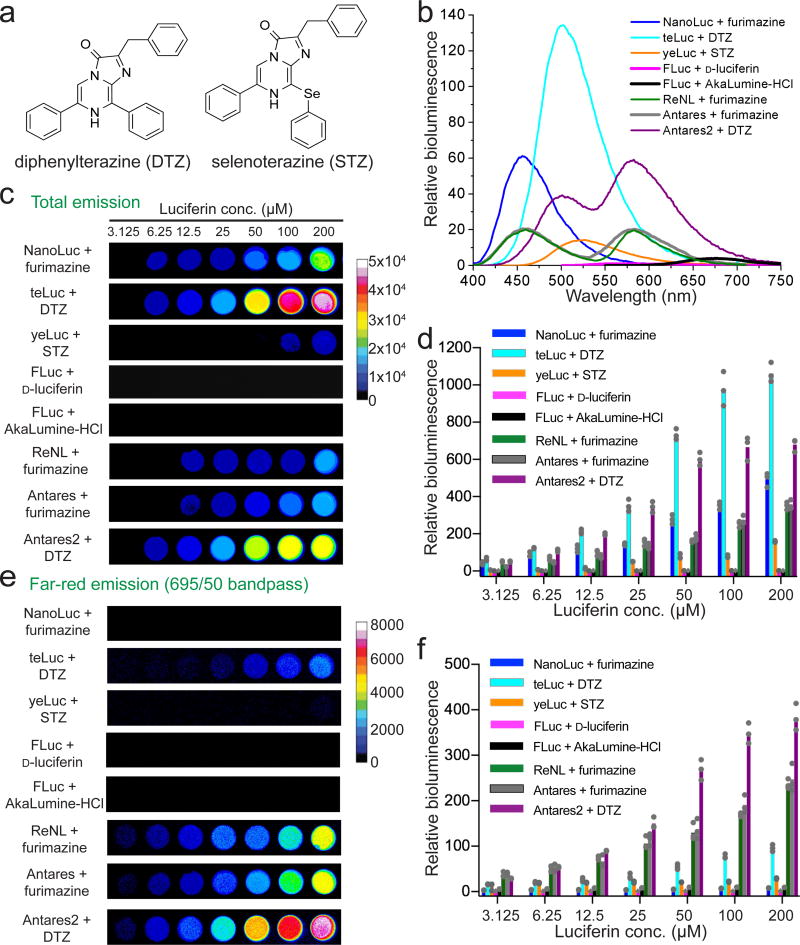
(a) Chemical structures of diphenylterazine (DTZ) and selenoterazine (STZ). (b) Bioluminescence emission of purified luciferases (1 nM) with their corresponding luciferin substrates (30 µM). The spectra were normalized to peak emission of FLuc/d -luciferin. (c–f) Representative pseudocolored images (c,e) and quantifications (d,f) of luciferase-expressing HEK 293T cells in the presence of various luciferins. Images were acquired without a filter (c) or with a 695±25 nm NIR emission filter (e). Panels d and f are quantification results for Panels c and e, respectively. All values were normalized to the intensities of FLuc/d -luciferin (50 µM) under the same imaging conditions. The graphs show mean values and individual data points of three independent measurements.
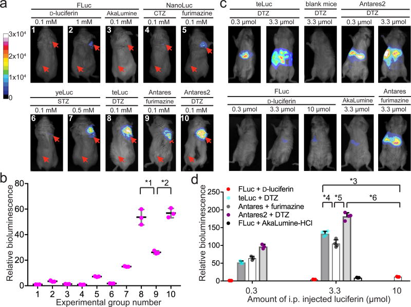
(a,b) Representative bioluminescence images (a) and quantitative analysis (b) of BALB/c mice with subcutaneously injected luciferase-expressing HEK 293T cells and 100 µL luciferin substrates at the indicated concentrations. The group numbers in panel b are aligned with those in panel a. Two injection sites (one for luciferase-expressing cells and one for empty vehicle controls) for each mouse are illustrated with red arrows. Intensity values were normalized to the intensity of FLuc/d -luciferin (0.1 mM) acquired under the same condition. The graphs show mean values and individual data points of three independent measurements. (c,d) Representative bioluminescence images (c) and quantitative analysis (d) of BALB/c mice, to which luciferase-coding plasmids were hydrodynamically delivered through tail vein injection, and luciferase substrates were intraperitoneally injected at 12 h post-plasmid injection. Intensity values were normalized to the intensity of FLuc/d -luciferin (0.3 µmol). Data are shown as individual data points and mean with s.d. (n = 4 for teLuc, Antares, and Antares2 with 3.3 µmol substrates, and n = 3 for all other groups). Unpaired two-tailed t-tests were used to compare teLuc/DTZ and Antares2/DTZ with FLuc/d -luciferin or Antares/furimazine (*1: P=0.0015; *2: P=0.0002; *3: P<0.0001; *4: P=0.0042; *5: P<0.0001; *6: P<0.0001), indicating the existence of a significant enhancement by teLuc and Antares2.
References
MeSH terms
Substances
Grants and funding
LinkOut - more resources
Full Text Sources
Other Literature Sources
Research Materials
