. 2018 Apr:14:72-81.
doi: 10.1016/j.redox.2017.08.011. Epub 2017 Aug 24.

PKR activation causes inflammation and MMP-13 secretion in human degenerated articular chondrocytes

Affiliations

PKR activation causes inflammation and MMP-13 secretion in human degenerated articular chondrocytes

Ching-Hou Ma et al. Redox Biol. 2018 Apr.

Abstract

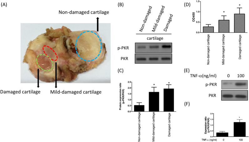
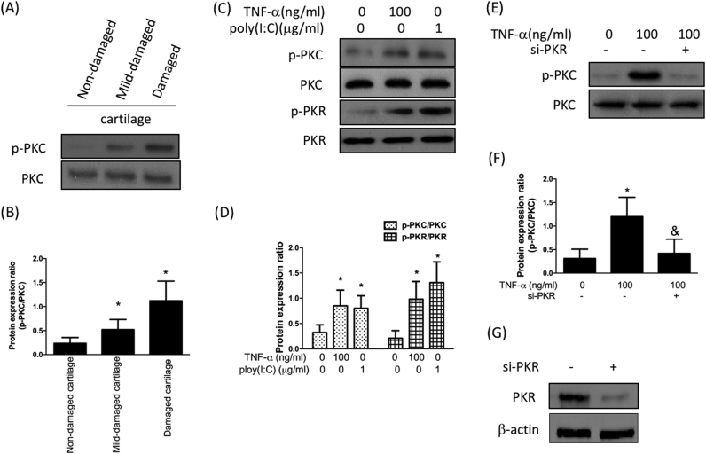
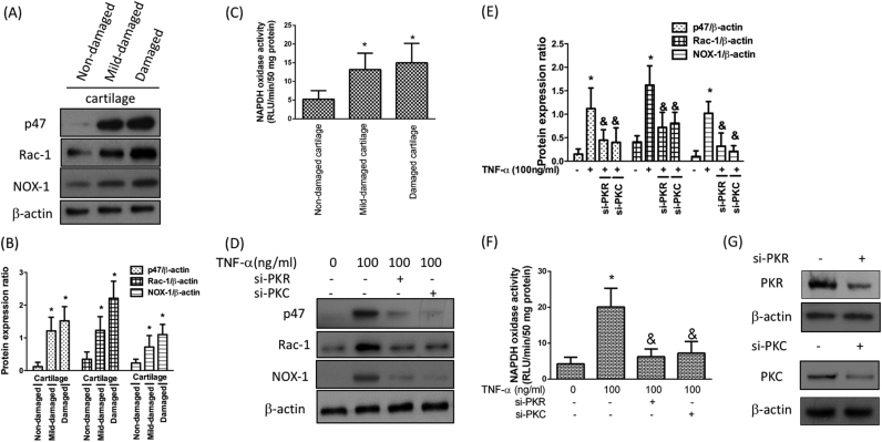
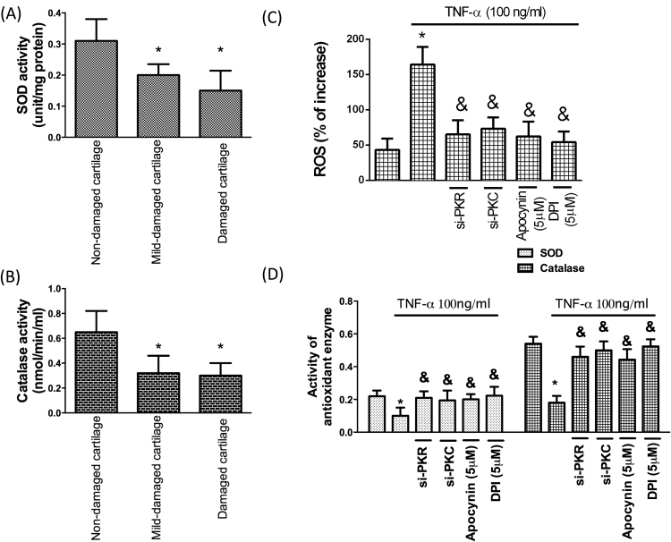
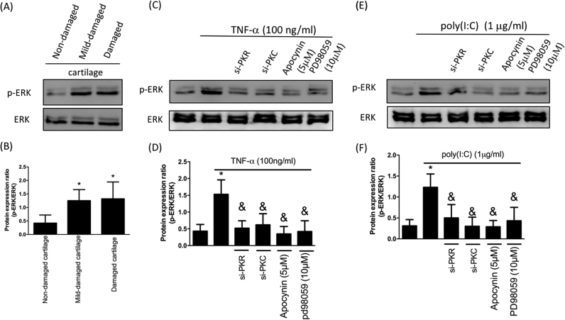
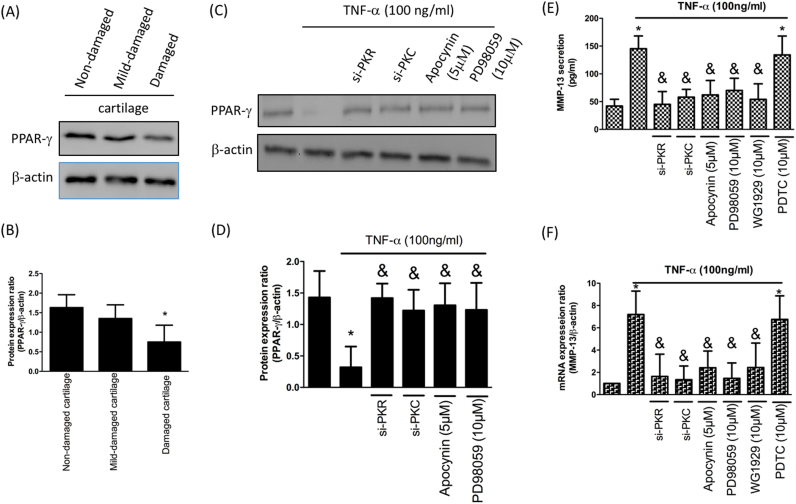
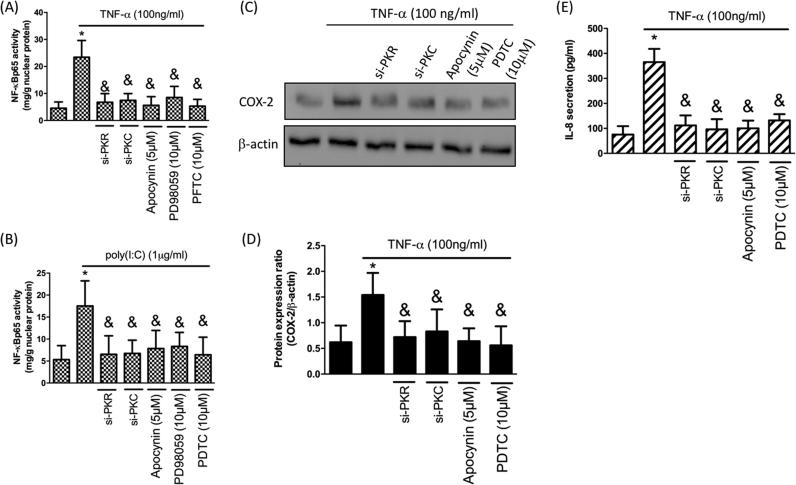
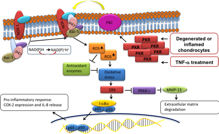
Osteoarthritis (OA) is a degenerative joint disease affecting a large population of people. Although the elevated expression of PKR (double stranded RNA-dependent protein kinase) and MMP-13 (collagenase-3) have been indicated to play pivotal roles in the pathogenesis of OA, the exact mechanism underlying the regulation of MMP-13 by PKR following inflammatory stimulation was relatively unknown. The purpose of this study was to determine the signaling pathway involved in the PKR-mediated induction of MMP-13 after TNF-α-stimulation. In this study, cartilages of knee joint were obtained from OA subjects who underwent arthroplastic knee surgery. Cartilages were used for tissue analysis or for chondrocytes isolation. In results, the upregulated expression of PKR was observed in damaged OA cartilages as well as in TNF-α-stimulated chondrocytes. Phosphorylation of PKC (protein kinase C) was found after TNF-α administration or PKR activation using poly(I:C), indicating PKC was regulated by PKR. The subsequent increased activity of NADPH oxidase led to oxidative stress accumulation and antioxidant capacity downregulation followed by an exaggerated inflammatory response with elevated levels of COX-2 and IL-8 via ERK/NF-κB pathway. Activated ERK pathway also impeded the inhibition of MMP-13 by PPAR-γ. These findings demonstrated that TNF-α-induced PKR activation triggered oxidative stress-mediated inflammation and MMP-13 in human chondrocytes. Unraveling these deregulated signaling cascades will deepen our knowledge of OA pathophysiology and provide aid in the development of novel therapies.

Keywords: Human chondrocyte; MMP-13; Osteoarthritis; Oxidative stress; PKR.


Figures

fx1
Graphical abstract
Fig. 1
Fig. 1
Upregulation of PKR following cartilage inflammation. A. Representative image of cartilage from patient with total knee replacement showing non-damaged, mid-damaged and damaged regions; Protein expression (B) and ratio (C) of p-PKR to total PKR; (D) Kinase activity of p-PKR from three different regions; protein expression (E) and ratio (F) of p-PKR to total PKR after exogenous administration of TNF-α for 12 h in human chondrocytes. (n = 3; * p < .05 compared to non-damaged cartilage or control group).
Fig. 2
Fig. 2
Increased expression of PKC after cartilage inflammation is due to PKR upregulation Protein expression (A) and the ratio (B) of p-PKC to total PKC from three different regions; Protein expression (C) and quantification (D) of PKR as well as PKC activation by addition of TNF-α and poly(I:C), which is known to activate PKR. Protein expression (E) and the ratio (F) of p-PKC to total PKC after treatment of TNF-α with or without the addition of si-PKR. (G)Western blotting confirming PKR knockdown efficiency. (n = 3; * p < .05 compared to non-damaged cartilage or no treatment control group; & p < .05 compared to TNF-α-treated group).
Fig. 3
Fig. 3
Activation of NADPH oxidase (NOX) under the inflammatory condition is mediated by increased level of PKR or PKC. Protein expression (A) and quantification (B) of NADPH oxidase cytosolic subunits, including p47 and Rac-1, as well as NOX1; (C) Activity of NOX from three different regions; The protein expression levels (D) and quantification of NOX subunits and isoform (E) in TNF-α-stimulated chondrocytes in the presence of si-PKR or si-PKC. The activity of NOX was tested by NADPH oxidase activity assay (F). (G) Western blotting confirming PKR and PKC knockdown efficiency. (n = 3; * p < .05 compared to non-damaged cartilage or no treatment control group; & p < .05 compared to TNF-α only group).
Fig. 4
Fig. 4
Accumulated oxidative stress by inflammation in cartilage is reversed by inhibition of PKR/PKC/NOX pathway Activities of the antioxidant enzymes, (A) SOD and (B) catalase, in three different regions of cartilage; (C) TNF-α-induced ROS in chondrocytes was interfered by si-PKR or si-PKC; (D) Activities of antioxidant enzymes (SOD and catalase) following TNF-α exposure with si-PKR, si-PKC or two NOX inhibitors (apocynin and DPI). (n = 3; * p < .05 compared to non-damaged cartilage or no treatment control group; & p < .05 compared to TNF-α only group).
Fig. 5
Fig. 5
Inflammation-induced p-ERK is downregulated by modulation of PKR/PKC/NOX pathway. Protein expression (A) and ratio (B) of p-ERK to total ERK in three different regions; Protein expression (C) and ratio (D) of p-ERK to total ERK after TNF-α treatment with si-PKR, si-PKC or NOX inhibitor apocynin; Protein expression (E) and ratio (F) of p-ERK to total ERK following poly(I:C) treatment with si-PKR, si-PKC or NOX inhibitor apocynin. (n = 3; * p < .05 compared to non-damaged cartilage or no treatment control group; & p < .05 compared to TNF-α only or poly(I:C) only group).
Fig. 6
Fig. 6
Mitigation of PPAR-γ is via activation of PKR/PKC/NOX/ERK pathway, leading to reduction of MMP-13. Protein expression (A) and quantification (B) of PPAR-γ in three different regions; Protein expression (C) and quantification (D) of PPAR-γ by TNF-α stimulation in chondrocytes along with si-PKR, si-PKC, NOX inhibitor apocynin or ERK inhibitor PD98059; The up-regulation of MMP-13 secretion (E) and MMP-13 mRNA expression (F) by TNF-α in chondrocytes was hindered by si-PKR, si-PKC, apocynin, PD98059 or PPAR-γ agonist GW1929. (n = 3; * p < .05 compared to non-damaged cartilage or no treatment control group; & p < .05 compared to TNF-α only group).
Fig. 7
Fig. 7
PKR-mediated elevation of COX-2 and IL-8 expression. N-FκB p65-activity in response to TNF-α (A) or poly(I:C)-induction (B) with si-PKR, si-PKC, apocynin or PD98059; Protein expression (C) and quantification (D) of COX-2 by TNF-α stimulation in chondrocytes with si-PKR, si-PKC, apocynin or NF-κB inhibitor PDTC; (E) IL-8 secretion after TNF-α treatment in chondrocytes with si-PKR, si-PKC, apocynin or PDTC. (n = 3; * p < .05 compared with no treatment control group; & p < .05 compared to TNF-α only or poly(I:C) only group).
Fig. 8
Fig. 8
Schematic diagram of PKR signaling pathways that are activated by inflammatory stimulation. Phosphorylation of PKC by PKR activation induces the cytosolic complex (such as p47 and Rac1) to translocate to the membrane and associate with the integral membrane components, resulting in activation of NOX1 followed by accumulation of reactive oxidase species (superoxide) and down-regulation of antioxidant enzymes activity (SOD and catalase). Subsequently, the up-regulated oxidative stress triggers the ERK pathway which leads to enhanced inflammatory responses (increased COX-2 and IL-8) via NF-κB. In addition, the activated ERK hinder the inhibition of PPAR-γ to MMP-13, allowing MMP-13-mediated ECM degradation/remodeling.

References

    1. Goldring S.R., Goldring M.B. Clinical aspects, pathology and pathophysiology of osteoarthritis. J. Musculoskelet. Neuron. Interact. 2006;6(4):376–378. - PubMed
    1. Goldring M.B., Goldring S.R. Osteoarthritis. J. Cell Physiol. 2007;213(3):626–634. - PubMed
    1. Malemud C.J., Islam N., Haqqi T.M. Pathophysiological mechanisms in osteoarthritis lead to novel therapeutic strategies. Cells Tissues Organs. 2003;174(1–2):34–48. - PubMed
    1. Aigner T., Fundel K., Saas J., Gebhard P.M., Haag J., Weiss T., Zien A., Obermayr F., Zimmer R., Bartnik E. Large-scale gene expression profiling reveals major pathogenetic pathways of cartilage degeneration in osteoarthritis. Arthritis Rheum. 2006;54(11):3533–3544. - PubMed
    1. Goldring M.B. Osteoarthritis and cartilage: the role of cytokines. Curr. Rheumatol. Rep. 2000;2(6):459–465. - PubMed

MeSH terms