. 2017 Sep 21;549(7672):399-403.
doi: 10.1038/nature23887. Epub 2017 Sep 6.

Palmitoylation-dependent activation of MC1R prevents melanomagenesis

Affiliations

Palmitoylation-dependent activation of MC1R prevents melanomagenesis

Shuyang Chen et al. Nature. .

Abstract

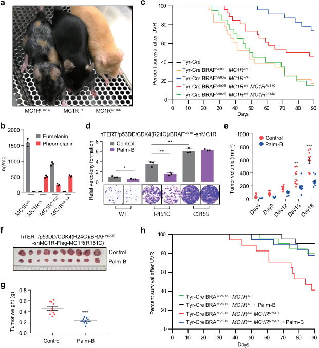
The melanocortin-1 receptor (MC1R), a G-protein-coupled receptor, has a crucial role in human and mouse pigmentation. Activation of MC1R in melanocytes by α-melanocyte-stimulating hormone (α-MSH) stimulates cAMP signalling and melanin production and enhances DNA repair after ultraviolet irradiation. Individuals carrying MC1R variants, especially those associated with red hair colour, fair skin and poor tanning ability (denoted as RHC variants), are associated with higher risk of melanoma. However, how MC1R activity is modulated by ultraviolet irradiation, why individuals with red hair are more prone to developing melanoma, and whether the activity of RHC variants might be restored for therapeutic benefit are unknown. Here we demonstrate a potential MC1R-targeted intervention strategy in mice to rescue loss-of-function MC1R in MC1R RHC variants for therapeutic benefit by activating MC1R protein palmitoylation. MC1R palmitoylation, primarily mediated by the protein-acyl transferase ZDHHC13, is essential for activating MC1R signalling, which triggers increased pigmentation, ultraviolet-B-induced G1-like cell cycle arrest and control of senescence and melanomagenesis in vitro and in vivo. Using C57BL/6J-Mc1re/eJ mice, in which endogenous MC1R is prematurely terminated, expressing Mc1r RHC variants, we show that pharmacological activation of palmitoylation rescues the defects of Mc1r RHC variants and prevents melanomagenesis. The results highlight a central role for MC1R palmitoylation in pigmentation and protection against melanoma.


Conflict of interest statement

The authors declare no competing financial interests.

Figures

Extended Data Figure 1
Extended Data Figure 1. MC1R is palmitoylated
(a) Fatty acids were dissolved in 100% ethanol (250 mM) mixed with 25 mM BSA and then added to serum-free DMEM/medium 254 at a final concertation of 500 μM. MC1R RHC-variant B16 cells were serum starved for 6 h. For the last 30 min cells were incubated with 1 μM α-MSH, followed by 100 J/m2 UVB treatment. Lastly cells were treated with indicated BSA-conjugated fatty acid medium for 3 h. Results were calculated as mean ± SD from three independent experiments (n=3). (b) A schematic showing the general process of protein palmitoylation. (c) MC1R RHC-variant B16 melanoma cells were treated as indicated in (a). 2-BP (25 μM) was added or not with palmitic acid for 3 h and cAMP content was determined. Results were calculated as mean ± SD from three independent experiments (n=3). (d-e) MC1R RHC-variant or WT B16 melanoma cells (d) and MC1R RHC-variant or WT HPMs (e) were treated as indicated in Figure 1a. 2-BP (25 μM) was added or not with palmitic acid for 3 h and cAMP content was determined. Results were calculated as mean ± SD from three independent experiments (n=3). (f) A schematic showing the general process of the acyl-biotin exchange (ABE) palmitoylation assay. Unmodified cysteines were irreversibly blocked by N-Ethylmaliemide (NEM), and palmitoylated cysteines were then exposed using HAM and sequentially biotinylated. Biotin levels were determined as a measurement for palmitoylation. (g) Flowchart of palmitoylated protein identification. NEM, N-Ethylmaleimide. HAM, hydroxylamine. 1×10⁸ MC1R RHC-variant HPMs were treated with palmitic acid as indicated in (a), and then processed as shown in flowchart. Streptavidin IP was performed and protein samples were separated by SDS-PAGE before staining by Coomassie brilliant blue. Palmitoylation-specific bands were excised for Mass Spectrometry for protein identification. (h) The peptide spectral counts of MC1R from Figure 1c. (i) NBA-palm of MC1R. The prediction shows two possible sites of MC1R palmitoylation. (j) A schematic illustration showing the palmitoylation site at MC1R C315 predicted by NBA-PALM and PEP-FOLD3, and a schematic showing the conserved C-terminal domain of MC1R. (k) Membrane topology of MC1R with indicated RHC, non-RHC and palmitoylation site mutants.
Extended Data Figure 2
Extended Data Figure 2. Palmitoylation of MC1R in melanocytes
(a) Endogenous MC1R protein is palmitoylated. B16 cells were incubated with 1 μM α-MSH for 3.5 h. Cells were then harvested for IP by specific anti-MC1R antibody, and then for ABE and IB analysis. (b) Exogenous MC1R protein is palmitoylated. B16 cells were infected with retrovirus encoding Flag-MC1R WT and incubated with 1 μM α-MSH for 3.5 h. Cells were then harvested for IP, ABE and IB analysis. (c) MC1R is palmitoylated at C315. B16 cells with stable depletion of endogenous MC1R by shRNA were infected with the indicated Flag-MC1R encoding retroviral constructs. Cells were treated with 1 μM α-MSH for 3.5 h and then harvested for IP, ABE and IB analysis. (d) MC1R RHC variants are defective in palmitoylation. B16 cells with stable depletion of endogenous MC1R by shRNA were infected with the indicated Flag-MC1R encoding retroviral constructs. Cells were treated with 1 μM α-MSH for 3.5 h and then harvested for IP, ABE and IB analysis. (e-f) R151C does not create a palmitoylation site. B16 cells (e) or HPMs (f) with stable depletion of endogenous MC1R by shRNA were infected with the indicated Flag-MC1R encoding retroviral constructs. Cells were treated with 1 μM α-MSH for 3.5 h and then harvested for IP, ABE and IB analysis. (g) UVB enhances MC1R palmitoylation. B16 cells were incubated with 1 μM α-MSH for 30 min followed by 100 J/m2 UVB irradiation. Cells were harvested for IP by specific anti-MC1R antibody, then for ABE and IB analysis 3 h after UVB exposure. (h) UVB irradiation enhances MC1R palmitoylation. B16 cells were infected with retroviral constructs encoding Flag-MC1R WT and pre-treated with 1 μM α-MSH for 30 min followed by 100 J/m2 UVB irradiation. Cells were harvested for IP, ABE and IB analysis 3 h after UVB exposure. (i) MC1R C315S mutant does not respond to UVB irradiation. B16 with stable depletion of MC1R by shRNA were infected with the indicated Flag-MC1R encoding retroviral constructs. Cells were pre-treated with 1 μM α-MSH for 30 min followed by 100 J/m2 UVB irradiation. Cells were harvested for IP, ABE and IB analysis 3 h after UVB exposure. (j-m) MC1R RHC variants are defective in palmitoylation after UVB. B16 or HPMs with stable depletion of MC1R by shRNA were infected with the indicated Flag-MC1R encoding retroviral constructs. Cells were pre-treated with 1 μM α-MSH for 30 min followed by 100 J/m2 UVB irradiation. Cells were harvested for IP, ABE and IB analysis 3 h after UVB exposure.
Extended Data Figure 3
Extended Data Figure 3. ZDHHC13 is a major PAT of MC1R
(a) HEK293 cells were co-transfected with constructs encoding Flag-MC1R and HA-ZDHHCs in 6-well plate. Cells lysates were harvested for IP by anti-Flag antibody, and then for ABE and IB analysis. IB for total MC1R and HA-ZDHHC proteins was shown. (b) B16 cells were infected with Flag-MC1R and indicated ZDHHC13 WT or C456S mutant encoding retroviral constructs. Cells were treated with 1 μM α-MSH for 3.5 h and then harvested for IP, ABE and IB analysis. (c) B16 cells with deletion of ZDHHC13 by selective shRNAs were pre-treated with 1 μM α-MSH for 30 min followed by 100 J/m2 UVB irradiation and harvested for IP, ABE and IB analysis 3 h after UVB exposure. (d) B16 cells with stable depletion of MC1R by shRNA were infected with the indicated Flag-MC1R encoding retroviral constructs, then cells were infected with shZDHHC13 and/or WT HA-ZDHHC13 expressing virus. Finally, cells were pre-treated with 1 μM α-MSH for 30 min followed by 100 J/m2 UVB irradiation and harvested for IP, ABE and IB analysis 3 h after UVB exposure. (e-f) B16 cells or HPMs with stable depletion of MC1R by shRNA were infected with the Flag-MC1R R151C or R151C+C315S double mutant encoding retroviral constructs, then cells were infected with WT HA-ZDHHC13 expressing virus. Finally, cells were pre-treated with 1 μM α-MSH for 3.5 h and harvested for IP, ABE and IB analysis. (g) HPMs were incubated with 1 μM α-MSH or vehicle for 30 min followed by 100 J/m2 UVB irradiation. Cells were harvested for IP by specific anti-MC1R antibody, then for ABE and IB analysis 3 h after UVB exposure. (h) HPMs were incubated with 1 μM α-MSH or vehicle for 30 min followed by 100 J/m2 UVB irradiation. Cells were harvested for IP by specific anti-MC1R antibody, then for IB analysis 3 h after UVB exposure. (i) A schematic showing the conserved SQ motif of ZDHHC13. (j) HPMs were infected with WT ZDHHC13 or ZDHHC13 S8A mutant encoding retroviral constructs, then cells were irradiated with 100 J/m2 UVB irradiation and harvested for IP and IB analysis 3 h after UVB exposure. (k) WT Flag-ATR or the kinase-dead (KD) Flag-ATR mutant transfected HEK293 cells were irradiated before Flag beads immunoprecipitation. Then immunoprecipitated WT ZDHHC13 or S8A mutant were incubated with immunoprecipitated WT ATR or the KD ATR mutant in kinase buffer. After reaction, proteins were collected for IB analysis. (l) HPMs were infected with WT ZDHHC13 or ZDHHC13 S8A mutant and Flag-MC1R encoding retroviral constructs, then cells were irradiated with 100 J/m2 UVB irradiation and harvested for IP and IB analysis 3 h after UVB exposure. (m) HPMs cells with stable depletion of ZDHHC13 by shRNA were infected with the indicated ZDHHC13 and Flag-MC1R encoding retroviral constructs. Then cells were pre-treated with 1 μM α-MSH for 30 min followed by 100 J/m2 UVB irradiation and harvested for IP, ABE and IB analysis 3 h after UVB exposure.
Extended Data Figure 4
Extended Data Figure 4. Palmitoylation is essential for MC1R function
(a) B16 and HPMs with stable depletion of MC1R by shRNA were infected with the indicated Flag-MC1R encoding retroviral constructs. After 3 h, cells were pre-treated with 1 μM α-MSH for 30 min followed by 100 J/m2 UVB irradiation. Cells were harvested for a cAMP immunoassay. These data were compiled from three independent experiments. Data are represented as mean ± SD. (b-c) B16 and HPMs with stable depletion of MC1R by shRNA were infected with the indicated Flag-MC1R encoding retroviral constructs. After 3 h, cells were pre-treated with 1 μM α-MSH for 30 min followed by 100 J/m2 UVB irradiation. Total RNA were collected for reverse transcription and cDNA were then used for quantitative real time PCR (qRT-PCR) by specific primers targeting mouse/human MITF or TYR. Three independent experiments were quantified. Data are represented as mean ± SD. (d) HPMs with stable deletion of MC1R by shRNA were infected with the indicated Flag-MC1R encoding retro-viral constructs. Cells were pre-treated with 1 μM α-MSH for 30 min followed by 100 J/m2 UVB irradiation. Genomic DNA were extracted at the different time points as indicated and photoproducts were detected by ELISA. Cyclobutane pyrimidine dimer (CPD) or 6–4 pyrimidine photoproduct (6–4PP) antibodies were used. Three independent experiments were measured and calculated as mean ± SD. (e) B16 and HPMs with stable depletion of MC1R by shRNA were pre-treated with 1 μM α-MSH for 30 min followed by 25 J/m2 UVB irradiation. Cells were subjected to SA-β-gal staining assay 7 days after UVR. The quantification of the percentage of SA-β-gal positive cells and representative pictures were shown. Data shown correspond to one representative experiment out of three independent experiments. Data are represented as mean ± SD. (f) B16 and HPMs with stable depletion of MC1R by shRNA were infected with the indicated Flag-MC1R encoding retroviral constructs. Cells were pre-treated with 1 μM α-MSH for 30 min followed by 25 J/m2 UVB irradiation. Cells were subjected to SA-β-gal staining assay 7 days after UVR. The quantification of the percentage of SA-β-gal positive cells and representative pictures were shown. Data shown correspond to one representative experiment out of three independent experiments. Data are represented as mean ± SD. (g) hTERT/p53DD/CDK4(R24C)/BRAFV600E melanocytes with stable depletion of MC1R by shRNA were pre-incubated with 1 μM α-MSH for 30 min before being irradiated with 20 J/m2 UVB. Cell lysates were collected for IB analysis. (h) Cells generated in (g) were subjected to clonogenic survival assays 15 days after UVR. Crystal violet was used to stain colonies and the colony numbers were counted from three independent experiments. The relative colony numbers were calculated as mean ± SD. (i) Cells generated in (g) were seeded (10,000 cells per well) in 0.5% low-melting-point agarose in DMEM with 10% FBS, layered onto 0.8% agarose in DMEM with 10% FBS. The plates were cultured for 30 days where upon the colonies >50 μm were counted under a light microscope. The colony numbers were plotted as mean ± SD from three independent experiments. (j) MC1R-depleted hTERT/p53DD/CDK4(R24C)/BRAFV600E melanocytes were further infected with the indicated Flag-MC1R encoding retroviral constructs. Cells were pre-incubated with 1 μM α-MSH for 30min before being irradiated with 20 J/m2 UVB. Cell lysates were collected for IB analysis. (k) The cells generated as indicated were seeded (10,000 cells per well) in 0.5% low-melting-point agarose in DMEM with 10% FBS, layered onto 0.8% agarose in DMEM+10% FBS. The plates were cultured for 30 days where upon the colonies >50 μm were counted under a light microscope. The colony numbers were plotted as mean ± SD from three independent experiments.
Extended Data Figure 5
Extended Data Figure 5. Activating MC1R palmitoylation rescues the defect of MC1R RHC variants
(a) B16 and HPMs with stable depletion of MC1R by shRNA were infected with the indicated Flag-MC1R encoding retroviral constructs, then cells were infected with shZDHHC13 and/or WT HA-ZDHHC13 virus. Cells were pre-treated with 1 μM α-MSH for 30 min followed by 100 J/m2 UVB irradiation. After 3 h, cells were harvested for cAMP immunoassay. These data were compiled from three independent experiments. Data are represented as mean ± SD. (b) B16 and HPMs with stable depletion of MC1R by shRNA were infected with the indicated Flag-MC1R encoding retroviral constructs, then cells were infected with shZDHHC13 and/or WT HA-ZDHHC13 virus. Cells were pre-treated with 1 μM α-MSH for 30 min followed by 100 J/m2 UVB irradiation. After 3 h, total RNA was collected for reverse transcription and cDNA were then used for qRT-PCR by specific primers targeting mouse MITF. Three independent experiments were quantified. Data are represented as mean ± SD. (c) HPMs with stable depletion of MC1R by shRNA were infected with the indicated Flag-MC1R encoding retro-viral constructs, then cells were infected with shZDHHC13 and/or WT HA-ZDHHC13 virus. Cells were pre-treated with 1 μM α-MSH for 30 min followed by 100J/m2 UVB irradiation. Genomic DNA was extracted at the different time points indicated and photoproducts detected by ELISA using Cyclobutane pyrimidine dimer (CPD) or 6–4 pyrimidine photoproduct (6–4PP) antibodies. Three independent experiments were measured and calculated as mean ± SD. (d) B16 and HPMs with stable depletion of MC1R by shRNA were infected with the indicated Flag-MC1R encoding retroviral constructs, then cells were infected with shZDHHC13 and/or WT HA-ZDHHC13 virus. Cells were pre-treated with 1 μM α-MSH for 30 min followed by 25 J/m2 UVB irradiation. Cells were subjected to SA-β-gal staining assay 7 days after UVR. The quantification of the percentage of SA-β-gal positive cells and representative pictures were shown. Data shown correspond to one representative experiment out of three independent experiments. Data are represented as mean ± SD. (e) The cells generated as indicated were seeded (10,000 cells per well) in 0.5% low-melting-point agarose in DMEM with 10% FBS, layered onto 0.8% agarose in DMEM+10% FBS. Plates were cultured for 30 days where upon the colonies >50 μm were counted under a light microscope. Colony numbers were plotted as mean ± SD from three independent experiments.
Extended Data Figure 6
Extended Data Figure 6. MC1R variant and mutant transgenic mice
(a) Schematic diagrams of MC1R variant constructs. Transgenic mice were designed to express melanocyte-specific MC1R variants or mutants (controlled by the Tyr enhancer/promoter). (b) C57BL/6 MC1R variant or mutant transgenic mice. (c) Human transgene content in transgenic and control mice. Results were calculated as mean ± SD from three independent experiments. (d) Whole skins from C57BL/6 MC1R variant transgenic mice (8-12 weeks) were collected and stained with Dct antibody. Melanocytes were then isolated and quantified by FACS sorting. Results were calculated as mean ± SD from three independent experiments. (e) Frozen sections of skins from C57BL/6 MC1R variant transgenic mice (8-12 weeks) were stained with Dct antibody. The positive staining represents melanocytes. (f) Illustrations for UVB-induced melanoma development in Tyr-Cre-BRAFCA-MC1R variant mice. (g) H&E staining of histological sections and immunohistochemistry staining of S100 of representative cutaneous melanomas. Genotypes were showed as indicated.
Extended Data Figure 7
Extended Data Figure 7. Palm-B activates MC1R palmitoylation and rescues the defect of MC1R RHC variants
(a) HPMs were infected with retrovirus encoding Flag-MC1R WT and incubated with 1 μM α-MSH for 3.5 h. The medium was replaced with fresh medium with vehicle or 1 μM Palm-B and cells were treated with indicated time. Cells were harvested for IP, ABE and IB analysis. (b) B16 and HPMs with stable depletion of MC1R by shRNA were infected with the indicated Flag-MC1R encoding retroviral constructs. Cells were pre-treated with 1 μM α-MSH and 1 μM Palm-B for 30 min followed by 100 J/m2 UVB irradiation. Cells were harvested for IP, ABE and IB analysis 3 h after UVB exposure. (c) B16 and HPMs with stable depletion of MC1R by shRNA were infected with the indicated Flag-MC1R encoding retroviral constructs. Cells were pre-treated with 1 μM α-MSH and 1 μM Palm-B for 30 min followed by 100 J/m2 UVB irradiation. After 3 h, cells were harvested for cAMP immunoassay. These data were compiled from three independent experiments. Data are represented as mean ± SD. (d-e) B16 and HPMs with stable depletion of MC1R by shRNA were infected with the indicated Flag-MC1R encoding retroviral constructs. Cells were pre-treated with 1 μM α-MSH and 1 μM Palm-B for 30 min followed by 100 J/m2 UVB irradiation. After 3 h, total RNA were collected for reverse transcription and cDNA were then used for qRT-PCR by specific primers targeting mouse and/or human MITF or TYR. Three independent experiments were quantified. Data are represented as mean ± SD. (f) HPMs with stable depletion of MC1R by shRNA were infected with the indicated Flag-MC1R encoding retro-viral constructs. Cells were pre-treated with 1 μM α-MSH and 1 μM Palm-B for 30 min followed by 100 J/m2 UVB irradiation. Genomic DNA were extracted at the different time points indicated and photoproducts were detected by ELISA. Cyclobutane pyrimidine dimer (CPD) or 6–4 pyrimidine photoproduct (6–4PP) antibodies were used. Three independent experiments were measured and calculated as mean ± SD. (g) C57BL/6 mice or C57BL/6-MC1Re/e-MC1RR151C-tg mice were given a 10 mg/kg Palm-B or vehicle injection intraperitoneally 3 h before UVB irradiation (500 J/m2). 3 h after UVB, whole skins were collected and the lysates were subjected for IP, ABE and IB analysis. (h-i) C57BL/6 mice or C57BL/6-MC1Re/e-MC1RR151C-tg mouse were injected intraperitoneally with 10 mg/kg Palm-B 3 h before UVB irradiation (500 J/m2). Melanocytes were isolated by flow cytometry, then DNA were extracted and subjected to ELISA 3 h after UVB irradiation. Results were calculated as mean ± SD from three independent experiments. (j) B16 with stable depletion of MC1R by shRNA were infected with the indicated Flag-MC1R encoding retroviral constructs. Cells were pre-treated with 1 μM α-MSH and 1 μM Palm-B for 30 min followed by 25 J/m2 UVB irradiation. Cells were subjected to SA-β-gal staining assay 7 days after UVR. The quantification of the percentage of SA-β-gal positive cells and representative pictures are shown. Data shown correspond to one representative experiment out of three independent experiments. Data are represented as mean ± SD. (k) The cells generated as indicated were seeded (10,000 cells per well) in 0.5% low-melting-point agarose in DMEM with 10% FBS, layered onto 0.8% agarose in DMEM+10% FBS. The plates were cultured for 30 days where upon the colonies >50 μm were counted under a light microscope. Colony numbers were plotted as mean ± SD from three independent experiments.
Extended Data Figure 8
Extended Data Figure 8. Palm-B rescues the defect of MC1R R160W variant
(a) B16 and HPMs with stable depletion of MC1R by shRNA were infected with the indicated Flag-MC1R encoding retroviral constructs. Cells were pre-treated with 1 μM α-MSH and 1 μM Palm-B for 30 min followed by 100 J/m2 UVB irradiation. Cells were harvested for IP, ABE and IB analysis at 3 h after UVB exposure. (b) B16 and HPMs with stable depletion of MC1R by shRNA were infected with the indicated Flag-MC1R encoding retroviral constructs. Cells were pre-treated with 1 μM α-MSH and 1 μM Palm-B for 30 min followed by 100 J/m2 UVB irradiation. After 3 h, cells were harvested for cAMP immunoassay. These data were compiled from three independent experiments. Data are represented as mean ± SD. (c) B16 and HPMs with stable depletion of MC1R by shRNA were infected with the indicated Flag-MC1R encoding retroviral constructs. Cells were pre-treated with 1 μM α-MSH and 1 μM Palm-B for 30 min followed by 100 J/m2 UVB irradiation. After 3 h, total RNA was collected for reverse transcription and cDNA then used for qRT-PCR by specific primers targeting mouse and/or human MITF or TYR. Three independent experiments were quantified. Data are represented as mean ± SD. (d) HPMs with stable depletion of MC1R by shRNA were infected with the indicated Flag-MC1R encoding retroviral constructs. Cells were pre-treated with 1 μM α-MSH and 1 μM Palm-B for 30 min followed by 100 J/m2 UVB irradiation. Genomic DNA were extracted at the different time points indicated and photoproducts were detected by ELISA using anti-cyclobutane pyrimidine dimer (CPD) or 6–4 pyrimidine photoproduct (6–4PP) antibodies. Three independent experiments were measured and calculated as mean ± SD. (e) B16 and HPMs with stable depletion of MC1R by shRNA were infected with the indicated Flag-MC1R encoding retroviral constructs. Cells were pre-treated with 1 μM α-MSH and 1 μM Palm-B for 30 min followed by 25 J/m2 UVB irradiation. Cells were subjected to SA-β-gal staining assay 7 days after UVR. The quantification of the percentage of SA-β-gal positive cells and representative pictures are shown and correspond to one representative experiment out of three independent experiments. Data are represented as mean ± SD. (f) MC1R-depleted hTERT/p53DD/CDK4(R24C)/BRAFV600E melanocytes were further infected with the indicated Flag-MC1R encoding retro-viral constructs. Cells were pre-incubated with 1 μM α-MSH and 1 μM Palm-B for 30min before being irradiated with 20 J/m2 UVB, and then subjected to clonogenic survival assays 15 days after UVR. Crystal violet was used to stain colonies and the colony numbers were counted from three independent experiments. The relative colony numbers were calculated as mean ± SD. (g) The cells generated in (k) were seeded (10,000 cells per well) in 0.5% low-melting-point agarose in DMEM with 10% FBS, layered onto 0.8% agarose in DMEM+10% FBS. Plates were cultured for 30 days where upon the colonies >50 μm were counted under a light microscope. The colony numbers were plotted as mean ± SD from three independent experiments.
Figure 1
Figure 1. Palmitoylation of MC1R in melanocytes
a-b, MC1R RHC-variant HPMs exposed to α-MSH and UVB irradiated were treated with BSA-conjugated fatty acids. b, cells were exposed to palmitic acid +/− 2-BP. cAMP were calculated by three independent experiments shown as mean ± SD. c-f, HPMs (c), HPMs expressing WT, mutant or variant Flag-MC1R (d-f) were incubated with α-MSH and processed for ABE analysis. g-k, HPMs (g), HPMs expressing WT, mutant or variant Flag-MC1R (h-k) were treated with α-MSH, irradiated with UVB, and harvested for ABE analysis. Western blots shown were representative of three independent experiments. **p<0.01, ***p<0.001, unpaired student’s t-test. For gel source data, see Supplementary Figure 1.
Figure 2
Figure 2. ZDHHC13 is a major MC1R palmitoyl acyltransferase
a-b, B16 cells co-expressing Flag-MC1R and HA-ZDHHCs (a) and HPMs expressing Flag-MC1R and ZDHHC13 WT or C456S mutant (b) were incubated with α-MSH and processed for ABE analysis. c-f, HPMs expressing ZDHHC13 shRNAs (c), HPMs expressing Flag-MC1R together with shZDHHC13 and/or WT HA-ZDHHC13 (d), HPMs (e) and HPMs expressing Flag-MC1R (f) were pre-treated with α-MSH, UVB irradiated and harvested for IP, ABE and IB. g-i, HPMs expressing HA-ZDHHC13 (g), HPMs expressing shATR and HA-ZDHHC13 (h-i) were pre-treated with α-MSH, UVB irradiated and processed for IP and IB analysis. Western blots shown were representative of three independent experiments. For gel source data, see Supplementary Figure 1.
Figure 3
Figure 3. Activating MC1R palmitoylation rescues the defect of MC1R RHC variants
a-b, MC1R-depleted hTERT/p53DD/CDK4(R24C)/BRAFV600E melanocytes expressing Flag-MC1R (a) or cells further infected with shZDHHC13 and/or WT HA-ZDHHC13 virus (b) were pre-incubated with α-MSH, UVB irradiated and assayed for clonogenic survival. Results were calculated as mean ± SD from three independent experiments. c-e, Growth curves (c), dissected tumors (d) and tumor weight (e) for the xenograft experiments with indicated cells inoculated subcutaneously into each flank of nude mice (n=8). Visible tumors were measured at the indicated days. Error bars represent ±SEM. *p<0.05, **p<0.01, ***p<0.001, unpaired student’s t-test.
Figure 4
Figure 4. MC1R palmitoylation controls melanomagenesis
a, C57BL/6 MC1R variant transgenic mice. b, Eumelanin and pheomelanin content of whole skin from C57BL/6 MC1R variant transgenic mice. Data shown represent the mean ± SD of three independent experiments. c, Melanoma-free survival. Tyr-Cre n=15, Tyr-Cre-BRAFV600E-MC1Re/e n=23, Tyr-Cre-BRAFV600E-MC1R+/+ n=23, Tyr-Cre-BRAFV600E-MC1Re/e-MC1RR151C n=26, Tyr-Cre-BRAFV600E-MC1Re/e-MC1RC315S n=20. By Log-rank test, p=0.0001 (e/e & +/+), p=0.0179 (e/e & R151C), p=0.8943 (e/e & C315S), p=0.0232 (+/+ & R151C), p=0.0001 (+/+ & C315S), p=0.0233 (R151C & C315S). d, Indicated melanocytes were treated with α-MSH and Palm-B before UVB irradiation and assayed for clonogenic survival. Results were calculated as mean ± SD from three independent experiments. e-g, Growth curves (e), dissected tumors (f) and tumor weight (g) for subcutaneous xenograft experiments in nude mice (n=10) using indicated cells. Error bars represent ±SEM. h, Melanoma-free survival of Tyr-Cre-BRAFV600E-MC1R+/+ n=20, Tyr-Cre-BRAFV600E-MC1R+/+ + Palm-B n=20, Tyr-Cre-BRAFV600E-MC1Re/e-MC1RR151C n=17, Tyr-Cre-BRAFV600E-MC1Re/e-MC1RR151C + Palm-B n=18. By Log-rank test, p=0.0241 (R151C & R151C + Palm-B), p=0.3711 (MC1R+/+ & MC1R+/+ + Palm-B). *p<0.05, **p<0.01, ***p<0.001, unpaired student’s t-test.

Comment in

References

    1. Jackson IJ, Budd PS, Keighren M, McKie L. Humanized MC1R transgenic mice reveal human specific receptor function. Hum Mol Genet. 2007;16:2341–2348. - PubMed
    1. D’Orazio JA, et al. Topical drug rescue strategy and skin protection based on the role of Mc1r in UV-induced tanning. Nature. 2006;443:340–344. - PubMed
    1. Healy E, et al. Functional variation of MC1R alleles from red-haired individuals. Hum Mol Genet. 2001;10:2397–2402. - PubMed
    1. Palmer JS, et al. Melanocortin-1 receptor polymorphisms and risk of melanoma: is the association explained solely by pigmentation phenotype? Am J Hum Genet. 2000;66:176–186. - PMC - PubMed
    1. Raimondi S, et al. MC1R variants, melanoma and red hair color phenotype: a meta-analysis. Int J Cancer. 2008;122:2753–2760. - PubMed

Publication types