. 2017 Sep 4;17(1):148.
doi: 10.1186/s12870-017-1096-1.

The lysin motif-containing proteins, Lyp1, Lyk7 and LysMe3, play important roles in chitin perception and defense against Verticillium dahliae in cotton

Affiliations

The lysin motif-containing proteins, Lyp1, Lyk7 and LysMe3, play important roles in chitin perception and defense against Verticillium dahliae in cotton

Jun Xu et al. BMC Plant Biol. .

Abstract

Background: Lysin motif (LysM)-containing proteins are important pattern recognition receptors (PRRs) in plants, which function in the perception of microbe-associated molecular patterns (MAMPs) and in the defense against pathogenic attack. To date, the LysM genes have not been systematically analyzed in cotton or effectively utilized for disease resistance.

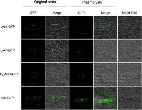
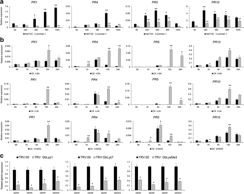
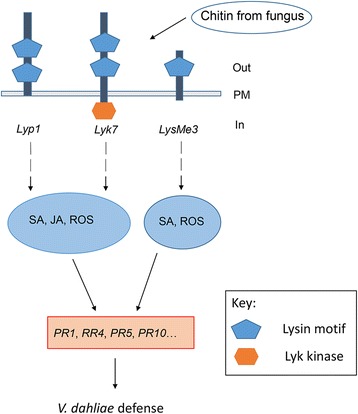
Results: Here, we identified 29, 30, 60, and 56 LysM genes in the four sequenced cotton species, diploid Gossypium raimondii, diploid G. arboreum, tetraploid G. hirsutum acc. TM-1, and G. barbadense acc. 3-79, respectively. These LysM genes were classified into four groups with different structural characteristics and a variety of expression patterns in different organs and tissues when induced by chitin or Verticillium dahliae. We further characterized three genes, Lyp1, Lyk7 and LysMe3, which showed significant increase in expression in response to chitin signals, V. dahliae challenge and several stress-related signaling compounds. Lyp1, Lyk7 and LysMe3 proteins were localized to the plasma membrane, and silencing of their expression in cotton drastically impaired salicylic acid, jasmonic acid, and reactive oxygen species generation, impaired defense gene activation, and compromised resistance to V. dahliae.

Conclusion: Our results indicate that Lyp1, Lyk7, and LysMe3 are important PRRs that function in the recognition of chitin signals to activate the downstream defense processes and induce cotton defense mechanisms against V. dahliae.

Keywords: Chitin signals; Cotton; Lysin motif; Pattern recognition receptors; Plasma membrane localization; Verticillium dahliae.


Conflict of interest statement

Ethics approval and consent to participate

Three cotton materials, including G. barbadense cv. Hai7124, G. hirsutum cv. Junmian 1 and G. hirsutum cv. Jinmian 19, were collected in this study. All necessary permits for using them were obtained from Nanjing Agricultural University, Jiangsu Province, China.

Consent for publication

Not applicable.

Competing interests

The authors declared that they had no competing interests.

Publisher’s Note

Springer Nature remains neutral with regard to jurisdictional claims in published maps and institutional affiliations.

Figures

Fig. 1
Fig. 1
Chromosomal distribution of LysM genes in G. raimondii. The chromosome numbers are shown at the top of each bar. The 29 LysM genes in G. raimondii were classified into four groups and marked on the linkage map. The names of the scaffolds from the genome are also indicated in brackets. The chromosome numbers from D1 to D6, and D8 to D13, were consistent with the newly-updated interspecific genetic map of allotetraploid cultivated cotton species [32]. The nomenclature of the LysM genes for each group was based on the order of the chromosomes in G. raimondii
Fig. 2
Fig. 2
Transcriptional profiling of LysM genes in different tissues and organs of G. hirsutum acc. TM-1. Roots, stems, leaves, petals, stamens, ovules at −3, 0, and 3 DPA, and fibers at 5, 10, 20, and 25 DPA were used for comparative transcriptome analysis. The expression data were converted to Log2 (FPKM) to calculate the expression levels of the LysM genes in TM-1. Differences in gene expression are shown in the colors indicated in the scale. The RNA-Seq data used here can be downloaded from http://www.ncbi.nlm.nih.gov/bioproject/PRJNA248163/
Fig. 3
Fig. 3
Induced expression of LysM genes by two PAMPs. Two-week-old seedlings were treated with either 200 mg/mL insoluble crab shell chitin or soluble chitin fragment N-acetylchitohexaose and sampled 0.5, 1, 2, 4, 6 h (h) after treatment, respectively. The induction of each gene was examined by qRT-PCR, and the 0 h expression levels were used as controls when calculating the level of induction. The data represent the mean ± SD of three samples from three independent tests at each time point. “*”: significant difference at P < 0.05; “**”: significant difference at P < 0.01
Fig. 4
Fig. 4
Expression patterns of the LysM genes in response to Verticillium dahliae in cotton. The expression patterns of LysM genes in response to Verticillium dahlia were investigated in G. barbadense cv. Hai7124 and G. hirsutum cv. Junmian 1, which show resistance and susceptibility to V. dahliae, respectively. qRT-PCR analysis showed differences in the expression of LysM genes in Hai7124 and Junmian 1 after inoculation with V. dahliae strain V991. The statistical analysis compared expression levels at different time points following treatment with those at 0 h. Error bars show the standard deviation of three biological replicates. “*”: significant difference at P < 0.05; “**”: significant difference at P < 0.01
Fig. 5
Fig. 5
Silencing of Lyp1 and Lyk7 significantly impaired the resistance to V. dahliae in G. barbadense cv. Hai7124. Lyp1 and Lyk7 were silenced in V. dahliae resistant Hai 7124 seedlings by VIGS, and about 2 weeks later, the seedlings were inoculated with V. dahliae at a concentration of 1 × 107 spores/mL. a Analysis of Lyp1 and Lyk7 expression levels. Total RNA was extracted from the leaves of the VIGS seedlings 14 d post-agroinfiltration, and transcription levels of Lyp1 and Lyk7 in the Lyp1 and Lyk7-silenced plants were compared with that of the control (TRV: 00 plants). Asterisks indicate statistically significant differences, as determined by Student’s t-tests (**P < 0.01). b Disease symptoms of the Lyp1 and Lyk7-silenced plants 20 and 25 days after V. dahliae inoculation; c The percentage of diseased leaves of the Lyp1 and Lyk7-silenced plants and controls after V. dahliae inoculation. These experiments were repeated using at least 20 seedlings per treatment. Error bars show the standard deviation of three biological replicates. Asterisks indicate statistically significant differences in the percentage of diseased leaves between treated plants and TRV: 00 controls, as determined by Student’s t-tests (*P < 0.05, **P < 0.01)
Fig. 6
Fig. 6
Increased susceptibility of the LysMe3- and LysMn6- silenced cotton plants to V. dahliae. LysMe3 and LysMn6 were silenced by VIGS in Hai7124 seedlings, and about 2 weeks later, the seedlings were inoculated with V. dahliae at a concentration of 1 × 107 spores/mL. a The expression levels of LysMe3 and LysMn6 were compared in the LysMe3-, LysMn6- silenced and TRV: 00 cotton plants. Asterisks indicate statistically significant differences, as determined by Student’s t-tests (**P < 0.01). b Phenotypes of the LysMe3-, LysMn6-silenced plants 20 and 25 days after V. dahliae inoculation. c The percentage of diseased leaves in the LysMe3-, and LysMn6-silenced plants and controls after V. dahliae inoculation. All experiments were repeated using at least 20 seedlings, and error bars show the standard deviation of three biological replicates. Asterisks indicate statistically significant differences in the percentage of diseased leaves between treated plants and TRV: 00 controls, as determined by Student’s t-test (*P < 0.05, **P < 0.01)
Fig. 7
Fig. 7
Subcellular localization of Lyp1, Lyk7, and LysMe3 proteins. The GFP, Lyp1-GFP, Lyk7-GFP, and LysMe3-GFP fusion proteins were transiently expressed in onion epidermal cells. GFP fluorescence was visualized by confocal microscopy, and 20% sucrose solution was used for the plasmolysis of the onion cells. Scale bars = 100 μm
Fig. 8
Fig. 8
Lyp1, Lyk7, and LysMe3 were involved in SA, JA, and ROS production. a Comparison of the expression levels of Lyp1, Lyk7 and LysMe3 in cotton plants treated with SA, JA, and ROS signal compounds and H2O-treated control plants. b The transcription levels of genes related to SA, JA, and ROS signaling pathways were analyzed in Lyp1, Lyk7, and LysMe3-silenced plants and TRV: 00 control plants by qRT-PCR. Error bars represent the standard deviation of three independent experiments with three technical replicates for each experiment. Asterisks indicate statistically significant differences, as determined by Student’s t-tests (*P < 0.05, **P < 0.01)
Fig. 9
Fig. 9
Lyp1, Lyk7, and LysMe3 activated downstream defense genes. a The expression of defense-related genes (PR1, PR4, PR5, and PR10) was analyzed in Hai7124 and Junmian 1 after inoculation with V. dahliae strain V991. The 0 h treatments of the Hai7124 and Junmian 1 acted as controls. Asterisks indicate statistically significant differences, as determined by Student’s t-tests (*P < 0.05, **P < 0.01). b The transcript levels of the four PR genes in cotton treated with SA, JA, and H2O2 were detected, with H2O treatment acting as a control. c The expression levels of the defense genes were compared in the TRV: 00 control and VIGS cotton plants. Error bars represent the standard deviation of three independent experiments with three technical replicates for each experiment. Asterisks indicate statistically significant differences, as determined by Student’s t-tests (*P < 0.05, **P < 0.01)
Fig. 10
Fig. 10
Model for Lyp1, Lyk7, and LysMe3 involvement in chitin perception and Verticillium dahliae defense. Lyp1, Lyk7, and LysMe3 were found to be membrane-anchored proteins and their expression was induced significantly by chitin signals. Lyp1, Lyk7, and LysMe3 not only activated downstream SA, JA, or ROS pathways, but also affected defense gene expression following V. dahliae infection. The exogenous application of SA, JA or ROS to cotton plants further promoted the upregulation of these defense genes. PM, plasma membrane

Similar articles

Cited by

References

    1. Zipfel C, Kunze G, Chinchilla D, Caniard A, Jones JD, Boller T, et al. Perception of the bacterial PAMP EF-Tu by the receptor EFR restricts Agrobacterium-mediated transformation. Cell. 2006;125(4):749–760. doi: 10.1016/j.cell.2006.03.037. - DOI - PubMed
    1. Zipfel C, Robatzek S, Navarro L, Oakeley EJ, Jones JD, Felix G, et al. Bacterial disease resistance in Arabidopsis through flagellin perception. Nature. 2004;428(6984):764–767. doi: 10.1038/nature02485. - DOI - PubMed
    1. Kaku H, Nishizawa Y, Ishii-Minami N, Akimoto-Tomiyama C, Dohmae N, Takio K, et al. Plant cells recognize chitin fragments for defense signaling through a plasma membrane receptor. Proc Natl Acad Sci U S A. 2006;103(29):11086–11091. doi: 10.1073/pnas.0508882103. - DOI - PMC - PubMed
    1. Jones JD, Dangl JL. The plant immune system. Nature. 2006;444(7117):323–329. doi: 10.1038/nature05286. - DOI - PubMed
    1. Baureithel K, Felix G, Boller T. Specific, high affinity binding of chitin fragments to tomato cells and membranes. Competitive inhibition of binding by derivatives of chitooligosaccharides and a nod factor of Rhizobium. J Biol Chem. 1994;269(27):17931–17938. - PubMed

LinkOut - more resources