Review
. 2017 Sep 4;10(1):43.
doi: 10.1186/s13041-017-0324-9.

The DLGAP family: neuronal expression, function and role in brain disorders

Affiliations
Review

The DLGAP family: neuronal expression, function and role in brain disorders

Andreas H Rasmussen et al. Mol Brain. .

Abstract

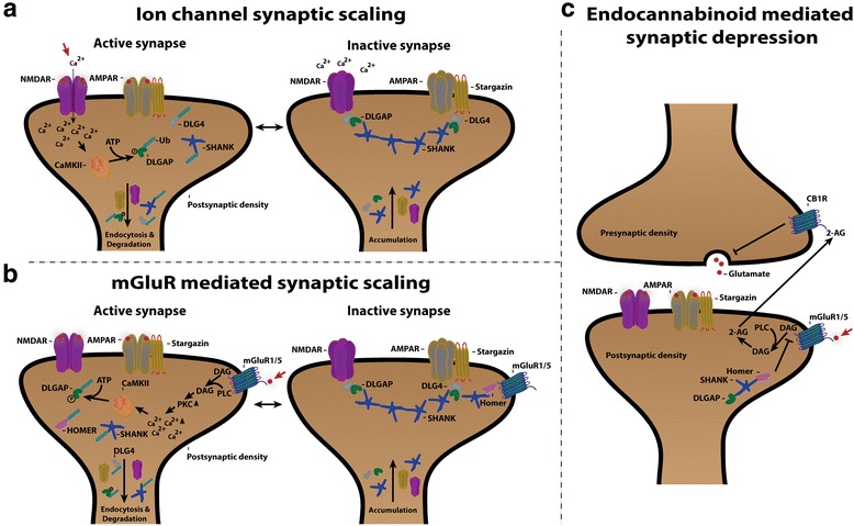
The neurotransmitter glutamate facilitates neuronal signalling at excitatory synapses. Glutamate is released from the presynaptic membrane into the synaptic cleft. Across the synaptic cleft glutamate binds to both ion channels and metabotropic glutamate receptors at the postsynapse, which expedite downstream signalling in the neuron. The postsynaptic density, a highly specialized matrix, which is attached to the postsynaptic membrane, controls this downstream signalling. The postsynaptic density also resets the synapse after each synaptic firing. It is composed of numerous proteins including a family of Discs large associated protein 1, 2, 3 and 4 (DLGAP1-4) that act as scaffold proteins in the postsynaptic density. They link the glutamate receptors in the postsynaptic membrane to other glutamate receptors, to signalling proteins and to components of the cytoskeleton. With the central localisation in the postsynapse, the DLGAP family seems to play a vital role in synaptic scaling by regulating the turnover of both ionotropic and metabotropic glutamate receptors in response to synaptic activity. DLGAP family has been directly linked to a variety of psychological and neurological disorders. In this review we focus on the direct and indirect role of DLGAP family on schizophrenia as well as other brain diseases.

Keywords: DLGAP1; DLGAP2; DLGAP3; DLGAP4; GKAP; PSD; SAPAP; Scaffold proteins; Schizophrenia; Synaptic scaling.


Conflict of interest statement

Authors’ information

AHR is a PhD student in the Cellular and Genetic Medicine Program. HBR is Associate Professor in the Department of Biomedical Sciences interested in molecular and cellular neurobiology, AS is Associate Professor in the Medical Genetics Program, Department of Cellular and Molecular Medicine. AS is interested in genomic organization, gene expression and gene regulatory mechanisms in the brain, DLGAP4 gene, cytogenetics.

Ethics approval and consent to participate

Not applicable.

Consent for publication

Not applicable.

Competing interests

The authors declare that they have no competing interests.

Publisher’s Note

Springer Nature remains neutral with regard to jurisdictional claims in published maps and institutional affiliations.

Figures

Fig. 1
Fig. 1
a Pictogram of chromosome 18, 8, 1 and 20 and the four DLGAP1–4 loci, respectively. DLGAP1 is located on chromosome 18 and transcript variants 1 to 9 are illustrated. DLGAP2 is found on chromosome 8 and has two transcript variants. The DLGAP3 gene is located on chromosome 1 and has one transcript variant. DLGAP4 is located on chromosome 20 and has 3 transcript variants. b Heatmap showing the homology in percentage between the longest isoform, isoform a, of DLGAP1–4 proteins respectively. The color key indicates the respective percentage to each colour. The heatmap is created in RStudio v.0.99.484 with data from a multiple alignment made with clustal omega v.1.2.1. DLGAP1 isoform a: 977 amino acids (NP_004737.2), DLGAP2 isoform a: 975 amino acids (NP_004736.2), DLGAP3 isoform a: 979 amino acids (NP_001073887.1) and DLGAP4 isoform a: 989 amino acids (NP_055717.2). c Phylogenetic tree of DLGAP1–4 protein conservation in following species; Homo sapiens (NP_004737.2, NP_004736.2, NP_001073887.1, NP_001035951.1), Macaca mulatta (AFE64413.1, AFJ72104.1, AFE64177.1), Bos taurus (NP_001179558.1, DAA17363.1, NP_001179367.1, AAI26739.1), Rattus norvegicus (NP_075235.3, NP_446353.2, NP_775161.2), Mus musculus (NP_808307.2, NP_766498.2, AAH57615.1, NP_001035953.1), Pongo abelii (XP_009251088.1, NP_001127321.1), Xenopus tropicalis (NP_001123829.1, NP_001106458.1, XP_012827022.1) and Danio rerio (NP_001189384.1, XP_009291347.1, NP_001038179.1, AAI33919.1). EV: Experimentally validated, CT: Conceptual Translation, P: Predicted from genomic sequence. The phylogenetic tree is created in RStudio v.0.99.484 with data from a multiple alignment made with clustal omega v.1.2.1. d Figure of multiple alignment of DLGAP1–4 GH1 domain and C-terminal. The sequence homology of the GH1 domain is 61 to 75% between DLGAP1 – DLGAP4. The alignment and data is generated in clustal omega v.1.2.1 and visualized in CLC sequence viewer v.7.6
Fig. 2
Fig. 2
a Pictogram of the DLGAP1–4 proteins with their respective domains. The DLGAP1–4 proteins have a 14 amino acid repeat domain (14-a.a. repeats) with 0 to 5 repeats depending on the DLGAP isoform, a Dynein light chain (DLC) domain and a GKAP homology domain 1 (GH1). b Illustration of how the four proteins interact. DLGAP binds via the C-terminal to the PDZ domain of SHANK. The 14-a.a. repeat domain of DLGAP binds the GK domain of DLG. Homer binds with the EVH1 domain to the proline rich domain of SHANK
Fig. 3
Fig. 3
DLGAPs are proteins of the postsynaptic density. The postsynaptic localization of DLGAPs in a cultured rat hippocampal neuron as revealed by a panDLGAP immunostaining. The excitatory postsynapses were visualized with an antibody directed against PSD-95 and MAP-2 used as a dendritic marker. Left scale bar, 20 μm; right scale bar, 3 μm
Fig. 4
Fig. 4
a Proposed model of the states in ion channel mediated synaptic scaling. In the active synapse calcium influx through the NMDA receptor activates CaMKII that utilizes ATP to phosphorylate DLGAP that causes dissociation from DLG4 and SHANK and prones them for ubiquitination (Ub). Subsequently, DLGAP, DLG4 and SHANK are degraded, which results in endocytosis of the AMPA and NMDA receptors. During synaptic inactivation the scaffold proteins DLGAP, DLG4 and SHANK accumulate together with the AMPA and NMDA receptors that are incorporated in the membrane. b Proposed model of mGluR mediated synaptic scaling displayed as an equilibrium between the active and the inactive synapse. The synaptic scaling is initiated by activation of mGluR1/5 by glutamate followed by PLC-mediated membrane release of DAG. The release of DAG leads to activation of PKC and release of intracellular Ca2+. The increase in calcium ions activates CaMKII that phosphorylates DLGAP and Homer. After phosphorylation DLGAP dissociates from DLG4 and SHANK, and Homer dissociates from mGluR1/5 and SHANK. DLGAP, DLG4, Homer and SHANK are then ubiquitinated (Ub) and degraded. With no intracellular bound scaffold proteins, NMDA receptors and AMPA receptors are internalised and removed from the synapse. In the inactive synapse after activation the scaffold proteins DLGAP, DLG4, Homer and SHANK accumulate together with AMPA receptors and NMDA receptors that are incorporated into the membrane. c Working model of endocannabinoid-mediated synaptic depression that is initiated by glutamate stimulation of mGluR1/5 that leads to PLC-mediated release of DAG. Then, DAG is converted to the endocannabinoid 2-AG that is transported to the synaptic cleft where it binds to the endocannabinoid receptor CB1R. After activation of CB1R, glutamate release from the presynapse to the synaptic cleft is halted, which leads to the depression. Presence of DLGAP in the PSD inhibits this endocannabinoid synaptic depression most likely via a DLGAP-SHANK-Homer- mGluR1/5

References

    1. Kornau HC, Schenker LT, Kennedy MB, Seeburg PH. Domain interaction between NMDA receptor subunits and the postsynaptic density protein PSD-95. Science. 1995;269:1737–1740. doi: 10.1126/science.7569905. - DOI - PubMed
    1. Naisbitt S, Kim E, Tu JC, Xiao B, Sala C, Valtschanoff J, et al. Shank, a novel family of postsynaptic density proteins that binds to the NMDA receptor/PSD-95/GKAP complex and cortactin. Neuron. 1999;23:569–582. doi: 10.1016/S0896-6273(00)80809-0. - DOI - PubMed
    1. Chen L, Chetkovich DM, Petralia RS, Sweeney NT, Kawasaki Y, Wenthold RJ, et al. Stargazin regulates synaptic targeting of AMPA receptors by two distinct mechanisms. Nature. Macmillian Magazines Ltd. 2000;408:936–943. - PubMed
    1. Steiner P, Higley MJ, Xu W, Czervionke BL, Malenka RC, Sabatini BL. Destabilization of the postsynaptic density by PSD-95 serine 73 Phosphorylation inhibits spine growth and synaptic plasticity. Neuron. 2008;60:788–802. doi: 10.1016/j.neuron.2008.10.014. - DOI - PMC - PubMed
    1. Kim E. GKAP, a novel synaptic protein that interacts with the Guanylate Kinase-like domain of the PSD-95/SAP90 family of channel clustering molecules. J Cell Biol. 1997;136:669–678. doi: 10.1083/jcb.136.3.669. - DOI - PMC - PubMed

Substances