. 2017 Sep 4;50(1):26.
doi: 10.1186/s40659-017-0130-y.

Neuron-derived CCL2 contributes to microglia activation and neurological decline in hepatic encephalopathy

Affiliations

Neuron-derived CCL2 contributes to microglia activation and neurological decline in hepatic encephalopathy

Li Zhang et al. Biol Res. .

Abstract

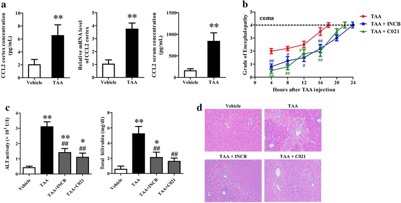
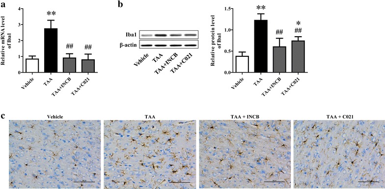
Background: CCL2 was up-regulated in neurons and involved in microglia activation and neurological decline in mice suffering from hepatic encephalopathy (HE). However, no data exist concerning the effect of neuron-derived CCL2 on microglia activation in vitro.

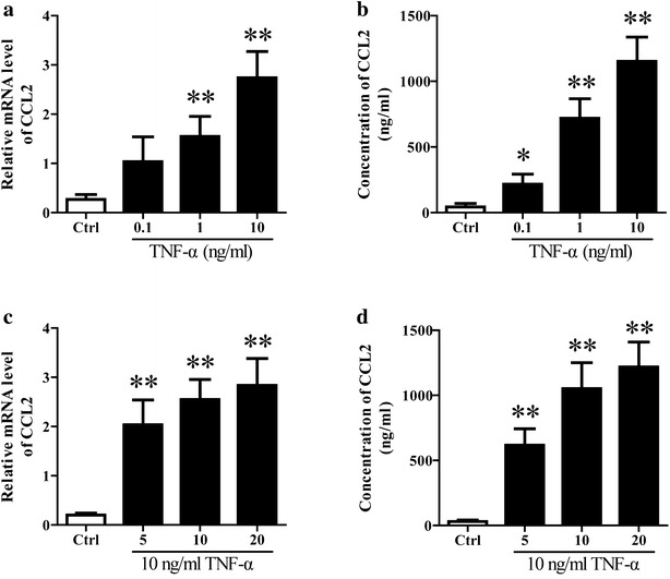
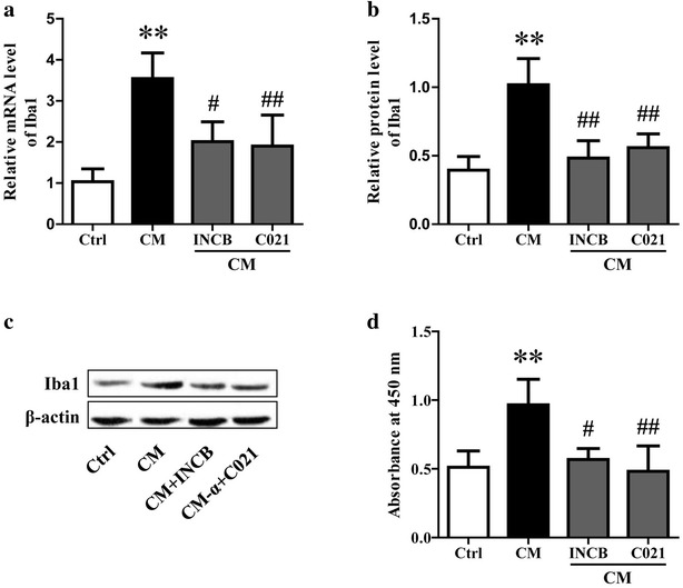
Methods: The rats were pretreated with CCL2 receptor inhibitors (INCB or C021, 1 mg/kg/day i.p.) for 3 days prior to thioacetamide (TAA) administration (300 mg/kg/day i.p.) for inducing HE model. At 8 h following the last injection (and every 4 h after), the grade of encephalopathy was assessed. Blood and whole brains were collected at coma for measuring CCL2 and Iba1 expression. In vitro, primary neurons were stimulated with TNF-α, and then the medium were collected for addition to microglia cultures with or without INCB or C021 pretreatment. The effect of the medium on microglia proliferation and activation was evaluated after 24 h.

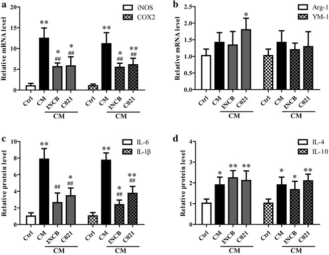
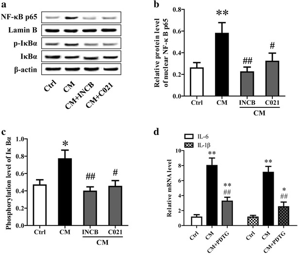
Results: CCL2 expression and microglia activation were elevated in the cerebral cortex of rats received TAA alone. CCL2 receptors inhibition improved neurological score and reduced cortical microglia activation. In vitro, TNF-α treatment induced CCL2 release by neurons. Medium from TNF-α stimulated neurons caused microglia proliferation and M1 markers expression, including iNOS, COX2, IL-6 and IL-1β, which could be suppressed by INCB or C021 pretreatment. The medium could also facilitate p65 nuclear translocation and IκBα phosphorylation, and NF-κB inhibition reduced the increased IL-6 and IL-1β expression induced by the medium.

Conclusion: Neuron-derived CCL2 contributed to microglia activation and neurological decline in HE. Blocking CCL2 or inhibiting microglia excessive activation may be potential strategies for HE.

Keywords: Chemokine CC motif ligand 2; Hepatic encephalopathy; Microglia; Neuron.


Figures

Fig. 1
Fig. 1
CCL2 blockage alleviated TAA-induced neurological decline and liver damage. At 8 h following the last injection of thioacetamide (TAA), and every 4 h after, the encephalopathy grade of rats from different group was assessed (b). Blood, whole brains and liver tissue were collected at coma and CCL2 expression and concentration in cerebral cortex and serum were determined using qRT-PCR and Elisa (a). Liver function was determined through measuring the serum levels of ALT and total bilirubin using commercially available kits (c). Liver damage was evaluated using HE staining and the original magnification was ×100 (d). **P < 0.01, *P < 0.05, vs vehicle; ## P < 0.01, # P < 0.05, vs TAA
Fig. 2
Fig. 2
CCL2 blockage alleviated TAA-induced microglia activation. Microglia activation in cerebral cortex collected at coma were evaluated through determination of Iba1 expression, a marker of microglia activation, using qRT-PCR (a), western blotting (b) and immunohistochemistry assays (c). The original magnification for immunohistochemical picture was ×200. **P < 0.01, *P < 0.05, vs vehicle; ## P < 0.01, # P < 0.05, vs TAA
Fig. 3
Fig. 3
TNF-α stimulation induced CCL2 expression and release in cultured neurons. The primary neurons isolated from neonatal rats were stimulated with different concentrations of TNF-α (0.1, 1 or 10 ng/ml) for 15 min or 10 ng/ml of TNF-α for 5, 10 or 20 min. Then the cells were washed with PBS and cultured in fresh medium. After 3 h, the CCL2 mRNA expression (a, c) and content in medium (b, d) were determined using qRT-PCR and ELISA respectively. **P < 0.01, *P < 0.05, vs Ctrl
Fig. 4
Fig. 4
Neuron-derived CCL2 induced the proliferation and activation of microglia. After stimulation with 10 ng/ml TNF-α for 10 min and washed with PBS, neurons were cultured in fresh medium for 3 h. Then the medium and lysates (CM) were collected and added into microglia cultures. To block CCL2, the microglia was pretreated with 5 μM CCL2 receptor inhibitor (INCB or C021) for 1 h before the addition of CM. After incubation for 24 h, the expression of Iba1 was determined using qRT-PCR (a) and western blotting (b, c). The proliferation of microglia was measured using CCK8 assay (d). **P < 0.01, *P < 0.05, vs Ctrl; ## P < 0.01, # P < 0.05, vs CM
Fig. 5
Fig. 5
Neuron-derived CCL2 induced the expression of M1 markers in microglia. As mentioned above, the medium and lysates (CM) of neurons stimulated with TNF-α were collected and added into microglia cultures. The microglia was pretreated with 5 μM CCL2 receptor inhibitor (INCB or C021) for 1 h before the addition of CM to block CCL2. After incubation for 24 h, the mRNA expression of iNOS, COX2, Arg-1 and YM-1 in microglia was determined using qRT-PCR (a, b), and the release of IL-6, IL-1, IL-1β, IL-4 and IL-10 were determined using Elisa assay (c, d). **P < 0.01, *P < 0.05, vs Ctrl; ## P < 0.01, # P < 0.05, vs CM
Fig. 6
Fig. 6
Neuron-derived CCL2 stimulated the activation of NF-κB in microglia. As previously described, the medium and lysates (CM) of neurons stimulated with TNF-α were collected and added into microglia cultures. The microglia was pretreated with 5 μM CCL2 receptor inhibitor (INCB or C021) for 1 h before the addition of CM to block CCL2. After incubation for 24 h, NF-κB activation was evaluated through determination of IκBα phosphorylation and NF-κB p65 nuclear translocation using western blotting (ac). d The cultured microglia was pretreated with 10 μM PDTG (NF-κB inhibitor) for 1 h prior to administration of CM. After cultured for 24 h, the mRNA expression of IL-6 and IL-1β was measured using qRT-PCR. **P < 0.01, *P < 0.05, vs Ctrl; ## P < 0.01, # P < 0.05, vs CM

References

    1. Butterworth RF. Pathogenesis of hepatic encephalopathy: new insights from neuroimaging and molecular studies. J Hepatol. 2003;39(2):278–285. doi: 10.1016/S0168-8278(03)00267-8. - DOI - PubMed
    1. Jiang W, Desjardins P, Butterworth RF. Cerebral inflammation contributes to encephalopathy and brain edema in acute liver failure: protective effect of minocycline. J Neurochem. 2009;109(2):485–493. doi: 10.1111/j.1471-4159.2009.05981.x. - DOI - PubMed
    1. Dennis CV, Sheahan PJ, Graeber MB, Sheedy DL, Kril JJ, Sutherland GT. Microglial proliferation in the brain of chronic alcoholics with hepatic encephalopathy. Metab Brain Dis. 2014;29(4):1027–1039. doi: 10.1007/s11011-013-9469-0. - DOI - PMC - PubMed
    1. Zemtsova I, Gorg B, Keitel V, Bidmon HJ, Schror K, Haussinger D. Microglia activation in hepatic encephalopathy in rats and humans. Hepatology. 2011;54(1):204–215. doi: 10.1002/hep.24326. - DOI - PubMed
    1. Semple BD, Kossmann T, Morganti-Kossmann MC. Role of chemokines in CNS health and pathology: a focus on the CCL2/CCR2 and CXCL8/CCR2 networks. J Cereb Blood Flow Metab. 2009;30(3):459–473. doi: 10.1038/jcbfm.2009.240. - DOI - PMC - PubMed

LinkOut - more resources