. 2017 Oct 15;144(20):3755-3765.
doi: 10.1242/dev.152611. Epub 2017 Sep 4.

Maternal Dead-end 1 promotes translation of nanos1 by binding the eIF3 complex

Affiliations

Maternal Dead-end 1 promotes translation of nanos1 by binding the eIF3 complex

Tristan Aguero et al. Development. .

Abstract

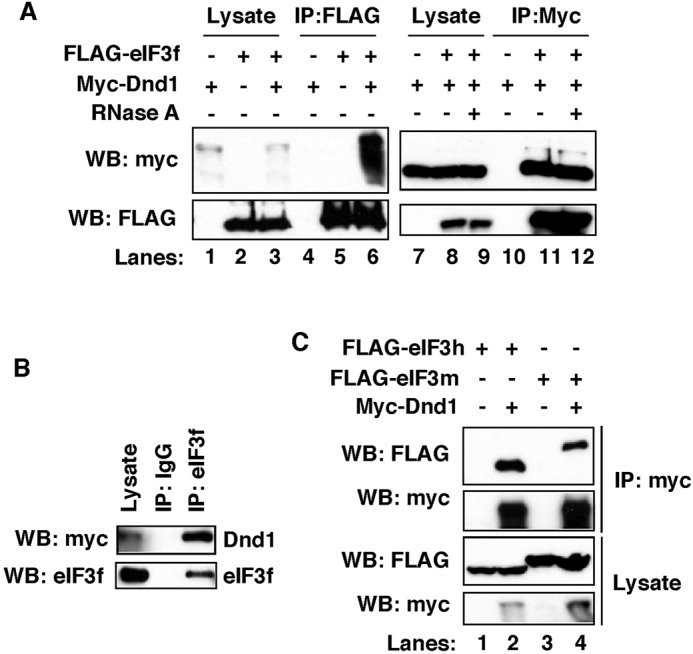
In the developing embryo, primordial germ cells (PGCs) represent the exclusive progenitors of the gametes, and their loss results in adult infertility. During early development, PGCs are exposed to numerous signals that specify somatic cell fates. To prevent somatic differentiation, PGCs must transiently silence their genome, an early developmental process that requires Nanos activity. However, it is unclear how Nanos translation is regulated in developing embryos. We report here that translation of nanos1 after fertilization requires Dead-end 1 (Dnd1), a vertebrate-specific germline RNA-binding protein. We provide evidence that Dnd1 protein, expression of which is low in oocytes, but increases dramatically after fertilization, directly interacts with, and relieves the inhibitory function of eukaryotic initiation factor 3f, a repressive component in the 43S preinitiation complex. This work uncovers a novel translational regulatory mechanism that is fundamentally important for germline development.

Keywords: Dnd1; Germline development; Nanos; Translation regulation; Xenopus.


Conflict of interest statement

Competing interestsThe authors declare no competing or financial interests.

Figures

Fig. 1.
Fig. 1.
Dnd1 is necessary and sufficient for nanos1 translation. (A) nanos1 (4 or 5 ng) was injected into oocytes with or without RNAs encoding Dnd1, Vasa, DeadSouth, Centroid and Dazl, each at 2 ng. Nanos1 protein was immunoprecipitated and analyzed by western blot. nanos1ΔTCE served as a positive control for nanos1 translation. The size difference between Nanos1ΔTCE and the wild-type Nanos1 is due to the deletion of the TCE, which is located immediately downstream of the translation initiation site. Experiments were repeated five times. (B) Quantification of band intensity of the western blot shown in A using ImageJ. (C) nanos1 RNA was added to wheat germ extracts with or without purified Dnd1 protein. Samples were analyzed for Nanos1 protein expression by western blot. nanos1ΔTCE served as a positive control. Experiments were repeated twice. (D) qPCR shows the levels of dnd1 and nanos1 in control and dnd1-depleted embryos (AS-oligo) at the 8-cell stage. Data are shown as mean±s.d. **P<0.01. Experiments were repeated three times. (E) Representative IF images show attenuation of endogenous Nanos1 protein expression by antisense depletion of maternal dnd1 (AS-oligo). Embryos were co-stained for Xiwi (green) and Nanos1 (red) at the 8- to 16-cell stage. Experiments were repeated twice.
Fig. 2.
Fig. 2.
Dnd1 expression pattern. (A) RT-PCR shows the expression of dnd1 RNA during early development. The experiment was performed twice. (B) Western blot showing expression of germline proteins during development. β-Tubulin was used as a loading control. Dnd1 was enriched by IP from 50 oocytes or embryos before western blotting. All other proteins were detected using protein extracts from the equivalent of one-eighth of an oocyte or embryo. The experiment was repeated three times. (C) Representative IF images show localization of endogenous Dnd1 protein (red) in an oocyte (top, n=7) and an 8-cell-stage embryo (bottom, n=7). A dnd1 knockdown oocyte (AS-oligo injected) served as a negative control for the specificity of Dnd1 staining (middle, n=10). Oocytes and embryos were hemi-sectioned. Images show the vegetal pole of oocytes or embryo. Asterisk marks the cleavage furrow of the embryo. Arrowhead indicates the germ plasm. Ov, ovulated; St, stage.
Fig. 3.
Fig. 3.
Dnd1 binds nanos1 RNA. (A) Double-filter nucleic acid-binding assay shows that recombinant Dnd1 protein binds nanos1 RNA. The experiment was repeated twice. (B) RIP assay followed by RT-PCR shows that endogenous Dnd1 protein selectively binds a subset of germline RNAs in stage 7 embryos. The experiment was performed three times. (C) RNAs were pulled down by GST-Dnd1 and measured by qPCR. Ratio between the pull down and 5% of RNA input is shown. GFP served as a negative control. Data are shown as mean±s.d. **P<0.01. The experiment was repeated four times. (D) nanos1-nanos1 3′UTR and nanos1-β-globin 3′UTR were injected into stage VI oocytes with or without dnd1 mRNA. Nanos1 protein was immunoprecipitated and analyzed by western blot for Nanos1 and Dnd1. The experiment was repeated twice.
Fig. 4.
Fig. 4.
Dnd1 physically interacts with eIF3f. (A) Anti-FLAG (lanes 4-6) or anti-Myc (lanes 10-12) antibodies were used to IP eIF3f or Dnd1 from cell lysates (lanes 9,12). Addition of RNaseA to lysates did not disrupt the Dnd1-eIF3f complex (lane 12). (B) Co-IP shows the interaction between myc-Dnd1 and endogenous eIF3f in HEK293T cells. IgG served as negative control. (C) Co-IP shows that myc-Dnd1 formed complexes with FLAG-eIF3 h and FLAG-eIF3m in HEK293T cells. Experiments were repeated three times.
Fig. 5.
Fig. 5.
Interaction between endogenous Dnd1 and eIF3f in Xenopus embryos. (A) Egg or embryo extracts were fractionated on 7-20% sucrose gradients. Gradient fractions were blotted with antibodies for Dnd1, eIF3f and eIF3c proteins. Experiments were repeated four times. (B) Fractions 1+2 and fractions 8+9+10 of embryo extracts from the sucrose gradients were pooled and immunoprecipitated with an anti-Dnd1 antibody. Endogenous eIF3f and Dnd1 proteins were monitored by western blot. Experiments were repeated three times.
Fig. 6.
Fig. 6.
Interaction between Dnd1 and eIF3f is essential for nanos1 translation. (A) FLAG-tagged eIF3f deletions were transfected into HEK293T cells along with myc-Dnd1. Lysates were immunoprecipitated with anti-FLAG antibody and analyzed by western blot. The minimal Dnd1-binding domain was mapped to residues 92-200 of eIF3f, which is indicated by the red asterisk. (B) Schematic summarizing experiments shown in A. +, Dnd1 binding; −, lack of Dnd1 binding. (C) Myc-Dnd1 and FLAG-eIF3f were transfected into HEK293T cells with increasing amounts of eIF3f92-200. Cell lysates were immunoprecipitated with an anti-myc antibody and analyzed by western blot. (D) Myc-Dnd1 and FLAG-eIF3h were transfected into HEK293T cells in the presence or absence of eIF3f92-200. Cell lysates were immunoprecipitated with an anti-myc antibody and analyzed by western blot. Experiments were repeated three times. (E) nanos1 RNA was injected into fertilized eggs alone or with eIF3f92-200, or with eIF3f92-200 and myc-dnd1 RNAs. At stage 11, Nanos1 protein was immunoprecipitated and analyzed by western blot. Non-specific band served as a loading control. The experiment was performed twice.
Fig. 7.
Fig. 7.
Interfering with the interaction between Dnd1 and eIF3f disrupts PGC development. (A-C) In situ hybridization of stage 33 embryos showing Xpat-expressing PGCs in uninjected control (A), eIF3f92-200-injected (B) and eIF3f92-200+dnd1-injected (C) embryos. Experiments were repeated three times. (D) Quantification of results shown in A-C. Two-tailed t-tests were performed. *P<0.05; n.s., non-significant.
Fig. 8.
Fig. 8.
Dnd1 relieves the inhibitory effect of eIF3f on nanos1 translation and PGC development. (A) nanos1 (4 ng) was translated alone or with FLAG-eIF3f (2 ng) in wheat germ extracts with enhanced translational efficiency (Promega WG+). Nanos1 protein was detected by western blot. eIF3f inhibited nanos1 translation. Addition of recombinant Dnd1 protein relieved the repressive activity of eIF3f in a dose-dependent manner. The experiment was repeated three times. (B) eIF3f represses nanos1 translation in vivo. nanos1 RNA was injected into fertilized eggs alone, or together with eIF3f or dnd1 RNA. At stage 11, Nanos1 protein was immunoprecipitated and analyzed by western blot. Experiments were performed twice. (C-E) In situ hybridization of stage 33 embryos showing PGCs by Xpat staining in uninjected control (C), eIF3f-injected (D) and eIF3f+dnd1-injected (E) embryos. Experiments were repeated three times. (F) Quantification of results shown in C-E. Two-tailed t-tests were performed. *P<0.05, **P<0.01. (G) Working model of Dnd1 function in regulating nanos1 translation. Before fertilization, very little Dnd1 protein is present. Translation of nanos1 RNA is blocked by TCE, a secondary structure within the ORF that prevents the preinitiation complex (PIC) from scanning and initiating translation. After fertilization, Dnd1 protein accumulates within the germ plasm and there binds to nanos1 RNA, altering the TCE structure. The PIC can now scan the nanos1 RNA. Meanwhile, Dnd1 binds with the eIF3 complex through the interaction with subunit eIF3f. This interaction blocks the repressive activity of eIF3f and promotes the translation of nanos1 RNA. The eIF3-Dnd1 complex is released from the 40S ribosomal subunit as translation proceeds.

References

    1. Ahringer J. and Kimble J. (1991). Control of the sperm-oocyte switch in Caenorhabditis elegans hermaphrodites by the fem-3 3’ untranslated region. Nature 349, 346-348. 10.1038/349346a0 - DOI - PubMed
    1. Asaoka-Taguchi M., Yamada M., Nakamura A., Hanyu K. and Kobayashi S. (1999). Maternal Pumilio acts together with Nanos in germline development in Drosophila embryos. Nat. Cell Biol. 1, 431-437. 10.1038/15666 - DOI - PubMed
    1. Berekelya L. A., Ponomarev M. B., Luchinskaya N. N. and Belyavsky A. V. (2003). Xenopus Germes encodes a novel germ plasm-associated transcript. Gene Expr. Patterns 3, 521-524. 10.1016/S1567-133X(03)00055-3 - DOI - PubMed
    1. Colozza G. and De Robertis E. M. (2014). Maternal syntabulin is required for dorsal axis formation and is a germ plasm component in Xenopus. Differentiation 88, 17-26. 10.1016/j.diff.2014.03.002 - DOI - PubMed
    1. Curtis D., Treiber D. K., Tao F., Zamore P. D., Williamson J. R. and Lehmann R. (1997). A CCHC metal-binding domain in Nanos is essential for translational regulation. EMBO J. 16, 834-843. 10.1093/emboj/16.4.834 - DOI - PMC - PubMed

Publication types

LinkOut - more resources