

Association Between RASSF1A Promoter Methylation and Testicular Germ Cell Tumor: A Meta-analysis and a Cohort Study
- PMID: 28871003
- PMCID: PMC5611522
- DOI: 10.21873/cgp.20046
Association Between RASSF1A Promoter Methylation and Testicular Germ Cell Tumor: A Meta-analysis and a Cohort Study
Abstract
Background: The RAS association domain family protein 1a (RASSF1A) is a prominent tumor suppressor gene showing altered promoter methylation in testicular germ cell tumors (TGCT). RASSF1A promoter hypermethylation might represent an early event in TGCT tumorigenesis. We investigated whether the RASSF1A promoter methylation in peripheral blood of TGCT patients can be associated with testicular cancer risk.
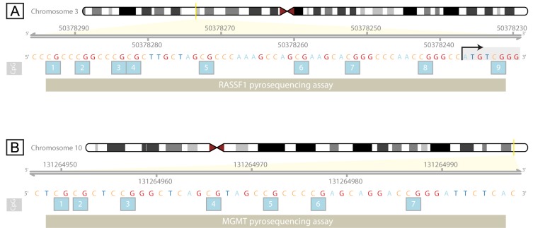
Materials and methods: Following a meta-analysis, we performed a cohort study including 32 testicular cancer patients and 32 healthy controls. Promoter methylation of the RASSF1A and O6-methylguanine-DNA-methyltransferase (MGMT) genes was analyzed using bisulfite pyrosequencing of DNA from peripheral blood.
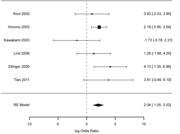
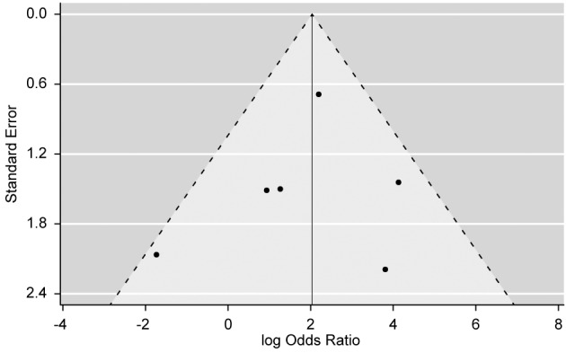
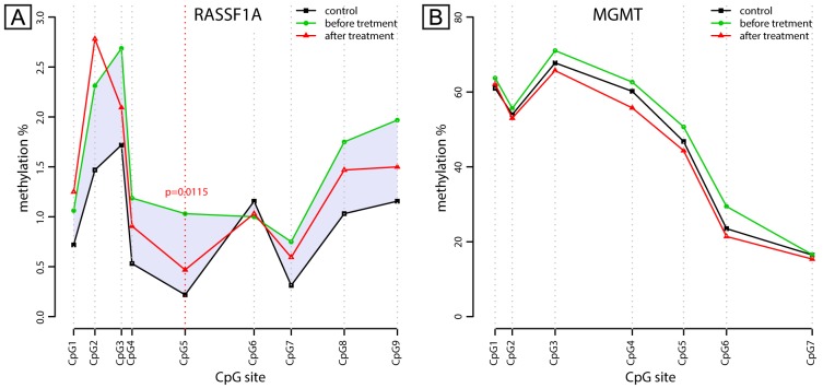
Results: Meta-analysis showed an odds ratio (OR) of 7.69 for RASSF1A promoter methylation as a risk factor for TGCT. Cohort study found altered methylation of the RASSF1A promoter in blood of TGCT patients. Methylation was higher in TGCT patients before BEP chemotherapy.
Conclusion: The meta-analysis indicates a role of the RASSF1A promoter hypermethylation from peripheral blood in TCGT. We confirmed that finding in our cohort study, which represents the first report of changed RASSF1A promoter methylation in peripheral blood TGCT.
Keywords: BEP chemotherapy; DNA methylation; MGMT; RASSF1; testicular cancer.
Copyright© 2017, International Institute of Anticancer Research (Dr. George J. Delinasios), All rights reserved.
Figures
References
-
- Ferlay J, Soerjomataram I, Dikshit R, Eser S, Mathers C, Rebelo M, Parkin DM, Forman D, Bray F. Cancer incidence and mortality worldwide: Sources, methods and major patterns in globocan 2012. Int J Cancer. 2015;136(5):E359–386. - PubMed
-
- Vasdev N, Moon A, Thorpe AC. Classification, epidemiology and therapies for testicular germ cell tumours. Int J Dev Biol. 2013;57(2-4):133–139. - PubMed
-
- Stevenson SM, Lowrance WT. Epidemiology and diagnosis of testis cancer. Urol Clin North Am. 2015;42(3):269–275. - PubMed
-
- Le Cornet C, Lortet-Tieulent J, Forman D, Beranger R, Flechon A, Fervers B, Schuz J, Bray F. Testicular cancer incidence to rise by 25% by 2025 in europe? Model-based predictions in 40 countries using population-based registry data. Eur J Cancer. 2014;50(4):831–839. - PubMed
-
- Illuminati G, Calio FG, Angelici AM, Pizzardi G, Pasqua R, Masci F, Vietri F. Outcome of resection and chemotherapy versus chemotherapy alone for retroperitoneal recurrence of testicular cancer involving the inferior vena cava: A retrospective cohort study of 22 consecutive patients. Anticancer Res. 2016;36(7):3483–3488. - PubMed
Publication types
MeSH terms
Substances
Supplementary concepts
LinkOut - more resources
Full Text Sources
Other Literature Sources
Medical
Research Materials