. 2017 Oct 4;37(40):9632-9644.
doi: 10.1523/JNEUROSCI.0643-16.2017. Epub 2017 Sep 4.

KLF9 and JNK3 Interact to Suppress Axon Regeneration in the Adult CNS

Affiliations

KLF9 and JNK3 Interact to Suppress Axon Regeneration in the Adult CNS

Akintomide Apara et al. J Neurosci. .

Abstract

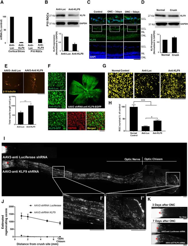
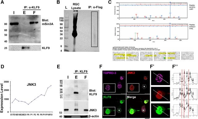
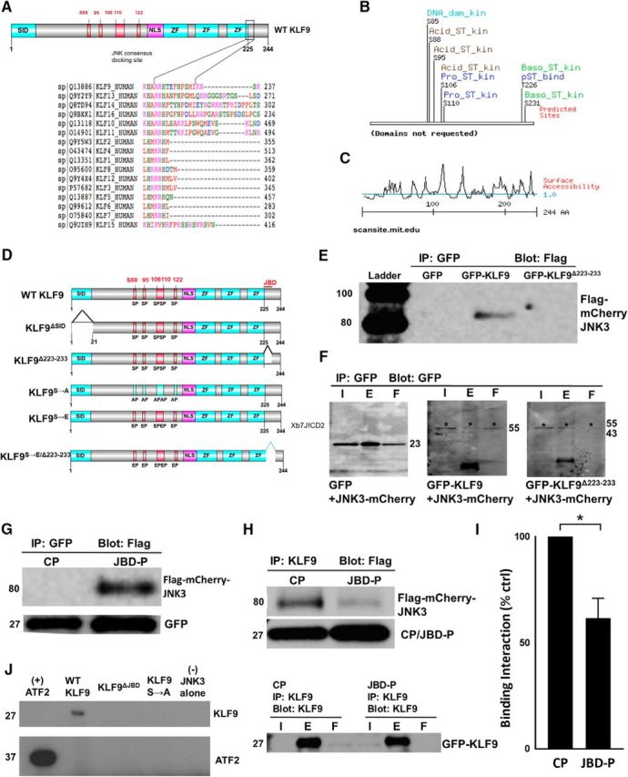
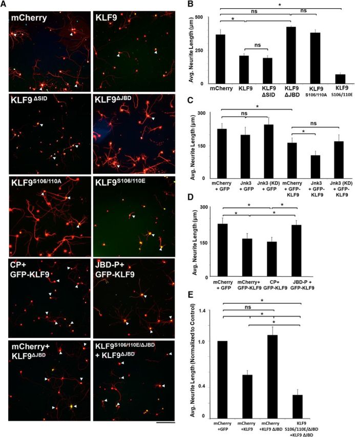
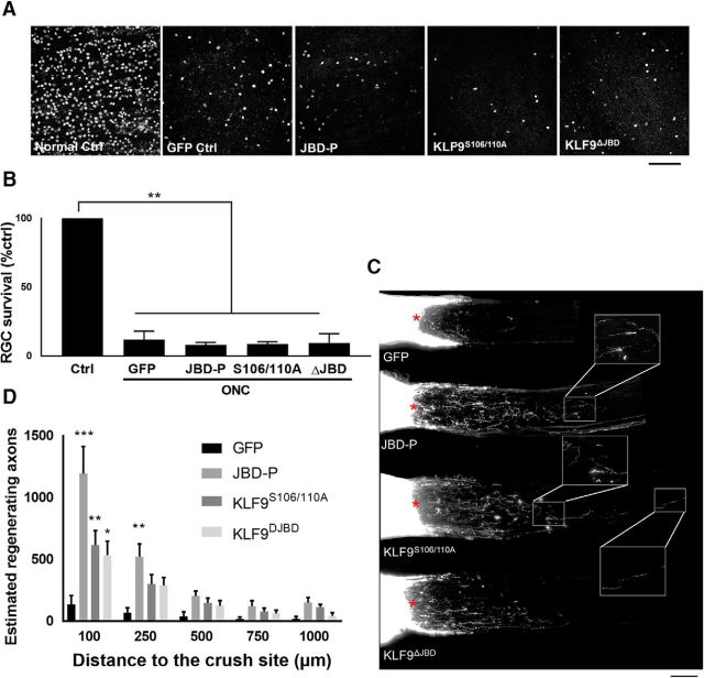
Neurons in the adult mammalian CNS decrease in intrinsic axon growth capacity during development in concert with changes in Krüppel-like transcription factors (KLFs). KLFs regulate axon growth in CNS neurons including retinal ganglion cells (RGCs). Here, we found that knock-down of KLF9, an axon growth suppressor that is normally upregulated 250-fold in RGC development, promotes long-distance optic nerve regeneration in adult rats of both sexes. We identified a novel binding partner, MAPK10/JNK3 kinase, and found that JNK3 (c-Jun N-terminal kinase 3) is critical for KLF9's axon-growth-suppressive activity. Interfering with a JNK3-binding domain or mutating two newly discovered serine phosphorylation acceptor sites, Ser106 and Ser110, effectively abolished KLF9's neurite growth suppression in vitro and promoted axon regeneration in vivo These findings demonstrate a novel, physiologic role for the interaction of KLF9 and JNK3 in regenerative failure in the optic nerve and suggest new therapeutic strategies to promote axon regeneration in the adult CNS.SIGNIFICANCE STATEMENT Injured CNS nerves fail to regenerate spontaneously. Promoting intrinsic axon growth capacity has been a major challenge in the field. Here, we demonstrate that knocking down Krüppel-like transcription factor 9 (KLF9) via shRNA promotes long-distance axon regeneration after optic nerve injury and uncover a novel and important KLF9-JNK3 interaction that contributes to axon growth suppression in vitro and regenerative failure in vivo These studies suggest potential therapeutic approaches to promote axon regeneration in injury and other degenerative diseases in the adult CNS.

Keywords: Jnk; KLFs; regeneration; survival.


Figures

Figure 1.
Figure 1.
Anti-KLF9 shRNA promotes RGC survival and axon regeneration at 2 weeks after optic nerve injury. A, AAV2-shRNA anti-KLF9 decreased KLF9 expression levels in cortical slice culture and purified P10 RGCs. B, Western blot for KLF9 in RGCs purified after intravitreal injection of AAV2-KLF9 shRNA or AAV2-anti-luceferase control. C, Immunohistochemistry of KLF9 in retinal slides in control and at 3 d and 5 d after optic nerve injury. D, Western blot for KLF9 in RGCs purified from P18 rats 3 d after optic nerve injury and uninjured (normal) control. E, KLF9 knockdown increased neurite growth in purified P8 RGCs. RGCs were treated with AAV2-shRNA anti-KLF9-GFP or AAV2-shRNA anti-Luciferase-GFP and cultured in growth media for 5 d. Neurites were labeled by β-III-tubulin staining (red). F, Images of the retinal segments showing GFP-positive virus transduced cells were co-labelled with RGC-specific marker RBPMS. G, Images of the retinal segments showing FG labeled RGCs. H, RGC quantification demonstrated significantly higher RGC density in anti-KLF9 group than the injury control group. **p < 0.01, one-way analysis of variance; N = 3–4 per group. I, Images of the optic nerve sections showing CTB-labelled regenerating axons at 2 weeks after optic nerve injury. Asterisks, lesion sites. High-magnification images of the boxed area in (I′) showed fibers extending contralaterally through the optic chiasm of an anti-KLF9 virus-treated animal (I′′). J, Significantly more fibers regenerated after KLF9 knockdown compared to control group. **p < 0.01, Student t-test; N = 3–4 per group. I′,I′′, High-magnification images of the boxed area in I. K, Images of the optic nerve sections showing CTB-labelled regenerating axons at 3 d and 1 week after optic nerve injury for AAV2-shRNA anti-KLF9-GFP or AAV2-shRNA anti-Luciferase-GFP. Asterisks, lesion sites. Error bars, SEM. Scale bars, 200 μm.
Figure 2.
Figure 2.
KLF9 is bound by JNK3 in neurons. A, mSin3a is present in the retina but does not bind KLF9. Retina lysate from P10 rats was immunoprecipitated for KLF9 and Western blotted for mSin3a or KLF9. B, Purified P0 rat RGCs virally transduced with flag-tagged KLF9 were lysed and immunoprecipitated for flag. The IP lane (box) was sent for MS analysis. C, JNK3 is identified from KLF9 IP/MS. MAPK10/JNK3 was detected with six independent peptides (identified from eight unique and 19 total spectra with >95% confidence, yellow) and with 13% coverage (oxidized residue in green). D, JNK3 mRNA expression (arbitrary units) in developing RGCs from a previously described microarray dataset (Wang et al., 2007) shows robust upregulation during development. E, Endogenous KLF9 co-IPs with JNK3. Retinas from P10 rats were immunoprecipitated for KLF9 and Western blotting for anti-KLF9, anti-JNK3, and anti-β-actin antibodies, as marked. F, Confocal nanoscopy imaging of GFP-KLF9- and Flag-JNK3-electroporated RGCs demonstrated nuclear colocalization. Asterisk indicates that not all cells were successfully cotransfected. Fluorescence intensity of KLF9, JNK3, and TOPRO-3 were calculated separately and compared along the line in the cell cotransfected with both proteins (F′, arrows). Intensities showed higher similarity between KLF9 and JNK3 thanTOPRO-3 (red lines). F′′, High-magnification images of the boxed area in F. Scale bar, 10 μm. I, input; E, eluent; F, flow-through.
Figure 3.
Figure 3.
Identification of JNK3-binding domain and potential serine/threonine phosphorylation sites of KLF9. A, KLF9 structural analysis revealing potential JNK3-binding domains. ClustalW2 alignment shows region spanning R223 to I233 near C terminus of KLF9, as well as similar regions on subfamily relatives KLF13, KLF14, and KLF16, as well as KLF10 and KLF11, closely conforming to the known consensus JNK3 “DEJL” docking domain. B, MIT Scansite surface analysis of KLF9 full-length protein reveals potential phospho-acceptor sites at S88, S95, S106, and S110. C, MIT Scansite-predicted surface accessibility of KLF9 protein (>1 indicates exposed residues; <1 indicates buried or inaccessible residues). D, Schematic of a subset of mutant constructs for KLF9. ZF, Zinc finger domains. E, F, R223–I233 of KLF9 is necessary for KLF9–JNK3 interaction in neurons. E18 hippocampal neurons were cotransduced with Flag-mCherry-JNK3 and GFP, GFP-KLF9, or GFP-KLF9 R223-I233 deletion mutant (GFP-KLF9 Δ223–233) constructs using lentivirus. Cells were immunoprecipitated for GFP and Western blotted for flag (E) or GFP (F). G, H, JBD-P of KLF9 is sufficient for KLF9/JNK3 interaction in neurons. E18 hippocampal neurons were virally cotransduced with Flag-mCherry-JNK3 and either CP or JBD-P constructs (G), immunoprecipitated for GFP and Western blotted for flag. Flag-mCherry-JNK3 was only detected with the JBD-P construct (G). H, I, JBD-P reduces KLF9–JNK3 interaction. E18 hippocampal neurons were virally cotransduced with GFP-KLF9, Flag-mCherry-JNK3, and the CP or JBD-P, immunoprecipitated for KLF9, and Western blotted for flag or GFP (I). Densitometry quantification of blot bands showed an average of 38% decrease in the amount of flag-mCherry-JNK3 pulled down by the JBD-P coexpression group compared with the CP condition (I). J, KLF9 incorporation of radiolabeled [γ-32P] ATP depends on JNK-binding domain (JBD) and identified serine residues. E18 hippocampal neurons were transduced with flag-tagged wild-type KLF9, KLF9ΔJBD, or KLF9S85/88/95/106/110A mutants. Cells were immunoprecipitated for flag. Eluents were combined with recombinant JNK3 and radiolabeled [ϒ-32] ATP in a standard in vitro kinase assay. Incorporation of radioactivity was only observed in the eluents from wild-type KLF9 transduced neurons. *p < 0.05, 2-tailed Student's t test, n = 3. Error bars indicate SD. I, Input; E, eluent; F, flow-through.
Figure 4.
Figure 4.
JNK3-binding domain and serines S106 and S110 are critical to KLF9's function in suppressing axon growth. A, Images of RGCs immunostained for GFP (transduced cells) and β-III tubulin (red). P0 RGCs were virally transduced with mCherry, KLF9 or mutants of KLF9 with GFP reporter. Scale bar, 50 μm. BE, Quantification of neurite growth of RGCs in different conditions. The KLF9ΔJBD but not the KLF9ΔSID deletion abolished wild-type KLF9's growth-suppressive effect on P0 (B) and P8 (data not shown) RGCs. JNK3 potentiated KLF9's neurite growth-suppressive activity on P0 RGCs; however, JNK3 alone or a kinase-dead mutant (KD) showed no effect (C). JBD-P but not CP abolished KLF9's suppressive effect on P0 RGCs cotransduced with KLF9 (D). KLF9S106/110A substitutions abolished wild-type KLF9's growth-suppressive effect, whereas KLF9S106/110E enhanced KLF9's suppressive effect on P0 (A) and P8 (data not shown) RGCs. Phosphomimic S106/S110E substitutions rescued KLF9 suppression on P0 RGC neurite growth even in the absence of the R223-I233 JNK3-binding domain (E). *p < 0.05, one-way ANOVA with Bonferroni/Dunn correction; n = 3–5. Error bars indicate SD., NS, Not significant.
Figure 5.
Figure 5.
JNK3 knock-out promoted RGC survival but not axon regeneration. A, Immunofluorescence for retinal ganglion cells using β-III tubulin (green) and phosphorylated JNK (red) in wild-type control retinal sections and after optic nerve injury in wild-type mice, Jnk2−/−, and Jnk2 and 3−/−. B, pJNK quantifications for control and optic nerve injury. C, Images of the retina showing RGC-specific markers RBPMS (green) and Brn3a (yellow) in JNK3 knock-out (KO) and age-matched wild-type (WT) mice before and 2 weeks after optic nerve crush. D, RGC quantification demonstrated similar RGC density in JNK3 KO and WT control mice before the optic nerve injury. Two weeks after optic nerve injury, however, RGC density in JNK3 KO mice was significantly higher than in WT animals (*p < 0.05, **p < 0.001, one-way ANOVA; n = 5 per group; NS, Not significant). E, Images of the optic nerve sections showing CTB-labeled axons 2 weeks after optic nerve injury. Asterisks indicate lesion sites. F, A similar number of regenerating fibers was observed in JNK3 KO and WT control animals. Error bars indicate SE. Scale bar, 200 μm.
Figure 6.
Figure 6.
Disrupting the KLF9–JNK3 interaction or expressing KLF9 S106/110A mutants enhance optic nerve axon regeneration 2 weeks after optic nerve injury. A, Images of Brn3a-positive cells in retinal segments from different groups. Scale bar, 100 μm. B, Quantification of RGC survival showed no significant differences among all groups. C, Images of merged optic nerve sections showing CTB-labeled regenerating axons. Scale bar, 200 μm. Asterisks indicate lesion sites. D, Significantly more regenerating fibers were observed in JBD-P, KLF9S106/110A, and KLF9ΔJBD treated animals than in control animals. *p < 0.05, **p < 0.01, one-way ANOVA; n = 4. Error bars indicate SEM.

References

    1. Abdelli S, Puyal J, Bielmann C, Buchillier V, Abderrahmani A, Clarke PG, Beckmann JS, Bonny C (2009) JNK3 is abundant in insulin-secreting cells and protects against cytokine-induced apoptosis. Diabetologia 52:1871–1880. 10.1007/s00125-009-1431-7 - DOI - PubMed
    1. Barnat M, Enslen H, Propst F, Davis RJ, Soares S, Nothias F (2010) Distinct roles of c-Jun N-terminal kinase isoforms in neurite initiation and elongation during axonal regeneration. J Neurosci 30:7804–7816. 10.1523/JNEUROSCI.0372-10.2010 - DOI - PMC - PubMed
    1. Barres BA, Silverstein BE, Corey DP, Chun LL (1988) Electrophysiological variation among retinal ganglion cells purified by panning. Neuron 1:791–803. 10.1016/0896-6273(88)90127-4 - DOI - PubMed
    1. Berkelaar M, Clarke DB, Wang YC, Bray GM, Aguayo AJ (1994) Axotomy ganglion results in delayed death and apoptosis of retinal cells in adult rats. J Neurosci 14:4368–4374. - PMC - PubMed
    1. Blackmore MG, Wang Z, Lerch JK, Motti D, Zhang YP, Shields CB, Lee JK, Goldberg JL, Lemmon VP, Bixby JL (2012) Krüppel-like Factor 7 engineered for transcriptional activation promotes axon regeneration in the adult corticospinal tract. Proc Natl Acad Sci U S A 109:7517–7522. 10.1073/pnas.1120684109 - DOI - PMC - PubMed

Publication types

MeSH terms

Substances