. 2017 Nov;2(11):1543-1557.
doi: 10.1038/s41564-017-0017-2. Epub 2017 Sep 4.

TRIM23 mediates virus-induced autophagy via activation of TBK1

Affiliations

TRIM23 mediates virus-induced autophagy via activation of TBK1

Konstantin M J Sparrer et al. Nat Microbiol. 2017 Nov.

Abstract

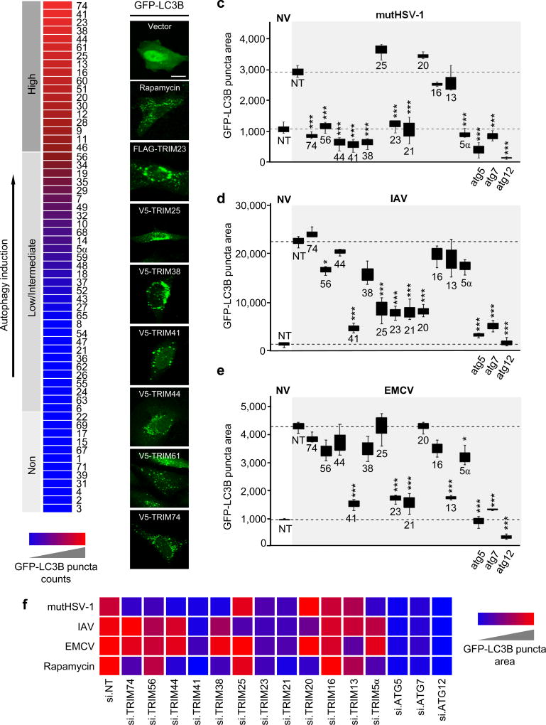
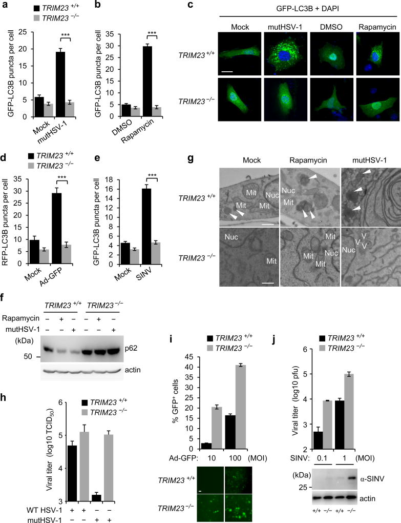
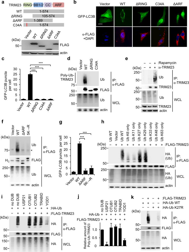
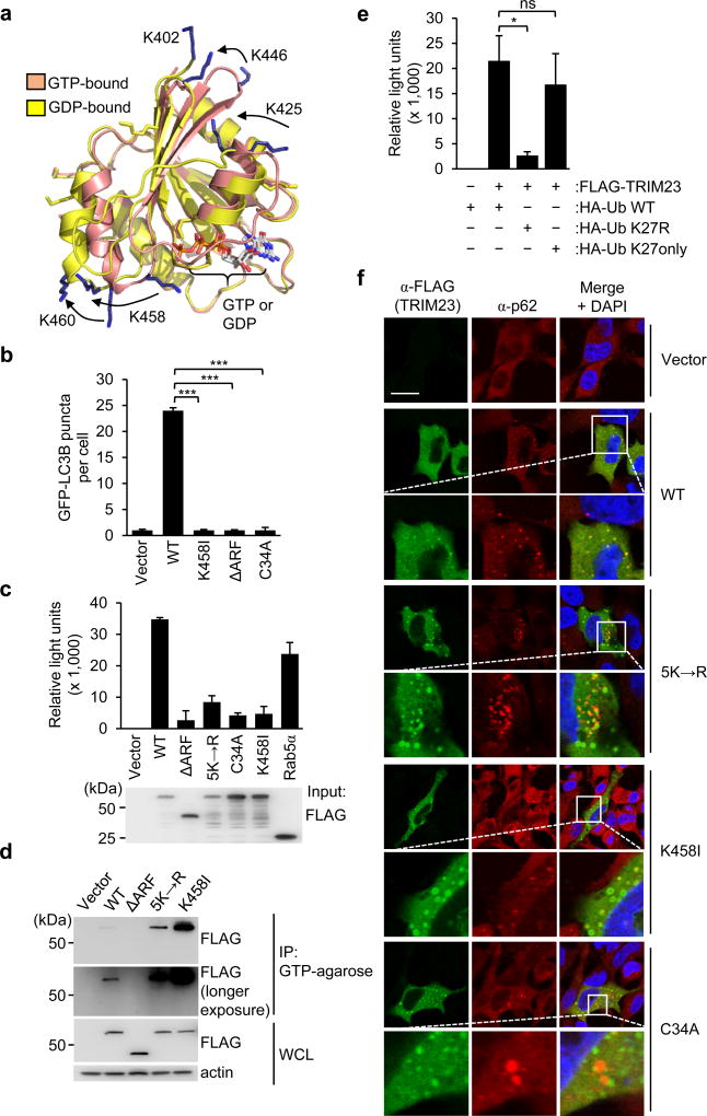
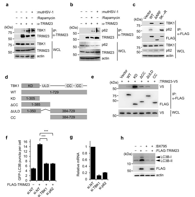
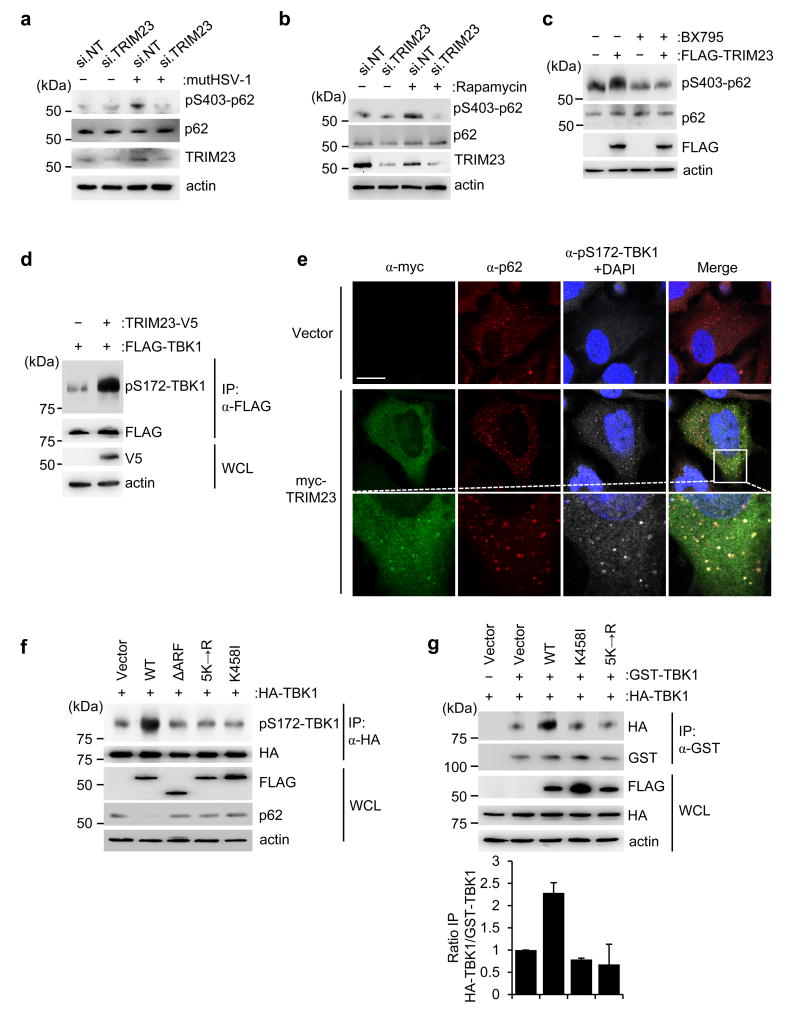
Autophagy and interferon (IFN)-mediated innate immunity are critical antiviral defence mechanisms, and recent evidence indicated that tripartite motif (TRIM) proteins are important regulators of both processes. Although the role of TRIM proteins in modulating antiviral cytokine responses has been well established, much less is known about their involvement in autophagy in response to different viral pathogens. Through a targeted RNAi screen examining the relevance of selected TRIM proteins in autophagy induced by herpes simplex virus 1 (HSV-1), encephalomyocarditis virus (EMCV) and influenza A virus (IAV), we identified several TRIM proteins that regulate autophagy in a virus-species-specific manner, as well as a few TRIM proteins that were essential for autophagy triggered by all three viruses and rapamycin, among them TRIM23. TRIM23 was critical for autophagy-mediated restriction of multiple viruses, and this activity was dependent on both its RING E3 ligase and ADP-ribosylation factor (ARF) GTPase activity. Mechanistic studies revealed that unconventional K27-linked auto-ubiquitination of the ARF domain is essential for the GTP hydrolysis activity of TRIM23 and activation of TANK-binding kinase 1 (TBK1) by facilitating its dimerization and ability to phosphorylate the selective autophagy receptor p62. Our work identifies the TRIM23-TBK1-p62 axis as a key component of selective autophagy and further reveals a role for K27-linked ubiquitination in GTPase-dependent TBK1 activation.


Conflict of interest statement

COMPETING FINANCIAL INTERESTS

The authors declare no competing financial interests.

Figures

Figure 1
Figure 1. TRIM proteins modulate viral-induced autophagy in a virus species-specific manner
a, Summary of the TRIM cDNA screen (shown in Supplementary Fig. 1a) in which 61 TRIM proteins were tested for their ability to induce GFP-LC3B puncta formation in transiently transfected HeLa cells. TRIM proteins were categorized into ‘high inducers’ of autophagy (red; defined as inducing more GFP-LC3B puncta than rapamycin stimulation), or ‘low/intermediate inducers’ and ‘non-inducers’ (magenta/blue; defined as inducing fewer GFP-LC3B puncta than rapamycin but more than empty vector transfection, or similar to vector transfection, respectively). b, Representative laser-scanning confocal microscopy images of GFP-LC3B puncta formation in HeLa cells for the top 7 hits from the TRIM cDNA screen shown in (a). Representative images for GFP-LC3B puncta formation induced by rapamycin treatment or empty vector transfection (controls) are also shown. Scale bar, 20 µm. c–e, GFP-LC3B puncta formation in HeLa cells transiently transfected with non-targeting control siRNA (NT) or siRNAs targeting the indicated TRIM proteins and subsequently infected with mutHSV-1 (MOI 4) for 8 h (c), IAV (MOI 5) for 14 h (d), or EMCV (MOI 100) for 6 h (e). Box and whisker plots show the distribution of the area of GFP-LC3B for 3 individual images, each containing ~30 cells. f, Summary of the results of GFP-LC3B puncta formation from the RNAi screen using viral stimuli (c-e) or rapamycin treatment (Supplementary Fig. 1d). NV, no infection. *p<0.05, ***p<0.0005 (ANOVA test). Data are representative of one screen (a), or two (b) or three (c–f) independent experiments.
Figure 2
Figure 2. TRIM23 is essential for virus-induced autophagy
a,b, GFP-LC3B puncta formation in TRIM23 +/+ or TRIM23 −/− MEFs transiently transfected with GFP-LC3B and either left uninfected (Mock) or infected with mutHSV-1 (MOI 4) for 12 h, or treated with DMSO or rapamycin for 2 h. Results represent the mean GFP-LC3B puncta per cell ± SD (n=50). ***p<0.001 (Student’s t-test). c, Representative images of GFP-LC3B puncta formation for the experiments shown in (a,b). Scale bar, 20 µm. d,e, Quantification of RFP-LC3B or GFP-LC3B puncta in transiently transfected TRIM23 +/+ or TRIM23 −/− MEFs that were left uninfected (Mock), or infected with Ad-GFP (MOI 100) for 48 h or SINV (MOI 5) for 24 h. Results represent the mean RFP/GFP-LC3B puncta per cell ± SD (n=50). ***p<0.001 (Student’s t-test). f, Endogenous p62 protein levels in the whole cell lysates (WCLs) of TRIM23 +/+ and TRIM23 −/− MEFs that were either left uninfected, infected with mutHSV-1 (MOI 4) for 12 h, or stimulated with rapamycin for 2 h, determined by immunoblot (IB) with anti-p62. g, EM analysis of TRIM23 +/+ and TRIM23 −/− MEFs that were mock-treated, stimulated with rapamycin for 2 h, or infected with mutHSV-1 (MOI 4) for 12 h. Arrows indicate autophagolysosomes. Nuc, nucleus; Mit, mitochondrion; V, virus particle. Scale bar, 500 nm. h, Replication of WT HSV-1 and mutHSV-1 (both MOI 0.1) in TRIM23 +/+ and TRIM23 −/− MEFs, determined by TCID50 assay at 48 hours postinfection (h.p.i.). Results are expressed as mean ± SD (n=2). i, Replication of Ad-GFP (MOI 10 and 100) in TRIM23 +/+ and TRIM23 −/− MEFs, determined by analyzing GFP-positive cells using FACS (upper panel) or microscopy (lower panel) at 24 h.p.i. Results are expressed as mean ±SD (n=3). Scale bar, 20 µm. j, Replication of SINV (MOI 0.1 and 1) in TRIM23 +/+ and TRIM23 −/− MEFs, determined by plaque assay (upper panel), or IB of WCLs using anti-SINV antibody (lower panel) at 16 h.p.i. Results are expressed as mean ± SD (n=3). Data are representative of at least two independent experiments (a–f,h–j), or one representative image from multiple datasets (g).
Figure 3
Figure 3. K27-linked auto-ubiquitination of the ARF domain of TRIM23 is necessary for its autophagy function
a, TRIM23 domain structure and mutant constructs. Numbers indicate amino acids. Efficient expression of TRIM23 WT and mutants was confirmed in transiently transfected HEK293T cells by IB with anti-FLAG. RING, Really Interesting New Gene domain; BB1/2, B-Box 1 and 2; CC, coiled-coil; ARF, ADP-ribosylation factor domain. b, GFP-LC3B puncta formation in TRIM23 −/− MEFs transiently transfected with GFP-LC3B (green) and either empty vector, or the indicated FLAG-tagged TRIM23 constructs (red). DAPI, nuclei (blue). Scale bar, 20 µm. c, Quantification of GFP-LC3B puncta for (b). Results represent the mean GFP-LC3B puncta per cell ± SD (n=50). ***p <0.001 (Student’s t-test). d, Ubiquitination of FLAG-TRIM23 WT and mutants in transiently transfected HEK293T cells, determined by immunoprecipitation (IP) with anti-FLAG and IB with anti-ubiquitin (Ub) at 48 h post-transfection. e, Ubiquitination of endogenous TRIM23 in HEK293T cells that were either mock-treated or stimulated with rapamycin for 4 h, determined by IP with anti-TRIM23 and IB with anti-Ub. f, Ubiquitination of FLAG-tagged TRIM23 WT and mutants in transiently transfected HEK293T cells, assessed by IP with anti-FLAG and IB with anti-Ub at 48 h post-transfection. Asterisk indicates unspecific band. HC indicates antibody heavy chain. g, Quantification of GFP-LC3B puncta in GFP-LC3B-HeLa cells transiently transfected with empty vector or the indicated TRIM23 constructs (FLAG-tagged). Results represent the mean GFP-LC3B puncta per cell ± SD (n=30). h, Ubiquitination of FLAG-TRIM23 in transiently transfected HEK293T cells co-expressing HA-tagged WT ubiquitin (Ub WT) or the indicated Ub mutants, assessed by IP with anti-FLAG and IB with anti-HA at 48 h post-transfection. i, In vitro DUB restriction assay of FLAG-TRIM23 purified from transfected HEK293T cells co-expressing HA-Ub WT. TRIM23 protein was incubated with the indicated DUB enzymes, followed by IB with anti-HA. j, Polyubiquitination (poly-Ub) of TRIM23 from (i) was quantified by densitometry. Results represent the mean of two independent experiments ± SD. k, Ubiquitination of FLAG-TRIM23 in transiently transfected HEK293T cells that co-expressed either HA-tagged WT Ub, or the K27R Ub mutant. 48 h later, WCLs were subjected to IP with anti-FLAG, followed by IB with anti-HA. Data are representative of two (a–c,e,i–k) or three (d,f,g,h) independent experiments.
Figure 4
Figure 4. ARF ubiquitination is required for the GTP hydrolysis activity of TRIM23 and its localization to autophagosomes
a, Homology models of the TRIM23 ARF domain (residues 402–574) based upon the GTP- and GDP-bound forms of ARF6. The five lysines that comprise the main sites of auto-ubiquitination are shown as blue sticks, with positional differences indicated by arrows (GTP to GDP). The position of K402 is shown only for the GDP-bound form as this residue is absent from the GTP-bound homology model. The active site containing bound nucleotide is indicated on the lower right. b, Quantification of GFP-LC3B puncta in TRIM23 −/− MEF cells transiently transfected with GFP-LC3B together with empty vector, or the indicated FLAG-tagged TRIM23 constructs. 48 h later, cells were stained with anti-FLAG antibody, and GFP-LC3B puncta were quantified in cells positive for anti-FLAG-staining. Results represent the mean LC3B puncta per cell ± SD (n=30). ***p <0.001 (Student’s t-test). c, In vitro GTPase activity of purified FLAG-tagged TRIM23 WT and mutants, or Rab5α (positive control). Purified proteins were incubated with GTP in vitro, followed by measuring GTP hydrolysis using a luciferase-based readout. Results are expressed as mean ± SD (n=3). d, Binding of TRIM23 WT and mutants to GTP-coupled agarose. FLAG-tagged TRIM23 constructs were expressed in transiently transfected HEK293T cells. 48 h later, WCLs were incubated with GTP-agarose and proteins bound to GTP determined by IB with anti-FLAG antibody. e, In vitro GTPase activity of purified FLAG-tagged TRIM23 WT from cells co-expressing HA-tagged Ub WT, or the Ub mutants K27R or K27only. Purified TRIM23 WT was incubated with GTP in vitro, followed by measuring GTP hydrolysis activity as described in (c). Results are expressed as mean ± SD (n=3). *p <0.1 (Student’s t-test). ns, statistically not significant. f, Co-localization of endogenous p62 with FLAG-tagged TRIM23 WT or mutants in transiently transfected HeLa cells, determined by immunostaining with anti-FLAG (TRIM23, green) and anti-p62 (red), followed by confocal microscopy analysis. DAPI, nuclei (blue); Scale bar, 20 µm. Data are representative of one (a), or at least two (b–f) independent experiments.
Figure 5
Figure 5. TRIM23 interacts with TBK1 and p62
a, Binding of endogenous TBK1 to TRIM23 in HEK293T cells that were mock-treated, infected with mutHSV-1 (MOI 4) for 12 h, or treated with rapamycin for 2 h, determined by IP with anti-TRIM23 and IB with anti-TBK1. b, Binding of endogenous p62 to TRIM23 in HEK293T cells stimulated as in (a), assessed by IP with anti-TRIM23 and IB with anti-p62. c, Binding of endogenous p62 and TBK1 to FLAG-tagged TRIM23 WT or its mutants in transiently transfected HEK293T cells, assessed by IP with anti-FLAG and IB with anti-TBK1 and anti-p62 at 48 h post-transfection. d, Schematic representation of TBK1 domain structure and mutant constructs used for mapping studies. Numbers indicate amino acids. KD, kinase domain; ULD, ubiquitin-like domain; CC, coiled-coil domain. e, Binding of TRIM23-V5 to FLAG-tagged TBK1 WT or mutants in transiently transfected HEK293T cells, determined by IP with anti-FLAG and IB with anti-V5 at 48 h post-transfection. f, Quantification of GFP-LC3B puncta in HEK293T cells transiently transfected with GFP-LC3B together with empty vector or FLAG-TRIM23 and either non-targeting control siRNA (si.NT), or siRNAs targeting TBK1 or p62 (si.TBK1 or si.p62). Results represent the mean GFP-LC3B puncta per cell ± SD (n=30). ***p<0.001 (Student’s t-test). g, Representative knockdown efficiency of endogenous TBK1 and p62 for the experiment shown in (f), determined by qRT-PCR. Results represent the mean ± SD (n=3). h, Endogenous LC3B-I-to-II conversion in HEK293T cells that were transiently transfected with empty vector or FLAG-TRIM23 for 48 h and subsequently mock-treated or treated with BX795 for 4 h, assessed by IB with anti-LC3B. Data are representative of at least two (a–b,e–h) or three (c) independent experiments.
Figure 6
Figure 6. TRIM23 GTPase activates TBK1 to phosphorylate p62
a,b, Endogenous p62 phosphorylation at S403 in the WCLs of HEK293T cells that were transfected with si.NT or si.TRIM23 for 48 h and then either mock-treated, infected with mutHSV-1 (MOI 10) for 3 h (a), or treated with rapamycin for 4 h (b), determined by IB with anti-pS403-p62. Knockdown of endogenous TRIM23 was confirmed by IB with anti-TRIM23. c, Endogenous p62 phosphorylation at S403 in the WCLs of HEK293T cells that were transfected with empty vector or FLAG-TRIM23 for 44 h and then mock-treated or treated with BX795 for 4 h, determined by IB with anti-pS403-p62. d, S172 phosphorylation of FLAG-TBK1 in transiently transfected HEK293T cells that co-expressed empty vector or TRIM23-V5, determined by IP with anti-FLAG, followed by IB with anti-pS172-TBK1. e, Laser scanning confocal microscopy analysis of the co-localization of transiently transfected myc-TRIM23 with endogenous phospho-(p)-S172-TBK1 and p62 in HeLa cells, determined by immunostaining with anti-myc (green), anti-pS172-TBK1 (grey), and anti-p62 (red) antibodies at 48 h post-transfection. DAPI, nuclei (blue). Scale bar, 20 µm. f, S172 phosphorylation of HA-TBK1 in HEK293T cells that were co-transfected with empty vector or FLAG-tagged TRIM23 WT or mutants, determined by IP with anti-HA and IB with anti-pS172-TBK1 at 48 h post-transfection. g, Upper panel: TBK1 dimerization assessed by co-purifying HA-TBK1 and GST-TBK1 in transiently transfected HEK293T cells that also co-expressed either empty vector, or FLAG-tagged TRIM23 WT or mutants, determined by IP with anti-GST and IB with anti-HA at 48 h post-transfection. Lower panel: Dimerization of GST-TBK1 and HA-TBK1 was quantified by densitometry, and data shown represent the mean of two independent experiments ± SD. Data are representative of at least two independent experiments (a–g).

References

    1. Ozato K, Shin DM, Chang TH, Morse HC., 3rd TRIM family proteins and their emerging roles in innate immunity. Nature reviews. Immunology. 2008;8:849–860. doi: 10.1038/nri2413. - DOI - PMC - PubMed
    1. Versteeg GA, et al. The E3-ligase TRIM family of proteins regulates signaling pathways triggered by innate immune pattern-recognition receptors. Immunity. 2013;38:384–398. doi: 10.1016/j.immuni.2012.11.013. - DOI - PMC - PubMed
    1. Levine B, Kroemer G. Autophagy in the pathogenesis of disease. Cell. 2008;132:27–42. doi: 10.1016/j.cell.2007.12.018. - DOI - PMC - PubMed
    1. Stolz A, Ernst A, Dikic I. Cargo recognition and trafficking in selective autophagy. Nature cell biology. 2014;16:495–501. doi: 10.1038/ncb2979. - DOI - PubMed
    1. Randow F, Youle RJ. Self and nonself: how autophagy targets mitochondria and bacteria. Cell host & microbe. 2014;15:403–411. doi: 10.1016/j.chom.2014.03.012. - DOI - PMC - PubMed

MeSH terms