. 2017 Sep 4;7(1):10343.
doi: 10.1038/s41598-017-10396-w.

Lipidomic dysregulation within the lung parenchyma following whole-thorax lung irradiation: Markers of injury, inflammation and fibrosis detected by MALDI-MSI

Affiliations

Lipidomic dysregulation within the lung parenchyma following whole-thorax lung irradiation: Markers of injury, inflammation and fibrosis detected by MALDI-MSI

Claire L Carter et al. Sci Rep. .

Abstract

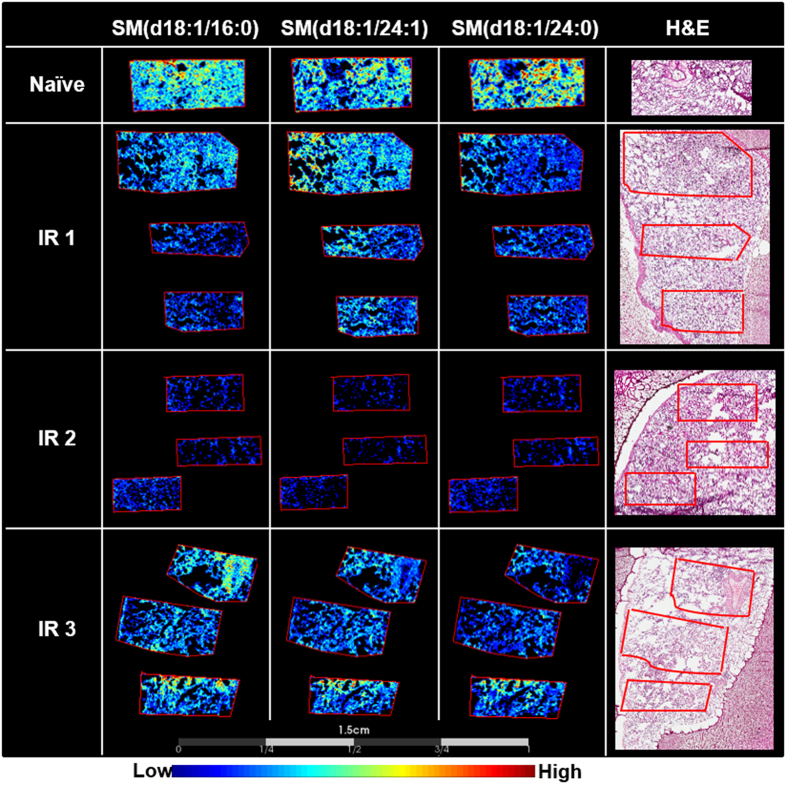
Radiation-induced lung injury (RILI) is a delayed effect of acute radiation exposure that can limit curative cancer treatment therapies and cause lethality following high-dose whole-thorax lung irradiation (WTLI). To date, the exact mechanisms of injury development following insult remain ill-defined and there are no FDA approved pharmaceutical agents or medical countermeasures. Traditionally, RILI development is considered as three phases, the clinically latent period, the intermediate acute pneumonitis phase and the later fibrotic stage. Utilizing matrix-assisted laser desorption ionization mass spectrometry imaging, we identified a number of lipids that were reflective of disease state or injury. Lipids play central roles in metabolism and cell signaling, and thus reflect the phenotype of the tissue environment, making these molecules pivotal biomarkers in many disease processes. We detected decreases in specific surfactant lipids irrespective of the different pathologies that presented within each sample at 180 days post whole-thorax lung irradiation. We also detected regional increases in ether-linked phospholipids that are the precursors of PAF, and global decreases in lipids that were reflective of severe fibrosis. Taken together our results provide panels of lipids that can differentiate between naïve and irradiated samples, as well as providing potential markers of inflammation and fibrosis.


Conflict of interest statement

The authors declare that they have no competing interests.

Figures

Figure 1
Figure 1
Masson’s trichrome stained sections of lung comparing the naïve and different disease pathologies of the irradiated (IR) animals. Representative Masson’s trichrome stained sections of lung taken from naïve (a), IR1 (b), IR2 (c), and IR3 (d). The naïve lung shows clear alveoli space and normal lung architecture. IR1 shows regions of inflammation and edema with normal appearing architecture and mild fibrosis as indicated by the blue stained collagen deposited in the interstitium. The lung section for IR2 is unrecognizable, there is no visible alveoli space, and the lung is flooded with immune infiltrate and alveolar epithelial cell hyperplasia, with severe levels of fibrosis as demonstrated by the blue stained collagen deposited in the interstitium of the whole lung section. IR3 shows regions of disease with some regions of clear alveolar space. Disease pathology again includes edema, immune infiltrate and collagen deposition. The scale bar for the higher magnification images is shown at the bottom of the figure for each sample.
Figure 2
Figure 2
Probabilistic latent semantic analysis (pLSA). 3D scores plot showing the first 3 components from the analysis of ROIs containing 7000 spectra from each of the naïve and the 3 irradiated (IR) samples. The plot shows 4 clear trends for naïve in black, IR1 in light blue, IR2 in dark blue, and IR3 in purple.
Figure 3
Figure 3
Decreases in heme b and lipids regardless of the pathology. MALDI-MS images showing the decreases in heme b, PC(14:0/16:0), PC(16:0/16:0), PC(16:0/16:1), and PC(36:5)/PC(38:8) in all IR samples compared to the naïve sample. The stained hematoxylin and eosin (H&E) section for each lung sample is shown to the right of the figure.
Figure 4
Figure 4
Localized increases in ether lipids are pathology specific. MALDI-MS images showing a region-specific increase in the ether lipids, 1-O-alkyl-2-acyl-sn-glycero-3-phosphocholine, in IR1 compared to the naïve, and IR2 and 3 samples. Representative H&E stained sections are shown on the right for each sample.
Figure 5
Figure 5
Reduction in the relative intensity of sphingomyelins 180 days post irradiation. MALDI-MS images of sphingomyelin species detected in naïve and irradiated lung samples taken 180 days post radiation. There is a clear decrease in the relative intensity of sphingomyelin species following radiation insult. The decrease is more prominent in the IR2 sample, followed by regional decreases in the IR1 and 3 samples. The stained hematoxylin and eosin (H&E) section for each lung sample is shown to the right of the figure.

References

    1. Rubin P, Johnston CJ, Williams JP, McDonald S, Finkelstein JN. A perpetual cascade of cytokines postirradiation leads to pulmonary fibrosis. International Journal of Radiation Oncology*Biology*Physics. 1995;33:99–109. doi: 10.1016/0360-3016(95)00095-G. - DOI - PubMed
    1. Garofalo M, et al. The Delayed Pulmonary Syndrome Following Acute High-dose Irradiation: A Rhesus Macaque Model. Health Physics. 2014;106:56–72. doi: 10.1097/HP.0b013e3182a32b3f. - DOI - PubMed
    1. Marks LB, et al. Radiation-induced lung injury. Seminars in Radiation Oncology. 2003;13:333–345. doi: 10.1016/S1053-4296(03)00034-1. - DOI - PubMed
    1. McDonald S, Rubin P, Phillips TL, Marks LB. Injury to the lung from cancer therapy: Clinical syndromes, measurable endpoints, and potential scoring systems. International Journal of Radiation Oncology*Biology*Physics. 1995;31:1187–1203. doi: 10.1016/0360-3016(94)00429-O. - DOI - PubMed
    1. Jenkins P, Welsh A. Computed Tomography Appearance of Early Radiation Injury to the Lung: Correlation With Clinical and Dosimetric Factors. International Journal of Radiation Oncology*Biology*Physics. 2011;81:97–103. doi: 10.1016/j.ijrobp.2010.05.017. - DOI - PubMed

Publication types

LinkOut - more resources