. 2017 Sep 4;7(1):10313.
doi: 10.1038/s41598-017-10875-0.

HIV-1 matrix protein p17 misfolding forms toxic amyloidogenic assemblies that induce neurocognitive disorders

Affiliations

HIV-1 matrix protein p17 misfolding forms toxic amyloidogenic assemblies that induce neurocognitive disorders

Yasmin Zeinolabediny et al. Sci Rep. .

Abstract

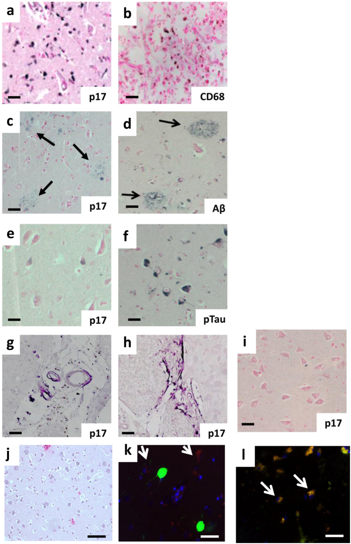
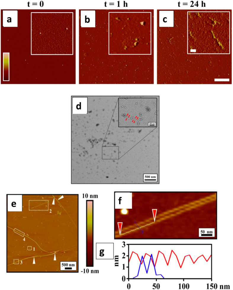
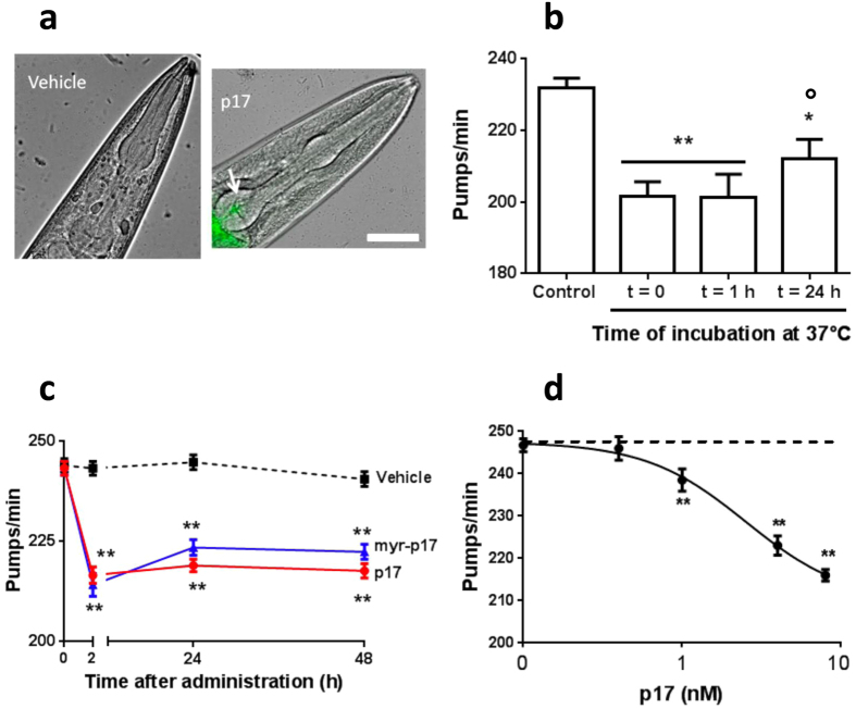
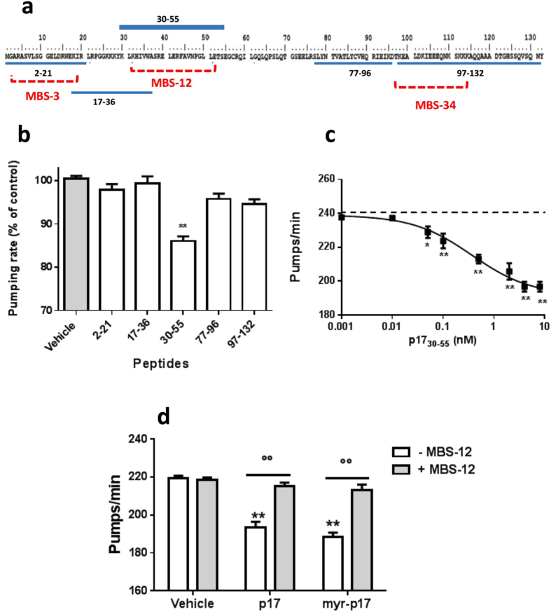
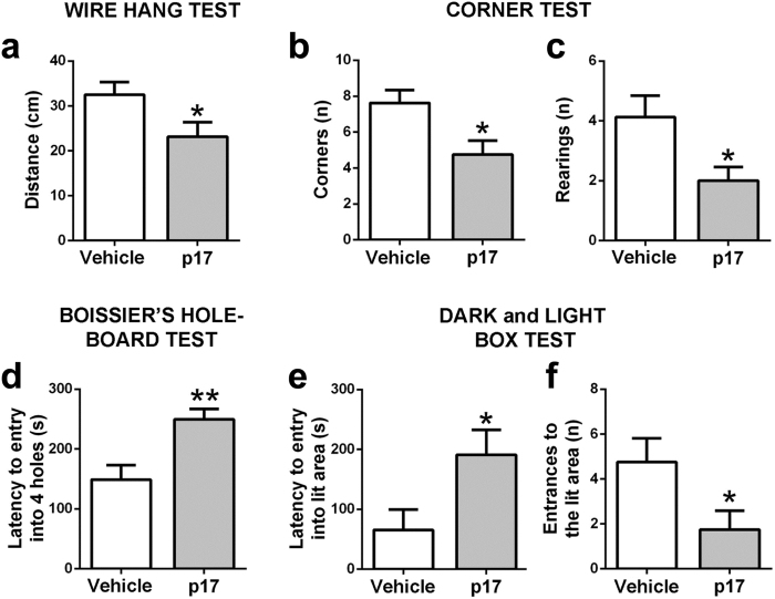
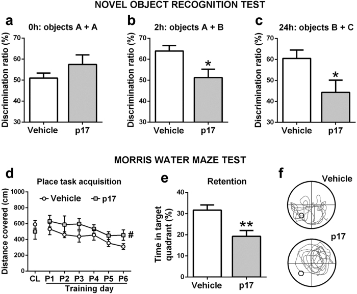
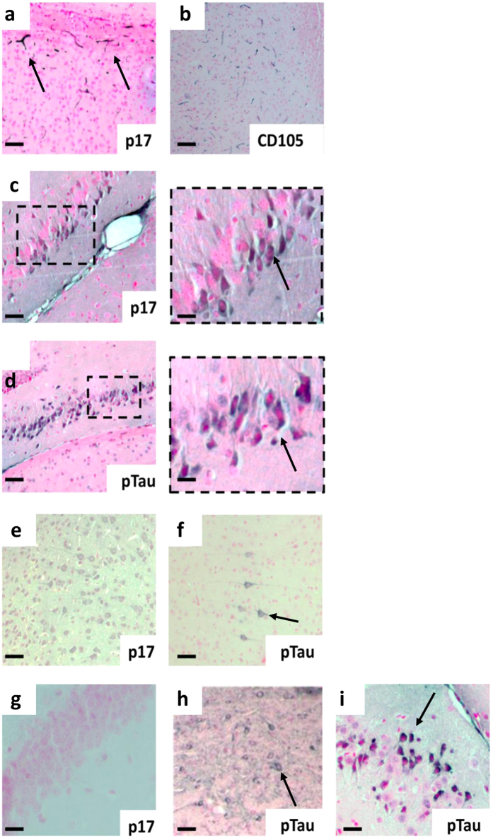
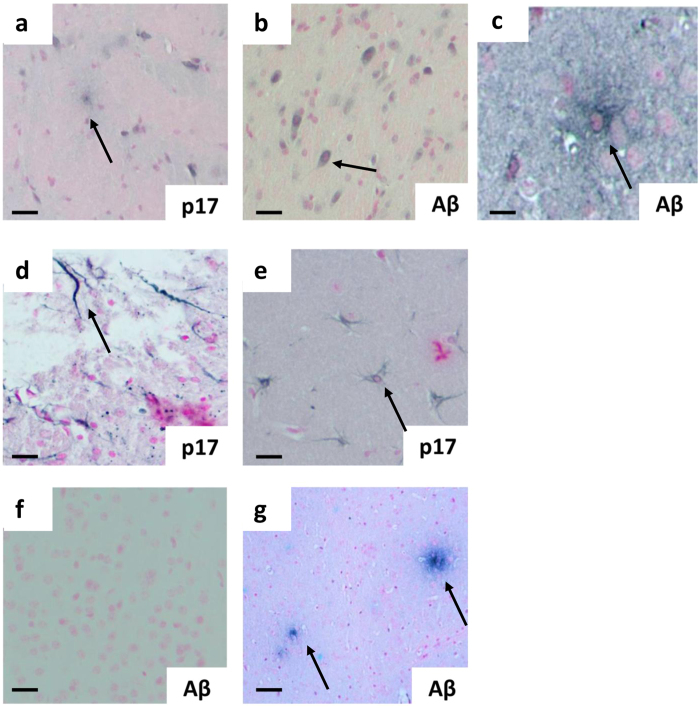
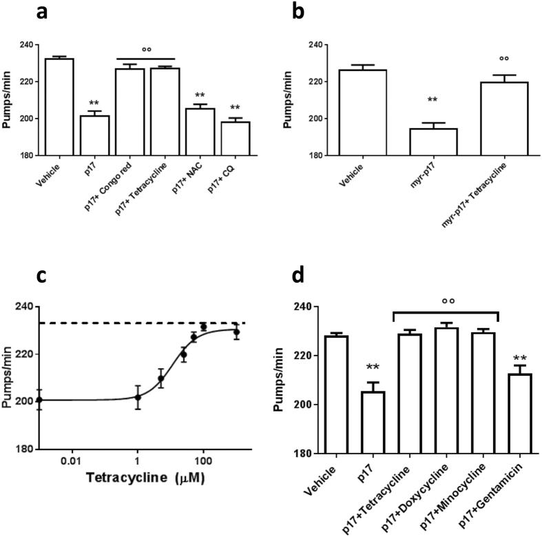
Human immunodeficiency virus type-1 (HIV-1)-associated neurocognitive disorder (HAND) remains an important neurological manifestation that adversely affects a patient's quality of life. HIV-1 matrix protein p17 (p17) has been detected in autoptic brain tissue of HAND individuals who presented early with severe AIDS encephalopathy. We hypothesised that the ability of p17 to misfold may result in the generation of toxic assemblies in the brain and may be relevant for HAND pathogenesis. A multidisciplinary integrated approach has been applied to determine the ability of p17 to form soluble amyloidogenic assemblies in vitro. To provide new information into the potential pathogenic role of soluble p17 species in HAND, their toxicological capability was evaluated in vivo. In C. elegans, capable of recognising toxic assemblies of amyloidogenic proteins, p17 induces a specific toxic effect which can be counteracted by tetracyclines, drugs able to hinder the formation of large oligomers and consequently amyloid fibrils. The intrahippocampal injection of p17 in mice reduces their cognitive function and induces behavioral deficiencies. These findings offer a new way of thinking about the possible cause of neurodegeneration in HIV-1-seropositive patients, which engages the ability of p17 to form soluble toxic assemblies.


Conflict of interest statement

The authors declare that they have no competing interests.

Figures

Figure 1
Figure 1
Human HIV-positive brains showed p17-positive staining in inflammatory and neurodegenerative regions. Representative immunohistochemistry of brain sections showing the staining for (a) p17 (N-DAB stain, gray-black) and (b) CD68-positive macrophages localized to the same region in serial sections as did (DAB stain, brown), (c) p17 (N-DAB) and (d) β-amyloid (Aβ, DAB stain) positive plaques (black arrows). (e) P17-positive (N-DAB) and (f) phosphorylated tau (p-tau, DAB stain) positive cortical neurons from the same region. (g) Medium sized cortical blood vessels were positive for p17 and (h) p17-positive fibril structures within the cortex. (i) Shows negative expression of p17 in the cortical region from a non-HIV positive individual and (j) a negative control, where the p-17 primary antibody was replaced with PBS during immunohistochemical staining. (k) Representative immunofluorescent co-localization of p17 (TRITC-red) and p-tau (FITC-green) and (l) β-amyloid (Aβ, FITC-green) in adjacent neurons within the cortex. Nuclei were counter-stained with DAPI (blue). A similar pattern for p17 where yellow/orange demarcates overlapping staining (white arrows) was evident. Representative images are shown from three patient tissue samples used in this immunohistochemical study.(aj) Scale bar = 50 µm and (k,l) = 20 µm.
Figure 2
Figure 2
Atomic force microscopy (AFM) and transmission electron microscopy (TEM) analysis of p17. (ac) p17 at 4 μM in 10 mM PB (pH 7.4) was incubated at 37 °C for different times (t = 0, t = 1 h and t = 24 h), sampled and left to adsorb on freshly cleaved mica. AFM images were obtained in taping mode and are shown as amplitude data (Z range: −10/+50 mV). Scale bar = 2 µm, inset = 200 nm. (d) TEM micrographs of freshly diluted p17 (4 μM in 10 mM PB pH 7.4). The insets in panel show details of the micrograph at high resolution. The red arrows point to annular structures and rosary-like structures of p17 as an indication of oligomerization process progression. The micrographs are representative of a minimum of 10 different areas. Scale bar = 500 nm, inset = 100 nm. (eg) Atomic force microscopy in height of p17 incubated at 37 °C for 24 h shows the presence of globular structures (1), protofibrils (2) and fibrils (3 and 4). The white arrows indicate the presence of twisted protofibrillar subunits. Sample areas marked by dashed rectangles in E3 are reported with a high magnification in (f) Moreover, the high profile of fibril analysis, between the positions marked by red arrows, is reported (g). (g) Sample marked between blue and purple triangles indicate the high profile of a single filament of ~15– 20 nm of diameter and ~2 nm in height.
Figure 3
Figure 3
Effect of p17 on C. elegans pharyngeal motility. (a) Representative images of the p17 localisation as overlays of Atto-fluorescence and light microscopy in worms. Scale bar = 50 µm. Worms fed for 2 h vehicle (10 mM PB, pH 7.4) or 4 nM p17-Atto. (b) p17 (4 μM in 10 mM PB, pH 7.4) was incubated at 37 °C. At different times (t = 0, t = 1 h and t = 24 h) aliquots were taken, diluted to 4 nM with PB (pH 7.4), and administered to worms. Control worms fed PB (pH 7.4) incubated for 24 h at 37 °C. Pharyngeal pumping was scored by counting the number of pharyngeal contractions in one minute (pumps/minute). *P < 0.05 and **P < 0.01 vs. Control, °P < 0.05 vs. Time 0 and 1 h, one-way ANOVA and Bonferroni post hoc test. (c) p17 and myr-p17 (at 4 μM in 10 mM PB, pH 7.4) were diluted to 4 nM with PB (pH 7.4), and administered to nematodes as described above. The pharyngeal pumping was scored before plating (t = 0) and at different times after plating. Control worms received vehicle alone (Vehicle). (d) Dose-response effect of 0.4–8 nM of p17. Control worms received vehicle alone (Vehicle, dotted line). Data are mean ± SE (N = 30 worms/group). **P < 0.01 vs. Vehicle, one-way ANOVA and Bonferroni post hoc test.
Figure 4
Figure 4
Effect of different epitopes of p17 on its toxicity. (a) Amino acid sequence of p17 and of peptides homologous to different residues (blue). In red the epitopes recognised by the different antibodies. (b) Effect of different p17 peptides on pharyngeal pumping. Nematodes (100 worms/100 µL) were incubated for 2 h with 4 nM peptides. Control worms received 10 mM PB (pH 7.4) (vehicle). Mean ± SE, **P < 0.01 vs. Vehicle, one-way ANOVA and Bonferroni post hoc test. (c) Dose-response effect of 0.01–8 nM p1730–55 peptide. Control worms received vehicle alone (vehicle, dotted line). (d) MBS-12 antibody at 6 ng/µL in 10 mM PB (pH 7.4) was co-incubated for 30 min with 4 nM p17 or myr-p17 before administering to the worms. MBS-12 (6 ng/µL) and 10 mM PB (pH 7.4) alone were used as positive and negative controls, respectively (vehicle). Mean ± SE (n = 20 worms/group). **Pp < 0.01 vs. vehicle and °°P < 0.01 vs. the corresponding p17 or myr-p17, two-way ANOVA and Bonferroni post hoc test.
Figure 5
Figure 5
p17-injected mice showed significant deterioration of behavior compared to control mice. (a) p17-injected mice (p17) showed lower fine motor coordination as they travelled a significantly shorter distance in the wire hang test in respect to control animals injected with vehicle (Vehicle). (bf) p17-injected mice showed behavioral and psychological symptoms of dementia–like, namely neophobia, indicated by (b) lower corner explorations and (c) lower number of rearings, (d) lower exploratory curiosity with higher latency to explore the whole 4 holes in the Boissier’s hole-board test, and (e) higher anxiety indicated by increased latency to enter to the lit compartment and (f) lower number of explorations in the dark and light box test, than Vehicle animals. Mean ± SE (n = 8 mice/group). *P < 0.05 and **P < 0.01 vs. Vehicle, unpaired Student’s t-test.
Figure 6
Figure 6
Learning and memory impairment in p17-injected mice. Cognition was tested with the novel object recognition test (NOR) and Morris water maze test (MWM). (ac) In the NOR, p17-injected animals (p17) lost the ability to discriminate a novel object from a familial one. Vehicle = Control animals injected with vehicle. (a) p17 mice explored similarly to Vehicle mice one of two identical “A” objects in “A + A” at time 0 h, despite higher variability in the former mice. (b) p17 mice showed lesser exploration of a novel object “B” than Vehicle mice when exposed to objects “A + B” 2 h later. (c) p17 mice also spent less exploration time in a novel object “C” than Vehicle mice when exposed to objects “B + C” 24 h later. (df) In the MWM, p17-injected animals had deficiencies in spatial learning and memory. (d) p17 mice showed lower abilities than Vehicle mice throughout six days of training to find the position of a hidden scape platform by relaying on distinctive landmarks around a circular pool (CL = cue learning at day 0, P = place task learning at days 1–6). (e) p17 mice spent less time swimming in the pool quadrant where the platform had been located than Vehicle mice when the platform was removed for the probe trial of retention. (f) The computer generated tracks showed that p17 mice swam at random, whilst Vehicle mice swam preferentially in the platform quadrant (representative tracks of Vehicle and p17 mice; circle indicates the previous platform position) during the retention trial. Mean ± SE, n = 7 mice/group in (a–c) and n = 8 mice/group in (d,e). Chance performance is 50% in (a-c) and 25% in (e). *P < 0.05 and **P < 0.01 vs. Vehicle, unpaired Student’s t-test in (b,c,e), # P < 0.05 effect of factor treatment in two-way repeated measures ANOVA in (d).
Figure 7
Figure 7
Histological localization of p17 in the mouse hippocampus and local cortical regions. Representative immunohistochemistry of brain sections showing the staining for (a) p17 (N-DAB stain, gray-black) into the CA1 region localized to cortical microvessels and (b) CD105 (DAB)-positively stained vessels in the same location in a serial section. (c) P17 was observed in hippocampal neurons adjacent to the injection point (inset scale bar = 10 µm), (d) in the same region as phosphorylated tau (p-tau, DAB) (inset scale bar = 10 µm). (e) P17 was found to be expressed consistently within cortical neurons, some of which (f) were also p-tau-positive. (g) No staining was observed in none-injected controls. (h,i) P-tau positivity in sham-injected 3xTg mouse. Scale bars (ah) = 50 µm, (i) = 10 µm. Images show representative staining results from one or more of six animals in each grouping where sectioning throughout the whole bregma was carried out. See Supplementary Information for immunofluorecence and double immunohistochemistry staining patterns as well as primary antibody-negative controls.
Figure 8
Figure 8
p17 staining localizes to plaque and fibril-like cortical structures in p17-injected mice. Representative immunohistochemistry of brain sections showing the staining for (a) p17 (N-DAB stain, gray-black), (b) β-amyloid (Aβ, DAB stain) and (c) focus on a Aβ plaque in p17-injected cortical regions. (d) Fibril-like structures composed of p17 within the local cortex to injection site and (e) p17-positively stained astrocytes. (f) Lack of Aβ staining in a sham control-injected wild -type mouse and (g) positive staining of Aβ was observed in sham-injected 3xTg-positive mice. (ag) Scale bar = 50 µm apart from (c) = 10 µm. Images show representative staining results from one or more of six animals in each grouping where sectioning throughout the whole bregma was carried out. See Supplementary Information for immunofluorecence and double immunohistochemistry staining patterns as well as primary antibody-negative controls.
Figure 9
Figure 9
Tetracyclines protect C. elegans from the toxicity induced by p17. (a,b,d) N2 nematodes (100 worms/100 µL) were incubated for 2 h with 4 nM p17 or myr-p17 in the absence or presence of: 200 µM Congo red, 50 µM tetracyclines, 5 mM N-acetylcysteine (NAC), 25 µM clioquinol (CQ), or 50 µM gentamicin, in the absence of OP50 E. coli. Control worms were fed 10 mM PB (pH 7.4) (vehicle). Mean ± SE (N = 20 worms/group). **P < 0.01 vs. vehicle and °°P < 0.01 vs. p17 alone, one-way ANOVA and Bonferroni post hoc test. (c) Dose-response effect of tetracycline. Control worms received vehicle alone (dotted line). Mean ± SE (n = 20 worms/group).

References

    1. Elbirt D, et al. HIV-associated neurocognitive disorders (HAND) Isr Med Assoc J. 2015;17:54–59. - PubMed
    1. Letendre SL, Ellis RJ, Ances BM, McCutchan JA. Neurologic complications of HIV disease and their treatment. Top HIV Med. 2010;18:45–55. - PMC - PubMed
    1. Dahl V, et al. Low levels of HIV-1 RNA detected in the cerebrospinal fluid after up to 10 years of suppressive therapy are associated with local immune activation. AIDS. 2014;28:2251–2258. doi: 10.1097/QAD.0000000000000400. - DOI - PMC - PubMed
    1. Teo I, et al. Circular forms of unintegrated human immunodeficiency virus type 1 DNA and high levels of viral protein expression: association with dementia and multinucleated giant cells in the brains of patients with AIDS. J Virol. 1997;71:2928–2933. - PMC - PubMed
    1. Desplats P, et al. Molecular and pathologic insights from latent HIV-1 infection in the human brain. Neurology. 2013;80:1415–1423. doi: 10.1212/WNL.0b013e31828c2e9e. - DOI - PMC - PubMed

Publication types

MeSH terms

Substances