. 2017 Sep 4;7(1):10396.
doi: 10.1038/s41598-017-11220-1.

Targeting transferrin receptors at the blood-brain barrier improves the uptake of immunoliposomes and subsequent cargo transport into the brain parenchyma

Affiliations

Targeting transferrin receptors at the blood-brain barrier improves the uptake of immunoliposomes and subsequent cargo transport into the brain parenchyma

Kasper Bendix Johnsen et al. Sci Rep. .

Abstract

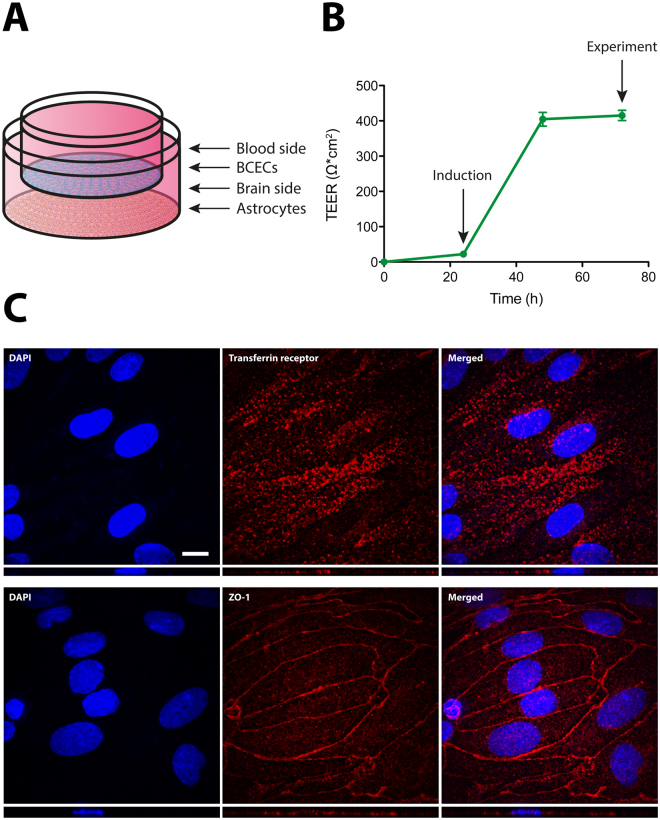
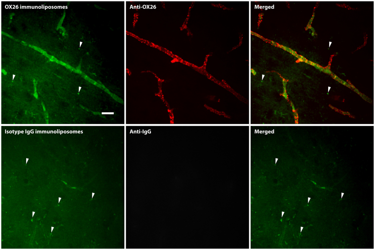
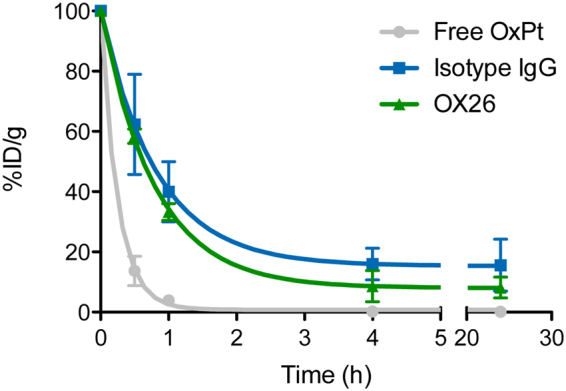
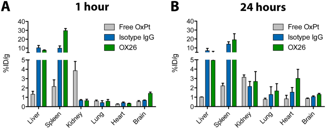
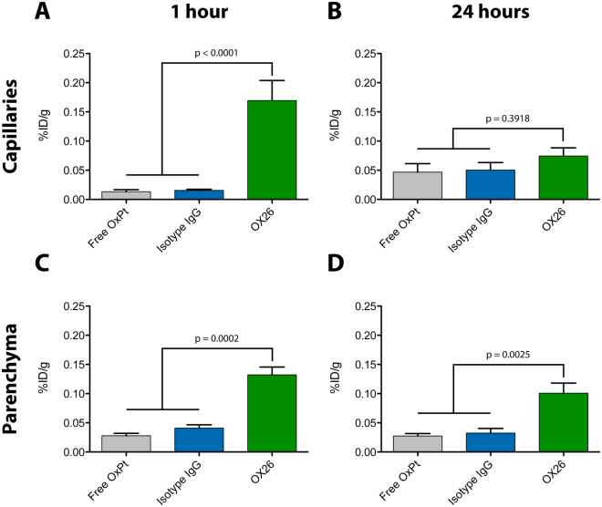
Drug delivery to the brain is hampered by the presence of the blood-brain barrier, which excludes most molecules from freely diffusing into the brain, and tightly regulates the active transport mechanisms that ensure sufficient delivery of nutrients to the brain parenchyma. Harnessing the possibility of delivering neuroactive drugs by way of receptors already present on the brain endothelium has been of interest for many years. The transferrin receptor is of special interest since its expression is limited to the endothelium of the brain as opposed to peripheral endothelium. Here, we investigate the possibility of delivering immunoliposomes and their encapsulated cargo to the brain via targeting of the transferrin receptor. We find that transferrin receptor-targeting increases the association between the immunoliposomes and primary endothelial cells in vitro, but that this does not correlate with increased cargo transcytosis. Furthermore, we show that the transferrin receptor-targeted immunoliposomes accumulate along the microvessels of the brains of rats, but find no evidence for transcytosis of the immunoliposome. Conversely, the increased accumulation correlated both with increased cargo uptake in the brain endothelium and subsequent cargo transport into the brain. These findings suggest that transferrin receptor-targeting is a relevant strategy of increasing drug exposure to the brain.


Conflict of interest statement

The authors declare that they have no competing interests.

Figures

Figure 1
Figure 1
Setup and characterization of the in vitro model of the BBB based on primary rat BCECs and astrocytes. (A) Primary BCECs derived from young rats were setup in Transwell co-culture with astrocytes, hereby yielding a polarized layer of BCECs to be used for uptake and transcytosis experiments. (B) TEER was measured continuously to evaluate the tightness of the in vitro BBB model. The TEER values were approximately 400 Ω*cm2 at the time of the experiments, and this value was stabile throughout the duration of any experiment. (C) After reaching high TEER values, the resulting tight BCEC monolayers expressed both transferrin receptors (upper panel) and the TJ-related protein, ZO-1 (lower panel). The positive staining for the transferrin receptors was found associated with the luminal membrane as well as in the cell cytoplasm, whereas the ZO-1 staining presented as a homogenous lining of the intercellular junctions, suggesting the presence of functional tight junctions. Scale bar depicts 10 µm. BBB: Blood-brain barrier. BCEC: Brain capillary endothelial cells. TEER: Transendothelial electrical resistance. DAPI: Diamino-phenylindole. TJ: Tight junction. ZO-1: Zonula occludens 1.
Figure 2
Figure 2
Flow cytometry evaluation of the association between immunoliposomes and BCECs in vitro. (A) BCECs were incubated with either stealth liposomes, isotype IgG, or OX26 immunoliposomes labelled with a fluorophore in the lipid membrane, and the treated cells were analyzed by flow cytometry to evaluate the association. OX26 immunoliposomes had a five-fold higher association compared to the isotype IgG immunoliposomes, whereas stealth liposomes had no association above the background of untreated cells (p < 0.0001). Due to the large difference in association between the two control liposomes, isotype IgG immunoliposomes was chosen as the most relevant control for subsequent experiments. (B) The association between BCECs and OX26 immunoliposomes was further characterized by co-incubation with free OX26 antibodies, which decreased the association significantly. Furthermore, incubation at 4°C also reduced the association, indicating an energy-demanding uptake mechanism (p < 0.0001). Data are presented as mean + SEM (n = 4–8), and the p-values depicted were derived from a one-way ANOVA with Tukey’s multiple comparisons post hoc test. MFI: Median fluorescence intensity.
Figure 3
Figure 3
Spinning disk confocal microscopy images of immunoliposome-treated, primary rat BCECs. Treatment with either OX26 (upper panel) or isotype IgG (lower panel) immunoliposome revealed a clear association between the fluorescently labelled immunoliposomes and the BCECs. Morphologically, the fluorescent signals were either particulate in the periphery of the cells (arrows) or clustered into larger structures in the perinuclear area (asterisks). Counterstaining with LysoTracker revealed that these larger structures were lysosomes that the endocytosed immunoliposomes had been sorted to. The smaller, particulate signal (arrows) did not co-localize with the lysosomes, suggesting these to be newly endocytosed immunoliposomes. Scale bar depicts 10 µm. DAPI: Diamino-phenylindole.
Figure 4
Figure 4
Transcytosis of oxaliplatin-loaded immunoliposomes across an in vitro model of the BBB. (A) Oxaliplatin-loaded immunoliposomes functionalized with either OX26 or isotype IgG antibodies were administrated to a monolayer of primary BCECs derived from young rats in Transwell co-culture with astrocytes. After incubation, the ‘BCEC’ and ‘brain’ fractions were isolated for subsequent ICP-MS analysis to quantify the platinum content. (B) In the BCEC fraction, OX26 immunoliposomes yielded a higher platinum content compared to the isotype IgG immunoliposomes (p = 0.0035). (C) In the brain fraction, the platinum content of the isotype IgG immunoliposome-treated group was significantly higher compared to that treated with OX26 immunoliposomes (p = 0.0017). Data are presented as mean + SEM (n = 7–8), and the p-values depicted were derived from a one-way ANOVA with Tukey’s multiple comparisons post hoc test. BCEC: Brain capillary endothelial cell. ICP-MS: Inductively-coupled plasma mass spectrometry. OxPt: Oxaliplatin.
Figure 5
Figure 5
Uptake of fluorescently labelled immunoliposomes in brain capillaries in vivo evaluated by spinning disk confocal microscopy. Young rats were injected with either OX26 (upper panel) or isotype IgG (lower panel) immunoliposomes to assess their potential of interacting with the capillaries of the brain. OX26 immunoliposomes (upper panel) showed a good association to the microvessel structures of the brain, although the fluorescent intensity of the immunoliposomes were very low. Counterstaining against the OX26 antibody revealed that the immunoliposome and ligand had accumulated in the brain capillaries, but no sign of transcytosis could be detected. Isotype IgG immunoliposomes (lower panel) had almost no association to the microvessel structures of the brain, which was also evident from the counterstaining against mouse IgG, which did not present with the same vessel pattern as the OX26 immunoliposomes. In both cases, artefacts from the paraformaldehyde fixation could easily be detected (arrows), which was not regarded as a transcytosed immunoliposome, since the same was observable in non-treated animals (data not shown). Scale bar depicts 20 µm.
Figure 6
Figure 6
Circulation properties of immunoliposomes and free oxaliplatin. Oxaliplatin-loaded immunoliposomes or free oxaliplatin were injected into young rats and allowed to circulate. Blood was sampled at various time points, and the platinum content quantified by ICP-MS. Free oxaliplatin had poor circulation properties with only a very small amount of platinum being present in the systemic circulation after 1 hour. Encapsulation in the immunoliposomes increased the circulation time of the platinum substantially, with indications of isotype IgG immunoliposomes residing the longest in the systemic circulation. Data are presented as mean ± SD (n = 3–5). %ID/g: Percentage of injected dose per gram. OxPt: Oxaliplatin.
Figure 7
Figure 7
Biodistribution of oxaliplatin 1 and 24 hours after administration. Oxaliplatin-loaded immunoliposomes or free oxaliplatin were intravenously injected into young rats and allowed to circulate. At the specified time points, the rats were sacrificed and their organs resected to be analyzed for their content of platinum by ICP-MS. (A) After 1 hour, there was a high liver and spleen accumulation of platinum in the groups treated with immunoliposomes, with OX26 immunoliposomes having a very high uptake in the spleen. Free oxaliplatin accumulated more in the kidneys compared to the immunoliposomal formulations, whereas the OX26 immunoliposomes had a high accumulation in the brain compared to free oxaliplatin and isotype IgG immunoliposomes. (B) After 24 hours, there was still high accumulation of immunoliposomes in the liver and spleen, but compared to the early time point, the other peripheral organs (i.e. kidney, lung and heart) showed greater accumulation of immunoliposomes. Data are presented as mean + SEM (n = 4–5). %ID/g: Percentage of injected dose per gram. OxPt: Oxaliplatin.
Figure 8
Figure 8
Uptake of oxaliplatin into different brain fractions after capillary depletion. Oxaliplatin-loaded immunoliposomes or free oxaliplatin were injected into young rats and allowed to circulate for the specified number of hours. The rats were sacrificed and their brains homogenized for subsequent capillary depletion. The isolated fractions from the capillary depletion (capillaries or parenchyma) were analysed for their content of platinum by ICP-MS. (A) After 1 hour, there was a pronounced platinum accumulation in the brain capillaries of rats treated with OX26 immunoliposomes as opposed to those treated with isotype IgG immunoliposomes or free oxaliplatin (p < 0.0001). (B) This difference in brain capillary accumulation between the different groups could not be detected after 24 hours (p = 0.3918). (C) After 1 hour, treatment with OX26 immunoliposomes facilitated a higher platinum content in the brain parenchyma compared to treatment with isotype IgG immunoliposomes or free oxaliplatin (p = 0.0002), (D) which seemed to stay residing after 24 hours (p = 0.0025). Data are presented as mean + SEM (n = 4–5), and the p-values depicted were derived from a one-way ANOVA with Tukey’s multiple comparisons post hoc test. %ID/g: Percentage of injected dose per gram. OxPt: Oxaliplatin.

References

    1. Abbott NJ. Blood-brain barrier structure and function and the challenges for CNS drug delivery. J. Inherit. Metab. Dis. 2013;36:437–449. doi: 10.1007/s10545-013-9608-0. - DOI - PubMed
    1. Zlokovic BV. The blood-brain barrier in health and chronic neurodegenerative disorders. Neuron. 2008;57:178–201. doi: 10.1016/j.neuron.2008.01.003. - DOI - PubMed
    1. Saraiva C, et al. Nanoparticle-mediated brain drug delivery: Overcoming blood-brain barrier to treat neurodegenerative diseases. J Control Release. 2016;235:34–47. doi: 10.1016/j.jconrel.2016.05.044. - DOI - PubMed
    1. Johnsen KB, Moos T. Revisiting nanoparticle technology for blood-brain barrier transport: Unfolding at the endothelial gate improves the fate of transferrin receptor-targeted liposomes. J Control Release. 2016;222:32–46. doi: 10.1016/j.jconrel.2015.11.032. - DOI - PubMed
    1. Leitner DF, Connor JR. Functional roles of transferrin in the brain. Biochim. Biophys. Acta. 2012;1820:393–402. doi: 10.1016/j.bbagen.2011.10.016. - DOI - PubMed

Publication types