. 2017 Aug 21:8:1584.
doi: 10.3389/fmicb.2017.01584. eCollection 2017.

Comparative Genomic Analysis of Neutrophilic Iron(II) Oxidizer Genomes for Candidate Genes in Extracellular Electron Transfer

Affiliations

Comparative Genomic Analysis of Neutrophilic Iron(II) Oxidizer Genomes for Candidate Genes in Extracellular Electron Transfer

Shaomei He et al. Front Microbiol. .

Abstract

Extracellular electron transfer (EET) is recognized as a key biochemical process in circumneutral pH Fe(II)-oxidizing bacteria (FeOB). In this study, we searched for candidate EET genes in 73 neutrophilic FeOB genomes, among which 43 genomes are complete or close-to-complete and the rest have estimated genome completeness ranging from 5 to 91%. These neutrophilic FeOB span members of the microaerophilic, anaerobic phototrophic, and anaerobic nitrate-reducing FeOB groups. We found that many microaerophilic and several anaerobic FeOB possess homologs of Cyc2, an outer membrane cytochrome c originally identified in Acidithiobacillus ferrooxidans. The "porin-cytochrome c complex" (PCC) gene clusters homologous to MtoAB/PioAB are present in eight FeOB, accounting for 19% of complete and close-to-complete genomes examined, whereas PCC genes homologous to OmbB-OmaB-OmcB in Geobacter sulfurreducens are absent. Further, we discovered gene clusters that may potentially encode two novel PCC types. First, a cluster (tentatively named "PCC3") encodes a porin, an extracellular and a periplasmic cytochrome c with remarkably large numbers of heme-binding motifs. Second, a cluster (tentatively named "PCC4") encodes a porin and three periplasmic multiheme cytochromes c. A conserved inner membrane protein (IMP) encoded in PCC3 and PCC4 gene clusters might be responsible for translocating electrons across the inner membrane. Other bacteria possessing PCC3 and PCC4 are mostly Proteobacteria isolated from environments with a potential niche for Fe(II) oxidation. In addition to cytochrome c, multicopper oxidase (MCO) genes potentially involved in Fe(II) oxidation were also identified. Notably, candidate EET genes were not found in some FeOB, especially the anaerobic ones, probably suggesting EET genes or Fe(II) oxidation mechanisms are different from the searched models. Overall, based on current EET models, the search extends our understanding of bacterial EET and provides candidate genes for future research.

Keywords: cytochrome c; extracellular electron transfer (EET); genomics; multicopper oxidase; neutrophilic Fe(II) oxidation; porin-cytochrome c complex (PCC).


Figures

Figure 1
Figure 1
Two generalized models proposed for Fe(II) oxidation at the cell outer surface through a dedicated Fe(II) oxidase (Cyt c or multicopper oxidase). Previously proposed EET proteins in Fe(II) oxidation/reduction and Mn(II) oxidation that fall into the two general models are indicated. Notably, the EET system involving PcoAB proteins was proposed for B. japonicum solely based on bioinformatic analysis without any experimental evidence.
Figure 2
Figure 2
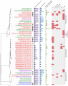
Phylogenetic tree constructed from FeOB genomes, with genome completeness, total number of Cyt c genes in the genome, the maximal number of heme-binding sites in a Cyt c, and putative EET genes indicated. Genomes labeled in red, green, and blue are microaerobic, anaerobic nitrate-dependent, and anaerobic phototrophic FeOB, respectively. Genomes labeled in purple are FeOB that can use both oxygen and nitrate as electron acceptors for Fe(II) oxidation. Experimentally verified obligate lithoautotrophic FeOB are labeled with a black circle, and FeOB that are likely capable of lithoautotrophic Fe(II) oxidation based on genome information and their closely related FeOB isolated from similar environments are labeled with a gray circle. Genomes containing alternative complex III (ACIII) are labeled with * on the left of the Cyc2 column.
Figure 3
Figure 3
Phylogenetic tree of Cyc2 in A. ferrooxidans (labeled in blue), Cyt572 in Leptospirillum sp. (labeled in green), and their homologs in the studied FeOB genomes, with their conserved N-terminal sequences aligned. Numbers in the bracket are IMG gene ID or GenBank accession numbers. The sequence numbering is according to the amino acid position in A. ferrooxidans ATCC 53993. Amino acids conserved in all sequences in the tree are indicated with *, and the conserved residues in the heme-binding motif are labeled with red *. Bootstrap values were calculated based on 1,000 replicates.
Figure 4
Figure 4
Gene clusters that contain OmpB/MofA homologs in G. sulfurreducens (A) and metal oxidizers (B), or other extracellular/outer membrane multicopper oxidase (MCO) encoding genes (C). FeOB names are labeled in blue. IMG gene IDs (or GenBank accession number) for the MCO genes are shown in the parenthesis for locating these gene clusters. OmpB/MofA homologs are colored in dark brown and other extracellular/outer membrane MCO genes are colored in light brown. Genes in red are homologous to mofC and contain a Cyt c domain, with the number of heme-binding sites indicated; genes in blue are mofB homologs; genes in green encode putative copper chaperones belonging to an electron transport protein SCO1-SenC family. Horizontal lines below genes indicate predicted outer membrane/extracellular protein coding genes.
Figure 5
Figure 5
Gene clusters containing MtoAB or PioAB homologs in FeOB genomes. IMG gene IDs for the porin-coding genes are shown in the parenthesis for locating these gene clusters. Porin-coding genes are colored in green, with the number of transmembrane motifs indicated in the gene. Cyt c genes are colored in red, with the number of heme-binding sites indicated. Predicted cellular locations of encoded proteins are shown by different line types under the genes, with thin lines, dashed lines, and thick lines indicating inner membrane, periplasm, and outer membrane, respectively.
Figure 6
Figure 6
Gene clusters potentially encoding a novel porin-cytochrome c complex, “PCC3.” The names of bacteria known for metal oxidation are labeled in blue, with IMG gene IDs for the porin-coding gene indicated in the parenthesis. Porin-coding genes (green) have the number of transmembrane motifs indicated, and extracellular and periplasmic MHC genes (orange and red respectively) have the number of heme-binding sites indicated. The two vertical lines in the Leptothrix ochracea L-12 gene cluster indicate the two ends of the contig, and its porin-coding gene was wrongly split to two (2506527664-5) due to a sequencing error (homopolymer of A's), which caused a frame shift and a stop codon in the middle. A model for the electron flow between the extracellular donor to the inner membrane quinone pool by PCC3 was hypothesized, with proteins colored according to the colors of their encoding genes on the left. ED(red) and ED(ox) stand for the reduced and oxidized forms of the electron donor.
Figure 7
Figure 7
Gene clusters potentially encoding another novel porin-cytochrome c complex, “PCC4.” IMG gene IDs for the porin-coding genes are shown in the parenthesis for locating these gene clusters. Porin-coding genes (green) have numbers of transmembrane motifs indicated, and MHC genes (red) have numbers of heme-binding sites indicated. The vertical line indicates the end of a contig. In Oceanospirillum beijerinckii DSM 7166, the Fe-S cluster containing gene and the IMP gene were annotated as one gene in IMG, but could have been split into two as shown here. A horizontal line between the two gene clusters indicates the boundary of Subgroups I and II. A model for the electron flow between the extracellular donor to the inner membrane quinone pool by PCC4 was hypothesized, with proteins colored according to the colors of their encoding genes on the left. ED(red) and ED(ox) stand for the reduced and oxidized forms of the electron donor.
Figure 8
Figure 8
Sequence conservation logo generated from the alignment of a total of 47 IMP sequences from PCC3 and PCC4. The logo was generated using the WebLogo tool (v3.4) (Crooks et al., 2004), with the stack height indicating the degree of conservation. Conserved amino acid residues on the 3rd, 4th, and 5th transmembrane (TM) regions (TM3, TM4, and TM5, respectively) are labeled with *. The logo is not continuous, as only the conserved regions are shown. The symbols under the conserved amino acids indicate the functions of that residue according to Zhang et al. (2013).

Similar articles

Cited by

References

    1. Bagos P. G., Liakopoulos T. D., Spyropoulos I. C., Hamodrakas S. J. (2004). PRED-TMBB: a web server for predicting the topology of beta-barrel outer membrane proteins. Nucleic Acids Res. 32, W400–W404. 10.1093/nar/gkh417 - DOI - PMC - PubMed
    1. Barco R. A., Emerson D., Sylvan J. B., Orcutt B. N., Jacobson Meyers M. E., Ramirez G. A., et al. (2015). New insight into microbial iron oxidation as revealed by the proteomic profile of an obligate iron-oxidizing chemolithoautotroph. Appl. Environ. Microbiol. 81, 5927–5937. 10.1128/AEM.01374-15 - DOI - PMC - PubMed
    1. Beliaev A. S., Saffarini D. A. (1998). Shewanella putrefaciens mtrB encodes an outer membrane protein required for Fe(III) and Mn(IV) reduction. J. Bacteriol. 180, 6292–6297. - PMC - PubMed
    1. Benzine J., Shelobolina E., Xiong M. Y., Kennedy D. W., McKinley J. P., Lin X., et al. (2013). Fe-phyllosilicate redox cycling organisms from a redox transition zone in Hanford 300 Area sediments. Front. Microbiol. 4:388. 10.3389/fmicb.2013.00388 - DOI - PMC - PubMed
    1. Bewley K. D., Ellis K. E., Firer-Sherwood M. A., Elliott S. J. (2013). Multi-heme proteins: Nature's electronic multi-purpose tool. Biochimica et Biophysica Acta (BBA) - Bioenergetics 1827, 938–948. 10.1016/j.bbabio.2013.03.010 - DOI - PMC - PubMed

LinkOut - more resources