. 2017 Oct 11;17(20):3447-3461.
doi: 10.1039/c7lc00512a.

Skeletal muscle-on-a-chip: an in vitro model to evaluate tissue formation and injury

Affiliations

Skeletal muscle-on-a-chip: an in vitro model to evaluate tissue formation and injury

Gaurav Agrawal et al. Lab Chip. .

Abstract

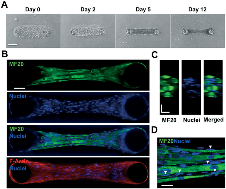
Engineered skeletal muscle tissues can be used for in vitro studies that require physiologically relevant models of native tissues. Herein, we describe the development of a three-dimensional (3D) skeletal muscle tissue that recapitulates the architectural and structural complexities of muscle within a microfluidic device. Using a 3D photo-patterning approach, we spatially confined a cell-laden gelatin network around two bio-inert hydrogel pillars, which induce uniaxial alignment of the cells and serve as anchoring sites for the encapsulated cells and muscle tissues as they form and mature. We have characterized the tissue morphology and strain profile during differentiation of the cells and skeletal muscle tissue formation by using a combination of fluorescence microscopy and computational tools. The time-dependent strain profile suggests the existence of individual cells within the gelatin matrix, which differentiated to form a multinucleated skeletal muscle tissue bundle as a function of culture time. We have also developed a method to calculate the passive tension generated by the engineered muscle tissue bundles suspended between two pillars. Finally, as a proof-of-concept we have examined the applicability of the skeletal muscle-on-chip system as a screening platform and in vitro muscle injury model. We studied the dose-dependent effect of cardiotoxin on the engineered muscle tissue architecture and its subsequent effect on the passive tension. This simple yet effective tool can be appealing for studies that necessitate the analysis of skeletal muscle structure and function, including preclinical drug discovery and development.


Conflict of interest statement

Conflicts of interest

There are no conflicts to declare.

Figures

Fig. 1
Fig. 1
3D photopatterning of support pillars and encapsulation of cells. (Left: Side view of full device; right: top view of central chamber) (A and B) To create support pillars, the bonded microfluidic chip was infused with acrylamide (Am) solution containing photoinitiator and was photopolymerized using a collimated UV light and a transparency photomask containing 100 μm diameter circle patterns. (C and D) After washing with PBS, a precursor solution composed of cells, GelMA, and photoinitiator was polymerized around the pillars using the same method as before, except with a capsule-shaped pattern. (E) PBS solution was used to wash the samples, and the device was perfused with maintenance media by using a syringe pump.
Fig. 2
Fig. 2
Characterization of various hydrogel structures within flow chamber. Z-stack images of the hydrogels, loaded with fluorescent microparticles, were obtained using a laser scanning confocal microscope (green: PAm pillars; red: GelMA hydrogel; magenta: PAm planar hydrogels). (A) X–Z cross-section and (B) X–Y planes at the specified Z locations are shown. (C) 3D renderings of each component and the composite structure are shown. Scale bars: (A) horizontal: 100 μm; vertical: 30 μm; (B) 100 μm.
Fig. 3
Fig. 3
Muscle tissue growth and characterization. (A) Brightfield images depict cell growth, attachment, and hydrogel compaction as a function of days in culture. (B) Immunofluorescent staining images taken from a spinning disk confocal microscope of day 12 samples for myosin heavy chain (MF20) (green), nuclei (blue), and the merged composite. This result suggests a highly matured muscle tissue composed of several multinucleated myotubes. F-actin staining (red) depicts cytoskeletal alignment (bottom). (C) Y–Z confocal sections of a microtissue stained for MF20 (green) and nuclei (blue) illustrate the three-dimensional, cylindrical morphology and fascicular structure of the engineered muscle tissue. (D) High magnification (100×) images of MF20 and nuclei depict the arrangement of nuclei on the periphery of myotubes (white arrows). Scale bars: (A) 150 μm; (B) 50 μm; (C) and (D) 20 μm.
Fig. 4
Fig. 4
Myotube alignment and muscle cell fusion. (A) Alignment score showing the deviation of the longitudinal myotube axis from the mean orientation axis for 5 experimental groups (1 : 15, 1 : 10, 1 : 5, 2 : 5, and 3 : 5) after 12 days of culture. Values are calculated as a percent of total myotubes per sample. 3–5 samples from 3 different chips were used per group. For each group, the plot illustrates the mean value along with standard deviation. (B) Fusion indices, calculated as the percentage of the total number of nuclei within myotubes relative to the total number of nuclei in the sample, for each experimental group, analyzed from confocal z-stacks of immunofluorescence staining for myosin heavy chain (MF20) and nuclei counterstain. 2000 nuclei from 3–5 samples examined from 3 different chips were used for each group. The plot shows the mean value alone with standard deviation. One-way ANOVA with Tukey’s multiple comparison test was used to assess statistical significance (*p < 0.1, **p < 0.01, ***p < 0.001).
Fig. 5
Fig. 5
Quantification of cell generated strains on the PAm hydrogel with culture time. (A) Heat map of tensile strain values, εxx, calculated from the deformations observed on the PAm hydrogel layer at culture days 2, 4, 8, and 12. The dotted lines display the contour of the cell-laden GelMA hydrogel. Negative and positive values indicate contraction and extension of the hydrogel. (B) Bar graph of binned values of εxx along the long axis of cell-laden GelMA hydrogel. The locations of the bin centers are indicated in the x-axis of the plot and are shown pictorially in the inset containing the Brightfield image of the microtissue. (C) An overlay of X–Y confocal sections of fluorescent particles embedded in the PAm hydrogel surface at day 0 (green) and day 2 (red) in the left panel and at day 0 (green) and day 12 (red) in the right panel. The lack of overlap between green and red beads in the right panel indicates the inward shift of the two pillars at day 12 suggesting the contraction of the microtissue. Scale bars: 50 μm.
Fig. 6
Fig. 6
Quantification of passive tension generated by the engineered muscle using COMSOL. (A) 3D rendering of the finite element domain used for simulating the deformations in the PAm hydrogel caused by pillar displacement. (B) The magnitude of the displacement vectors shown as a heat map on the finite element domain. (C) The strain values for εxx, εyy and εxy on the surface of the PAm hydrogels obtained from finite element simulations (theoretical, top row) and empirically (experimental, bottom row). (D) Stress tensor component, τxz, and traction stress vector obtained from the finite element simulations shown as a heat map and vector field, respectively. (E) Histogram showing the distribution of the magnitudes of passive tension generated by 30 microtissues.
Fig. 7
Fig. 7
Dose-dependent response of engineered muscle strips to cardiotoxin (CTX). (A) F-actin and brightfield images of muscle strips subjected to 0 μM, 0.1 μM, and 0.5 μM CTX for 24 hours. (B) CTX administration results in a dose-dependent drop in passive tension due to cytoskeletal disruption.

References

    1. Loessner D, Stok KS, Lutolf MP, Hutmacher DW, Clements JA and Rizzi SC, Biomaterials, 2010, 31, 8494–8506. - PubMed
    1. Karlsson H, Fryknas M, Larsson R and Nygren P, Exp. Cell Res, 2012, 318, 1577–1585. - PubMed
    1. Dhiman HK, Ray AR and Panda AK, Biomaterials, 2005, 26, 979–986. - PubMed
    1. Kienhuis AS, Wortelboer HM, Hoflack JC, Moonen EJ, Kleinjans JC, van Ommen B, van Delft JH and Stierum RH, Drug Metab. Dispos, 2006, 34, 2083–2090. - PubMed
    1. Chen AA, Thomas DK, Ong LL, Schwartz RE, Golub TR and Bhatia SN, Proc. Natl. Acad. Sci. U. S. A, 2011, 108, 11842–11847. - PMC - PubMed

Publication types