. 2018 Mar 4;12(2):127-137.
doi: 10.1080/19336918.2017.1322253. Epub 2017 Sep 12.

Periodontal pathogens promote epithelial-mesenchymal transition in oral squamous carcinoma cells in vitro

Affiliations

Periodontal pathogens promote epithelial-mesenchymal transition in oral squamous carcinoma cells in vitro

A A Abdulkareem et al. Cell Adh Migr. .

Abstract

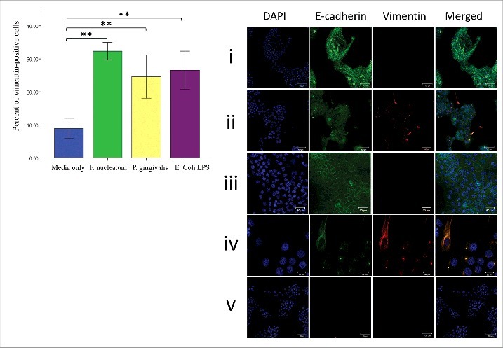
Epithelial-mesenchymal transition (EMT) is potentially involved in increasing metastasis of oral squamous cell carcinoma (OSCC). Periodontal pathogens are well-known for their ability to induce intense immune responses and here we investigated whether they are involved in inducing EMT. Cultures of OSCC cell line (H400) were treated separately with heat-killed periodontal pathogens F. nucleatum, or P. gingivalis or E. coli LPS for 8 d. EMT-associated features were assayed using sq-PCR and PCR-arrays, for EMT-related markers, and ELISAs for TGF-β1, TNF-α, and EGF. The migratory ability of cells was investigated using scratch and transwell migration assays. E-cadherin and vimentin expression was assessed using immunofluorescence while Snail activation was detected with immunocytochemistry. In addition, the integrity of the cultured epithelial layer was investigated using transepithelial electrical resistance (TEER). PCR data showed significant upregulation after 1, 5, and 8 d in transcription of mesenchymal markers and downregulation of epithelial ones compared with unstimulated controls, which were confirmed by immunofluorescence. Periodontal pathogens also caused a significant increase in level of all cytokines investigated which could be involved in EMT-induction and Snail activation. Exposure of cells to the bacteria increased migration and the rate of wound closure. Downregulation of epithelial markers also resulted in a significant decrease in impedance resistance of cell monolayers to passage of electrical current. These results suggested that EMT was likely induced in OSCC cells in response to stimulation by periodontal pathogens.

Keywords: cadherins; cytokines; invasiveness; mesenchymal cells; migration; neoplasm metastasis; oral keratinocytes.


Figures

Figure 1.
Figure 1.
Exposure of H400 cells to heat-killed F. nucleatum, P. gingivalis, and 20 μg/ml E. coli LPS produced changes in EMT-related indicators. Gel images and densitometric analysis showing fold changes relative to highest or lowest levels as determined for selected EMT-related genes using semi-quantitative RT-PCR analysis. F. nucleatum and P. gingivalis produced significant changes in (A) E-cadherin level after 24 hr cell stimulation and continued till day 8 (∼5-fold). Other EMT-related genes [(B) vimentin, (C) Twist, and (D) Snail-1] were upregulated up to 5-fold compared with unstimulated control in response to periodontal pathogens from days 1–8 (* = P< 0.05, ** = P< 0.001).
Figure 2.
Figure 2.
H400 cells were treated with media only (DMEM supplemented with 10% FCS), or heat inactivated periodontal pathogens F. nucleatum and P. gingivalis (100 bacteria per cell), and 20 μg/ml E. coli LPS over 8 d. Supernatant collected from each group after 1, 5, and 8 d and assayed using ELISA for (A) TGF-β1 which significantly increased with F. nucleatum and E. coli LPS (∼2 folds) at day 1, while P. gingivalis showed no difference compared with the control. Day 5 and 8 data showing significant upregulation for all test groups (∼6-folds). (B) EGF and (C) TNF-α, results showing similar pattern of stimulation to that of TGF-β1 in which P. gingivalis did not produce any statistical difference in the amount of EGF and TNF-α released in comparison with the control. At day 5 and 8 all bacterial groups significantly upregulated the level of both cytokines in the supernatant. The same picture is applied to the increase in gene transcription for the same cytokines (D, E, F), the only exception is the significant increased transcription of TNF-α associated with P. gingivalis after 24 h (* = P< 0.05, ** = P< 0.001).
Figure 3.
Figure 3.
Periodontal bacteria modulate vimentin and E-cadherin expression in H400 cells after 8-days culture. (A) Cells with vimentin positive staining were counted and expressed as a percentage. Analysis indicated a significant increase between cells stimulated with bacteria and unstimulated control in terms of vimentin expression. (B) Immunofluorescence staining indicates that F. nucleatum and P. gingivalis induce expression of vimentin (red) (ii) when compared with the control group treated with media only (i). Scale bars = 100 μm. Higher magnification showing that unstimulated control (iii) maintained normal E-cadherin distribution and negatively expressed vimentin. Scale bar = 50 μm. while stimulated cultures (iv) showed presence of vimentin-positive cells which either exhibit mesenchymal-like morphology and retained some characteristics of their parental origin by expressing E-cadherin or cluster of epithelial cells simultaneously expressing vimentin with downregulation of E-cadherin from periphery of cells. Scale bars = 20 μm. Negative controls (cultures treated with secondary antibodies only) were included to exclude possibility of unspecific staining (v). (** = P< 0.001).
Figure 4.
Figure 4.
Stimulation of H400 cells with periodontal bacteria resulted in significant increase in Snail-positive cells. (A) Immunocytochemical staining of H400 cell, growing on 22 × 22 mm coverslips, showing increased Snail expression. Activated cells manifested by dark brown discoloration of the nucleaus and/or cytoplasm (green arrow), while cells treated with media only mostly exhibit dark blue nucleaus (red arrow). Scale bars are(50 μm) (B) Analysis of percent of Snail-positive cells showing that P. gingivalis and F. nucleatum elicited the higher response (up to 70%) when compare with unstimulated control. (C) H400 cells were cultured on a 0.4 μm pore membrane insert and stimulated with periodontal pathogens. Resistance levels were stabilized in all groups after 4 days, from this point TEER measurements were recorded up to day 8. Analysis of data showed that resistance in response to bacterial stimulation was significantly decreased from day 4 to 8 in comparison with the media control group. Following 5 d of exposure to periodontal pathogens, cells in stimulated cultures exhibited fibroblast-like morphology while unstimulated control maintained normal epithelial sheet architecture (D). Scale bars = 50 μm. (* = P< 0.05, ** = P< 0.001).
Figure 5.
Figure 5.
(A) Analysis of transwell migration assay for H400 cells stimulated over 8 d with periodontal pathogens. Data demonstrated that a higher number of cells had migrated through the membrane in comparison with control. Confluent monolayers of H400 cells were disrupted by scratching with a 10 μm pipette tip following 8 d of exposure to heat-inactivated F. nucleatum and P. gingivalis and 20 μg/ml E. coli LPS. (B) Images were captured immediately following scratching and then after 12 and 24 hr culture. Scale bars are 100 μm (C) Measurements of the wound width at 12 hr indicated no significant difference, however at 24 hr scratch closure was significantly increased in all cultures exposed to bacterial components in comparison with unstimulated control. Data is shown as mean + SD. Experiments were performed in triplicate. (* = P< 0.05, ** = P< 0.001).

References

    1. Jemal A, Bray F, Center MM, Ferlay J, Ward E, Forman D. Global cancer statistics. CA: A Cancer J Clinicians 2011; 61:69-90; PMID:21296855 - PubMed
    1. Choi S, Myers JN. Molecular pathogenesis of oral squamous cell carcinoma: implications for therapy. J Dental Res 2008; 87:14-32; PMID:18096889; https://doi.org/10.1177/154405910808700104 - DOI - PubMed
    1. Feller L, Lemmer J. Oral squamous cell carcinoma: epidemiology, clinical presentation and treatment. J Cancer Therapy 2012; 3:263-8; https://doi.org/10.4236/jct.2012.34037 - DOI
    1. Marsh D, Suchak K, Moutasim KA, Vallath S, Hopper C, Jerjes W, Upile T, Kalavrezos N, Violette SM, Weinreb PH, et al.. Stromal features are predictive of disease mortality in oral cancer patients. J Pathol 2011; 223:470-81; PMID:21294121; https://doi.org/10.1002/path.2830 - DOI - PubMed
    1. Jechlinger M, Grunert S, Beug H. Mechanisms in epithelial plasticity and metastasis: insights from 3D cultures and expression profiling. J Mammary Gland Biol Neoplasia 2002; 7:415-32; PMID:12882526; https://doi.org/10.1023/A:1024090116451 - DOI - PubMed

Publication types

MeSH terms

LinkOut - more resources