. 2017 Sep 5;12(9):e0182434.
doi: 10.1371/journal.pone.0182434. eCollection 2017.

N-terminally truncated POM121C inhibits HIV-1 replication

Affiliations

N-terminally truncated POM121C inhibits HIV-1 replication

Hideki Saito et al. PLoS One. .

Abstract

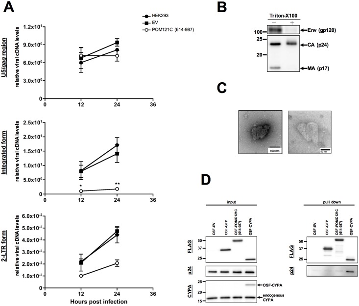
Recent studies have identified host cell factors that regulate early stages of HIV-1 infection including viral cDNA synthesis and orientation of the HIV-1 capsid (CA) core toward the nuclear envelope, but it remains unclear how viral DNA is imported through the nuclear pore and guided to the host chromosomal DNA. Here, we demonstrate that N-terminally truncated POM121C, a component of the nuclear pore complex, blocks HIV-1 infection. This truncated protein is predominantly localized in the cytoplasm, does not bind to CA, does not affect viral cDNA synthesis, reduces the formation of 2-LTR and diminished the amount of integrated proviral DNA. Studies with an HIV-1-murine leukemia virus (MLV) chimeric virus carrying the MLV-derived Gag revealed that Gag is a determinant of this inhibition. Intriguingly, mutational studies have revealed that the blockade by N-terminally-truncated POM121C is closely linked to its binding to importin-β/karyopherin subunit beta 1 (KPNB1). These results indicate that N-terminally-truncated POM121C inhibits HIV-1 infection after completion of reverse transcription and before integration, and suggest an important role for KPNB1 in HIV-1 replication.


Conflict of interest statement

Competing Interests: The authors have declared that no competing interests exist.

Figures

Fig 1
Fig 1. N-terminally truncated POM121C blocks HIV-1 infection in human cells.
(A) HEK293 cells were transduced with a VSV-G-pseudotyped retroviral vector expressing a human T-cell-derived cDNA library. After several passages, the cells were infected with high titer HIV-1 capable of expressing HSV-TK (VSV-G/ NL4-3TK) and subjected to lethal selection with gancyclovir to eliminate infected cells. After repeated infection and selection, inserted cDNAs isolated from gancyclovir-resistant cells were amplified by PCR. The cDNAs were subcloned again into a retroviral vector and expressed in HEK293 cells to verify that they acted as anti-HIV factors. (B) Upper panel: Structures of POM121C mRNA and the isolated POM121C cDNA, which was linked to random hexamer primers and BstXI adapters at both ends. Lower panel: schematic representation of the POM121C and POM121C (614–987) proteins. (C) Upper panels: Immunoblot analyses monitoring POM121C (614–987) expression in HEK293 (left panels), Jurkat (middle panels) or PHA-stimulated PBMCs (right panels). Lysates of parental cells (lane Parental), control vector-infected cells (lane EV), and cells stably expressing HA-tagged POMC121C (614–987) (lane POM121C [614–987]) were immunoblotted with anti-HA or anti-α-tubulin antibodies. Lower panels: Effects of POM121C (614–987) on viral infectivity. Parental, EV control and POM121C (614–987) cells were infected with VSV-G-pseudotyped NL4-3luc. Luciferase activity was measured 24 h after infection. Relative luciferase activities are shown as ratios (%) of the RLU of EV control cells with standard deviations calculated from at least three independent experiments. (D) Immunoblot analysis monitoring POMC121C (614–987) expression in MT4C5 cells stably expressing either EV or POM121C (614–987) (upper left panels). Parental, EV or POM121C (614–987) cells were infected with VSV-G-pseudotyped NL4-3luc (lower left panel) or replication-competent NL4-3luc-RC (lower middle panel). Relative luciferase activities are shown as ratios (%) of the RLU of EV control MT4C5 cells with standard deviations calculated from three independent experiments. MT4C5, EV or POM121C (614–987) cells were infected with the HIV-1NL4-3 strain (lower right panel). Viral production was monitored by measuring RT activity in the culture supernatants. Mean values and standard deviations in three independent experiments are shown. (E) Left panel: Immunoblot analysis monitoring HA-tagged POMC121C (614–987) expression in parental HeLa cells and those transduced with EV, POM121C (614–987) or POM121C (2–987) expression vectors. Right panel: Effects of POM121C (614–987) on VSV-G-pseudotyped HIV-1 infectivity in HeLa cells. Parental, EV, POM121C (614–987) or POM121C (2–987) HeLa cells were infected with VSV-G-pseudotyped NL4-3luc. Luciferase activity was measured 24 h after infection. Relative luciferase activities are shown as ratios (%) of the RLU of EV control cells with standard deviations calculated from three independent experiments. (F) Localization of POM121C (614–987) or POM121C (2–987) in HeLa cells stably expressing each of the proteins. Cells were multistained with anti-HA (red) to detect HA-tagged proteins and Hoechst33342 (blue) as nuclear counterstaining. Statistical significance was determined by unpaired two-tailed Student’s t test (C: panel PBMCs) or one-way analysis of variance (ANOVA) with Dunnett’s multiple comparison test (C: panels HEK293 and Jurkat, D, and E). ns, not significant (P>0.05); ***P<0.001.
Fig 2
Fig 2. POM121C (614–987) inhibits HIV-1 replication after reverse transcription.
(A) Quantitative DNA-PCR analysis of viral cDNA metabolism after VSV-G/NL4-3luc infection of HEK293-derived cells. Total DNA was extracted at the indicated times (12 and 24 h) and analyzed for the amount of viral cDNAs with a primer set recognizing the U5-gag region (top panel), integrated form (middle panel) or 2-LTR form (bottom panel). In cells exposed to heat-inactivated virus, the amount of viral DNA was below the level of detection in the real-time PCR assay. Mean values and standard deviations in three independent experiments are shown. (B) Immunoblot analysis of HIV-1 virions before or after stripping the envelope. Concentrated virions were subjected to step-gradient centrifugation in the absence (-) or presence (+) of 0.1% of Triton-X100. The pellets were immunoblotted using anti-HIV-1 env (gp120) or HIV-1-positive pooled serum from infected individuals (subtype B). (C) Electron micrographs showing envelope-stripped virions of HIV-1. TEM images of negatively stained naked HIV-1 cores prepared from HIV-1NL4-3 virions (left panel, lower magnification; right panel, higher magnification). Representative fields are shown. Bar scales indicate 100 nm (left panel) and 50 nm (right panel). (D) Immunoblot analyses showing OSF-tagged proteins bound to naked HIV-1 cores. Input naked HIV-1 cores and purified Strep-tagged proteins were immunoblotted using anti-FLAG (input, top panel), anti-p24 (input, middle panel), or anti-CYPA used as an internal control (input, bottom panel). Bound protein complexes were pulled-down and analyzed by immunoblotting with anti-FLAG or anti-p24 antibodies (right panels). Statistical significance was determined by one-way analysis of variance (ANOVA) with Dunnett’s multiple comparison test (A: middle panel). *P<0.05, **P<0.01.
Fig 3
Fig 3. POM121C (614–987) inhibits nuclear import and integration in a Gag-dependent manner.
(A) Immunoblot analysis of HEK293 cells expressing POM121C (614–987) or mCPSF6-358HA. Whole-cell lysates were immunoblotted using antibodies against HA or GAPDH (used as an internal control). (B) Effects of POM121C (614–987) or mCPSF6-358 on infection with HIV-1. Parental, EV, POM121C (614–987) or mCPSF6-358 cells were infected with VSV-G/NL4-3-luc (WT), VSV-G/NL4-3-luc CA N74D (CA N74D) or VSV-G/NL4-3-luc IN D116G (IN D116G). Luciferase activity was measured 24 h after infection. Relative luciferase activities are shown with standard deviations calculated from three independent experiments. (C) Relative levels of viral cDNA synthesized after VSV-G/NL4-3 CA N74D-luc or VSV-G/NL4-3 IN D116G-luc infection. Total DNA was isolated from a portion of the cells at the indicated times after infection. Experiments were done as in Fig 2A. Mean values and standard deviations in three independent experiments are shown. (D) Effects of POM121C (614–987) or mCPSF6-358 on infection with HIV-1, MLV or HIV/MLV chimeric virus, MHIV-mMA12CA. Parental, EV, POM121C (614–987) or mCPSF6-358 cells were infected with VSV-G/LAI-luc [HIV-1 (LAI)], VSV-G/MLV-luc (MLV) or VSV-G/MHIV-mMA12CA-luc (MHIV-mMA12CA). Luciferase activity was measured 48 h after infection. Relative luciferase activities are shown with standard deviations calculated from three independent experiments. Statistical significance was determined by one-way analysis of variance (ANOVA) with Dunnett’s multiple comparison test (B and D). ns, not significant (P>0.05); *P<0.05, ***P<0.001.
Fig 4
Fig 4. The C-terminal α-helix structure is required for HIV-1 inhibition.
(A) Schematic presentation of POM121C mutants. The black boxes indicate the domain of a prospective helical-structure. The white boxes indicate HA-epitope tag. (B and D) Immunoblot analysis of HEK293 cells expressing truncated forms of POM121C. Whole-cell lysates were immunoblotted using anti-HA or anti-α-tubulin antibodies. (C and E) Effects of POM121C mutants on viral infectivity. HEK293 cells stably expressing the indicated POM121C mutants were infected with VSV-G/NL4-3luc. Luciferase activity was measured 24 h after infection. Relative luciferase activities are shown as ratios (%) of the RLU of EV control cells with standard deviations calculated from three independent experiments. Statistical significance was determined by one-way analysis of variance (ANOVA) with Dunnett’s multiple comparison test (C and E). ns, not significant (P>0.05); *P<0.05, ***P<0.001.
Fig 5
Fig 5. GST-fused POM121C (801–987) inhibits HIV-1 infection.
(A) Schematic presentation of HA-tagged GST-POM121C (801–987). (B) Immunoblot analysis of HEK293 cells expressing HA-tagged GST or GST-POM121C (801–987) (upper panel). Whole-cell lysates were immunoblotted using anti-HA or anti-α-tubulin antibodies. Effects of GST-POM121C (801–987) on viral infectivity (lower panel). HEK293 cells stably expressing HA-tagged GST, POM121C (614–987) or GST-POM121C (801–987) were infected with VSV-G/NL4-3luc. Luciferase activity was measured 24 h after infection. The mean luciferase value from EV control cells was arbitrarily set as 100% and standard deviations in three independent experiments are shown. (C) Relative amounts of viral cDNA synthesized after VSV-G/NL4-3luc infection. Total DNA was isolated from the cells 12 h after infection. Viral cDNA synthesis was quantified by real-time PCR with a primer set recognizing the U5/gag region (upper panel) or 2-LTR form (lower panel) as described in EXPERIMENTAL PROCEDURES. In cells exposed to heat-inactivated virus, viral DNA was below the level of detection by real-time PCR assay. Mean values and standard deviations in three independent experiments are shown. (D) Purified GST or GST- POM121C (801–987) complexes were subjected to SDS-PAGE and processed for Silver Staining. The arrows indicate three different gel portions analyzed by mass spectrometry. Statistical significance was determined by one-way analysis of variance (ANOVA) with Dunnett’s multiple comparison test (B: lower panel, and C). ns, not significant (P>0.05); *P<0.05, **P<0.01, ***P<0.001.
Fig 6
Fig 6. POM121C mutant binding to KPNB1 correlates with inhibition of HIV-1 infection.
(A) Immunoblot analysis showing GST-POM121C (801–987) bound to KPNB1 in HEK293 cells. Cell lysates were analyzed by immunoblotting either directly (left panels: input) or following GST pull-down experiments. Purified GST or GST-POM121C (801–987) complexes were immunoblotted using anti-GST or anti-KPNB1 antibodies. (B) Schematic presentation of POM121C mutants fused with HA-tagged GST. (C) Immunoblot analysis of HEK293 cells expressing truncated forms of POM121C mutants fused with HA-tagged GST. Whole-cell lysates were immunoblotted using anti-HA or anti-α-tubulin antibodies. (D) Effect of GST-POM121C mutants on viral infectivity. HEK293 cells stably expressing GST or the indicated GST-POM121C mutants were infected with VSV-G/NL4-3luc. Luciferase activity was measured 24 h after infection. Relative luciferase activities are shown as ratios (%) of the RLU of EV control cells with standard deviations calculated from three independent experiments. (E) Cell lysates were analyzed by immunoblotting with anti-HA and anti-KPNB1 antibodies either directly (left panels: input) or following GST pull-down experiments (right panels: GST pull-down). (F) Immunoblot analysis of HEK293 cells expressing truncated forms of GST-POM121C mutants. Experiments were done as described in (C). (G) Effects of the indicated GST-POM121C mutants on viral infectivity. Experiments were done as described in (D). Relative luciferase activities are shown as ratios (%) of the RLU of EV control cells with standard deviations calculated from three independent experiments. (H) GST pull-down analysis showing GST-POM121C (801–987) bound to intracellular KPNB1. Cell lysates were analyzed by immunoblotting either directly (left panels: Input) or subsequent to GST pull-down (right panels) with anti-HA (lower panels) or anti-KPNB1 (upper panels) antibodies. Statistical significance was determined by one-way analysis of variance (ANOVA) with Dunnett’s multiple comparison test (D and G). ns, not significant (P>0.05); *P<0.05, **P<0.01, ***P<0.001.
Fig 7
Fig 7. POM121C (614–987) does not affect the late phase of HIV-1 replication.
(A) Effects of POM121C (614–987) on HIV-1 infection at the late stages. HEK293, EV or POM121C (614–987) cells were co-transfected with pNL4-3luc together with pGL4.84-EF1α-hRlucCP (Renilla-luc). Two days after transfection, luciferase activity was determined by the Dual-Luciferase assay system. The RLU of each firefly luciferase activity relative to renilla luciferase activity is given. The results are shown as an average of three independent experiments with standard deviations. (B) Immunoblot analysis of viral proteins in HEK293-derived cells transiently transfected with the proviral plasmid, pNL4-3. EV or POM121C (614–987) cells were transfected with 1.0 μg of pNL4-3. The cells were harvested 48 h post-transfection and lysates were subjected to immnoblot analyses with anti-HIV-1 p24 (top panel; the upper arrow indicated precursor Gag [PrGag], the lower arrow indicated CA), anti-HA (middle panel) and anti-CYPA (bottom panel). One representative set of results from three independent experiments is shown. (C) The amounts of p24 antigen in the supernatants of transfected cells were quantified with HIV-1 CA (p24) ELISA. Mean values and standard deviations are shown from three independent experiments. Statistical significance was determined by one-way analysis of variance (ANOVA) with Dunnett’s multiple comparison test (A), or unpaired two-tailed Student’s t test (C). ns, not significant (P>0.05).

Similar articles

Cited by

References

    1. Goff SP. Host factors exploited by retroviruses. Nat Rev Microbiol. 2007;5(4):253–63. Epub 2007/02/28. doi: 10.1038/nrmicro1541 . - DOI - PubMed
    1. Brass AL, Dykxhoorn DM, Benita Y, Yan N, Engelman A, Xavier RJ, et al. Identification of host proteins required for HIV infection through a functional genomic screen. Science. 2008;319(5865):921–6. Epub 2008/01/12. doi: 10.1126/science.1152725 . - DOI - PubMed
    1. Hutten S, Walde S, Spillner C, Hauber J, Kehlenbach RH. The nuclear pore component Nup358 promotes transportin-dependent nuclear import. J Cell Sci. 2009;122(Pt 8):1100–10. Epub 2009/03/21. doi: 10.1242/jcs.040154 . - DOI - PubMed
    1. Woodward CL, Prakobwanakit S, Mosessian S, Chow SA. Integrase interacts with nucleoporin NUP153 to mediate the nuclear import of human immunodeficiency virus type 1. J Virol. 2009;83(13):6522–33. Epub 2009/04/17. doi: 10.1128/JVI.02061-08 ; - DOI - PMC - PubMed
    1. Ciuffi A, Bushman FD. Retroviral DNA integration: HIV and the role of LEDGF/p75. Trends Genet. 2006;22(7):388–95. doi: 10.1016/j.tig.2006.05.006 - DOI - PubMed

MeSH terms