. 2017 Sep 5;12(9):e0183912.
doi: 10.1371/journal.pone.0183912. eCollection 2017.

CD18 deficiency improves liver injury in the MCD model of steatohepatitis

Affiliations

CD18 deficiency improves liver injury in the MCD model of steatohepatitis

Andrew A Pierce et al. PLoS One. .

Abstract

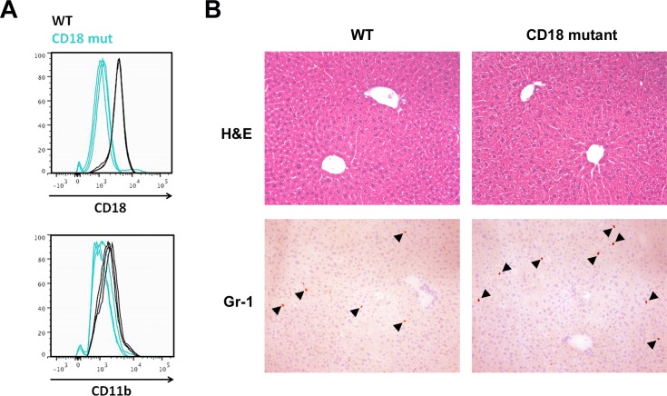
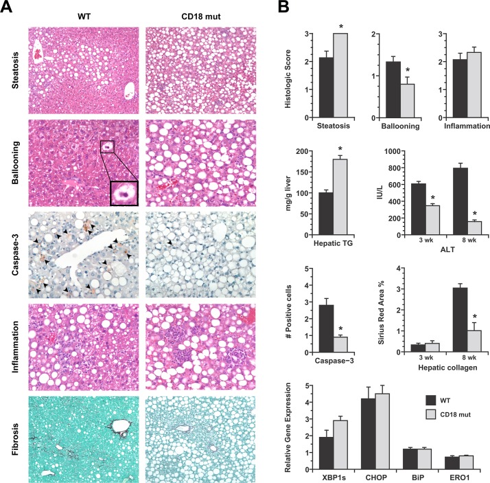
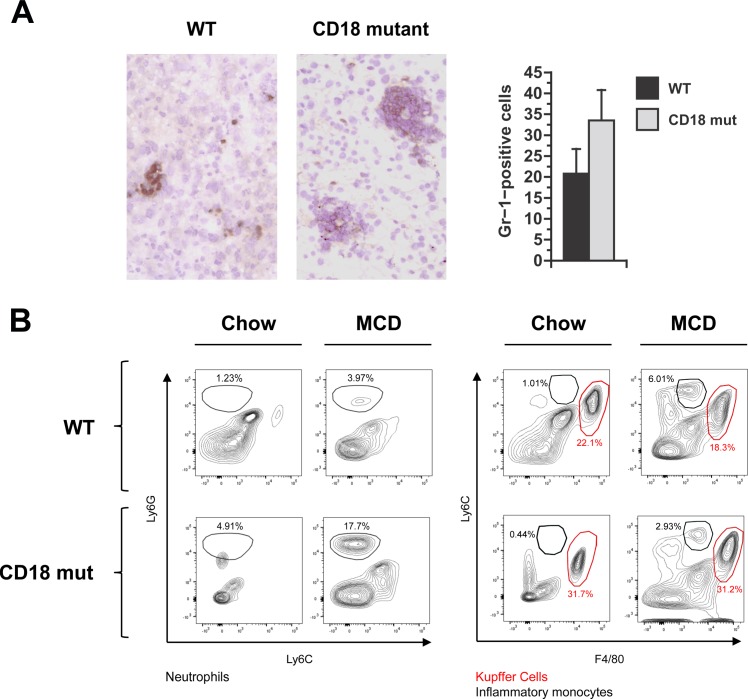
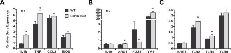
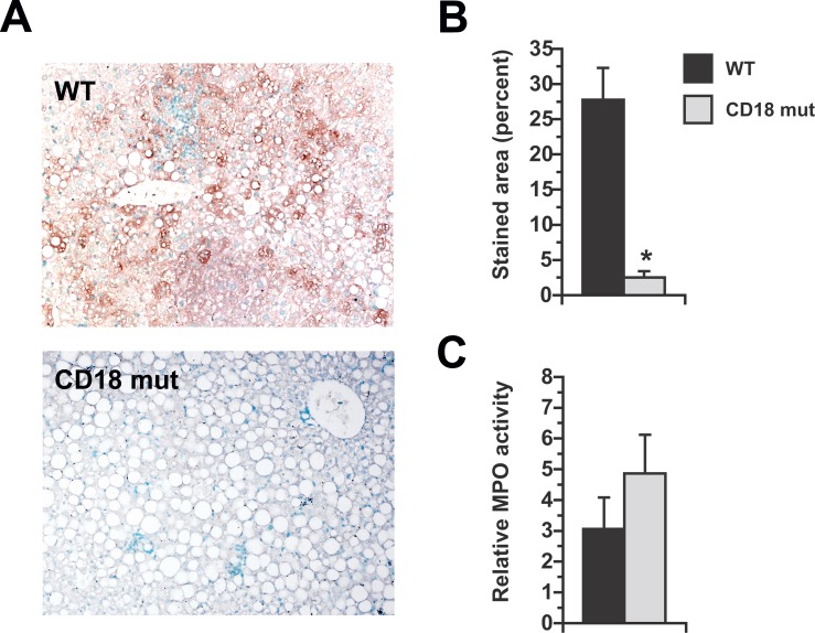
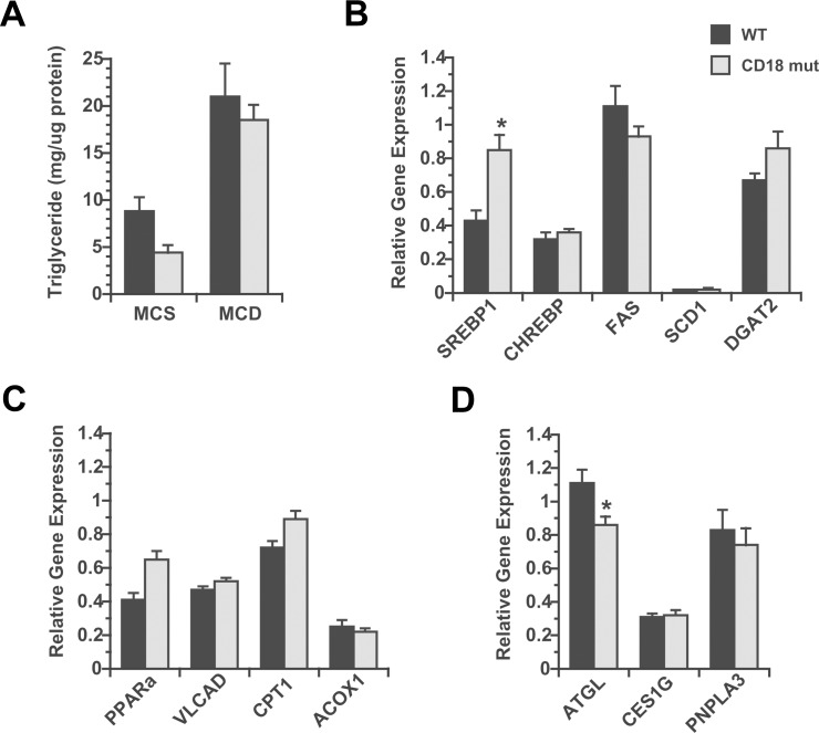
Neutrophils and macrophages are important constituents of the hepatic inflammatory infiltrate in non-alcoholic steatohepatitis. These innate immune cells express CD18, an adhesion molecule that facilitates leukocyte activation. In the context of fatty liver, activation of infiltrated leukocytes is believed to enhance hepatocellular injury. The objective of this study was to determine the degree to which activated innate immune cells promote steatohepatitis by comparing hepatic outcomes in wild-type and CD18-mutant mice fed a methionine-choline-deficient (MCD) diet. After 3 weeks of MCD feeding, hepatocyte injury, based on serum ALT elevation, was 40% lower in CD18-mutant than wild-type mice. Leukocyte infiltration into the liver was not impaired in CD18-mutant mice, but leukocyte activation was markedly reduced, as shown by the lack of evidence of oxidant production. Despite having reduced hepatocellular injury, CD18-mutant mice developed significantly more hepatic steatosis than wild-type mice after MCD feeding. This coincided with greater hepatic induction of pro-inflammatory and lipogenic genes as well as a modest reduction in hepatic expression of adipose triglyceride lipase. Overall, the data indicate that CD18 deficiency curbs MCD-mediated liver injury by limiting the activation of innate immune cells in the liver without compromising intrahepatic cytokine activation. Reduced liver injury occurs at the expense of increased hepatic steatosis, which suggests that in addition to damaging hepatocytes, infiltrating leukocytes may influence lipid homeostasis in the liver.


Conflict of interest statement

Competing Interests: The authors have declared that no competing interests exist.

Figures

Fig 1
Fig 1. Comparative features of WT and CD18-mutant mice fed chow diets.
(A) CD18 and CD11b expression measured by flow cytometry in leukocytes isolated from the livers of chow-fed WT and CD18-mutant (CD18 mut) mice. The fluorescence intensity of CD18 mut granulocytes was 70% lower than that measured in WT granulocytes, n = 3 per group. (B) Liver histology and Gr-1 immunohistochemistry in WT and CD18 mut mice. Arrowheads mark Gr-1-positive cells. For cell counts, see text. Original magnification 10X.
Fig 2
Fig 2. Comparative features of WT and CD18-mutant mice after MCD feeding.
(A) Photomicrographs illustrate several features of liver disease in WT and CD18-mutant (CD18 mut) mice after MCD feeding for 3–8 wk. Steatosis (H&E, 3 wk, 10X) is more severe in CD18 mut livers whereas ballooning (H&E, 3 wk, 10X) is more evident in WT livers. WT livers also have more cells staining positively for cleaved caspase-3 (20X). Hepatic inflammation is visible in both WT and CD18 mut livers, although cells are diffusely distributed in WT livers and clustered in CD18 mut livers. Hepatic fibrosis (Sirius red, 8 wk, 10X) is more advanced in WT livers than CD18 mut livers. (B) Graphs illustrate comparative histologic scores for steatosis, ballooning and inflammation in WT and CD18 mut mice at 3 wk, hepatic triglyceride concentration at 3 wk, and serum ALT measurements at 3 wk and 8 wk. Additional histograms illustrate quantitation of caspase-3-positive cells (# cells per 20X field) and quantitation of hepatic fibrosis (Sirius red, percent area). Final histograms show hepatic mRNA levels for several ER stress markers in WT and CD18 mut liver, normalized to chow-fed WT liver. XBP1s, X-box protein-1 spliced form; CHOP, CEBP-homologous protein; BiP, binding immunoglobulin protein; ERO1, ER oxidoreductin. Values represent mean ± SE for n = 10–15. * P < 0.05 vs. WT.
Fig 3
Fig 3. Hepatic inflammation in WT and CD18-mutant mice in response to MCD feeding.
(A) Gr-1 staining and quantitation of Gr1-positive cells in WT and CD18 mut liver at 3 wk. Photomicrographs illustrate that Gr-1-positive cells are abundant in both WT and CD18-mutant (CD18 mut) mice after MCD feeding, although in different distributions. Original magnification 15X. Histograms illustrated Gr1-positive cell counts in WT and CD18 mut livers, performed as described in Methods. Values represent mean ± SE for n = 5. (B) Representative FACS plots of hepatic leukocytes from mice fed chow or MCD diets. MCD feeding enhances the proportion of neutrophils (Ly6Ghigh) and inflammatory monocytes (Ly6Chigh), while the lack of CD18 further enhances the accumulation of neutrophils but not inflammatory monocytes. Data are illustrative of n = 6 mice per group (24 per cohort), performed as 2 replicate experiments involving 12 mice each (3 per group).
Fig 4
Fig 4. Comparative expression of cytokine and inflammatory genes in WT and CD18-mutant mice after MCD feeding.
All data are normalized to the expression levels measured in WT mice on chow diets. (A) Hepatic expression of M1 inflammatory genes is up-regulated by MCD feeding in both WT and CD18 mut mice. M1 genes are induced as much or more in CD18-mutant mice than WT mice. M1 genes are induced as much or more in CD18-mutant mice than WT mice. CCL2, C-C chemokine ligand-2; INOS, inducible nitric oxidase synthase. (B) Hepatic expression of M2 inflammatory genes after MCD feeding for 3 wk. ARG1, arginase-1; FIZZ1, found in inflammatory zone protein; Ym1, chitinase 3-like-3. (C) Hepatic expression of IL-1 receptor-1 (IL1R1) is decreased, and Toll-like receptors (TLR) increased, by MCD feeding in WT and CD18-mutant mice. TLR induction is not impaired, and in some instances enhanced, in CD18-mutant mice. Values represent mean ± SE for n = 8–14. *P < 0.05 vs. WT.
Fig 5
Fig 5. Chlorotyrosine-protein adduct formation in the livers of MCD-fed mice.
(A) Immunohistochemical staining for chlorotyrosine-protein adducts in WT and CD18 mut mice fed MCD diets for 3 wk. Original magnification 15X. (B) Morphometric quantitation of adduct-stained area. (C) Liver myeloperoxidase (MPO) activity normalized to the level in chow-fed WT mice. Values represent mean ± SE for n = 5. *P < 0.05 vs. WT.
Fig 6
Fig 6. Analysis of hepatic lipid homeostasis in response to MCD in culture and in vivo.
(A) Hepatic lipid accumulation in primary hepatocytes from chow-fed WT and CD18 mut mice, cultured in methionine-choline-sufficient (MCS) or methionine-choline-deficient (MCD) medium for 48 h. Values represent mean ± SE for n = 3. (B, C, D) Hepatic expression of genes pertinent to lipogenesis (B), fatty acid oxidation (C) and lipolysis (D) in WT and CD18 mut mice after 3 wk of MCD feeding in vivo, normalized to WT mice on chow diets. ACOX1, acyl-CoA oxidase-1; ATGL, adipose triglyceride lipase; CPT1, carnitine palmitoyl transferase-1; DGAT2, diacylglycerol transferase-2; FAS, fatty acid synthase; PNPLA3, patatin-like phospholipase domain containing 3; PPARa, peroxisome proliferator activated receptor-α; SCD1, stearoyl-CoA desaturase-1; VLCAD, very long chain acyl dehydrogenase. Values represent mean ± SE for n = 8–14. * P < 0.05 vs. WT.

References

    1. Browning JD, Szczepaniak LS, Dobbins R, Nuremberg P, Horton JD, Cohen JC, et al. Prevalence of hepatic steatosis in an urban population in the United States: impact of ethnicity. Hepatology. 2004;40(6):1387–95. doi: 10.1002/hep.20466 - DOI - PubMed
    1. Clark JM, Brancati FL, Diehl AM. The prevalence and etiology of elevated aminotransferase levels in the United States. Am J Gastroenterol. 2003;98(5):960–7. doi: 10.1111/j.1572-0241.2003.07486.x - DOI - PubMed
    1. Ruhl CE, Everhart JE. Trunk fat is associated with increased serum levels of alanine aminotransferase in the United States. Gastroenterology. 2010;138(4):1346–56, 56 e1-3. doi: 10.1053/j.gastro.2009.12.053 - DOI - PMC - PubMed
    1. Baffy G. Kupffer cells in non-alcoholic fatty liver disease: the emerging view. J Hepatol. 2009;51(1):212–23. doi: 10.1016/j.jhep.2009.03.008 - DOI - PMC - PubMed
    1. Miura K, Seki E, Ohnishi H, Brenner DA. Role of toll-like receptors and their downstream molecules in the development of nonalcoholic Fatty liver disease. Gastroenterology research and practice. 2010;2010:362847 doi: 10.1155/2010/362847 - DOI - PMC - PubMed

MeSH terms