. 2017 Sep 5;11(9):e0005913.
doi: 10.1371/journal.pntd.0005913. eCollection 2017 Sep.

Deleterious effect of Usutu virus on human neural cells

Affiliations

Deleterious effect of Usutu virus on human neural cells

Sara Salinas et al. PLoS Negl Trop Dis. .

Abstract

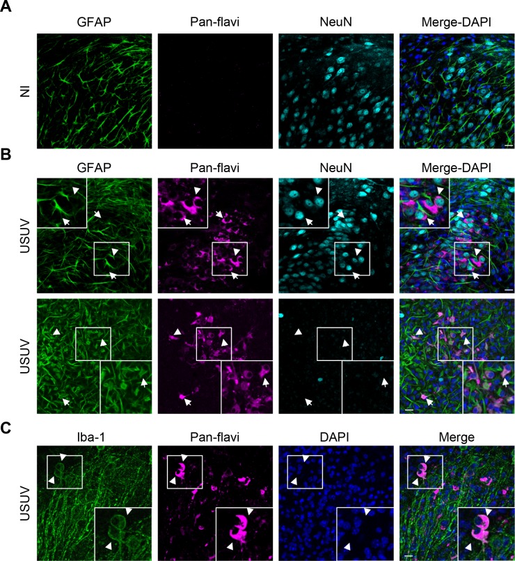
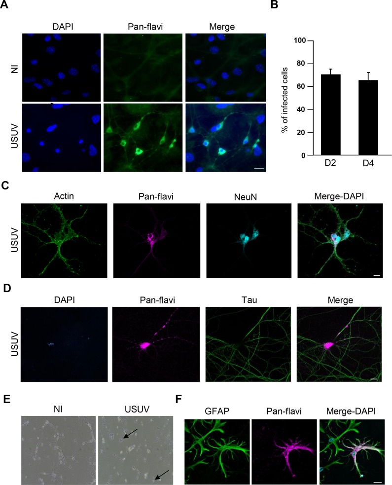
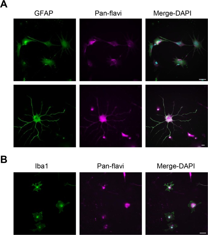
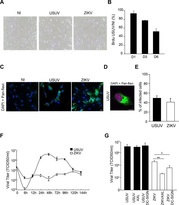
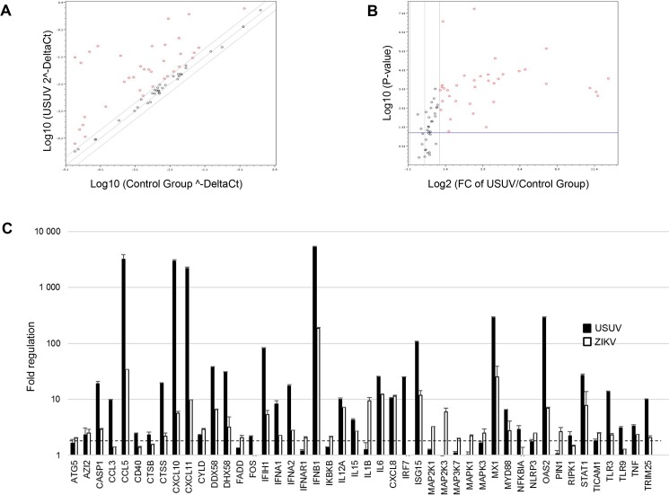
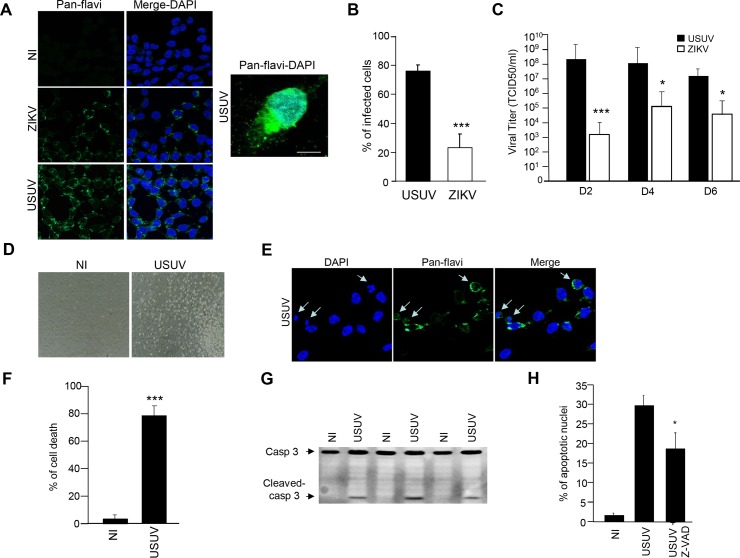
In the last decade, the number of emerging Flaviviruses described worldwide has increased considerably. Among them Zika virus (ZIKV) and Usutu virus (USUV) are African mosquito-borne viruses that recently emerged. Recently, ZIKV has been intensely studied due to major outbreaks associated with neonatal death and birth defects, as well as neurological symptoms. USUV pathogenesis remains largely unexplored, despite significant human and veterinary associated disorders. Circulation of USUV in Africa was documented more than 50 years ago, and it emerged in Europe two decades ago, causing massive bird mortality. More recently, USUV has been described to be associated with neurological disorders in humans such as encephalitis and meningoencephalitis, highlighting USUV as a potential health threat. The aim of this study was to evaluate the ability of USUV to infect neuronal cells. Our results indicate that USUV efficiently infects neurons, astrocytes, microglia and IPSc-derived human neuronal stem cells. When compared to ZIKV, USUV led to a higher infection rate, viral production, as well as stronger cell death and anti-viral response. Our results highlight the need to better characterize the physiopathology related to USUV infection in order to anticipate the potential threat of USUV emergence.


Conflict of interest statement

The authors have declared that no competing interests exist.

Figures

Fig 1
Fig 1. USUV infects efficiently organotypic murine brain slices.
Hippocampi slices obtained from 6 day old pups were infected with USUV (3×10⁵ infectious particles per slice). Five dpi, slices were fixed and subjected for indirect immunofluorescence using various neural cellular markers such as GFAP (astrocytes), NeuN (neurons) and Iba1 (microglia). (A) Non-infected (NI) slices did not show labeling by the anti-pan-flavivirus antibody, in contrast to USUV-infected samples (in magenta) (B and C). (B and C) White arrows show infected cells also expressing either NeuN (in cyan), GFAP or Iba1 (in green), indicating that USUV can infect and replicate in multiple types of neural cells in the murine brain. Nuclei are labeled with DAPI (in blue). Zoomed in panels of white boxes show co-labelling. Scale bars 20 μm.
Fig 2
Fig 2. USUV infects multiple murine neural cells ex vivo.
Primary murine hippocampal neurons were infected with USUV at a MOI of 2. (A) At 4 dpi, cells were fixed and labeled with a pan-flavivirus antibody. Strong labeling was observed in cells of neuronal morphology, both in soma and neurites. (B) Quantitative measurement showed that ~70% (+/- 4%) of cells of this quasi-pure culture were infected at day 2 and day 4 (n = 3 independent experiments). (C-D) USUV infects efficiently neurons ex vivo. Co-labeling from fixed USUV-infected cultures at 4 dpi demonstrates that USUV replicates in neuronal cell bodies (nuclei expressing NeuN) and axons (labeled with Tau). (E) USUV elicits neuronal toxicity. Bright light imaging of USUV-infected neurons at 8 dpi shows neurite damage. Arrows are showing dying cellular bodies. (F) Glia cells are infected by USUV. Cells expressing GFAP (astrocytes) were infected by USUV as shown by indirect IF studies at 4 dpi. Scale bars 20 μm.
Fig 3
Fig 3. USUV replicates in murine spinal glial cells ex vivo.
Spinal astrocytes and microglia are readily infected by USUV. Mixed cultures were infected with USUV at a MOI of 2 and fixed at 4 dpi. (A) USUV-infected cells labeled with the pan-flavivirus antibody also express GFAP, suggesting an efficient replication in spinal astrocytes. Rows show different pictures of the same experiment. Scale bar 10 μm. (B). USUV-infected cells labeled with the pan-flavivirus antibody express Iba1, suggesting an efficient replication in spinal microglial cells. Scale bar 20 μm.
Fig 4
Fig 4. Comparative infections between USUV and ZIKV in primary human astrocytes.
Primary human astrocytes were infected with USUV or ZIKV at a MOI of 2. (A) Bright light images of control and infected astrocytes at 4 dpi show sparser cells in USUV-infected condition. (B) Cellular proliferation is affected by USUV infection. Astrocytes pre-treated with BrdU were infected with USUV at MOI 2 and assayed for BrdU at 1, 3 and 6 dpi. (C) Mock, USUV- and ZIKV-infected cells were fixed at 4 dpi and labeled with the pan-flavivirus antibody (in green) by indirect immunofluorescence. Strong labeling was observed in cells. Nuclei are labeled with DAPI (in blue). (D) Typical flavivirus labeling (in green) is observed at high magnification. Nuclei are labeled with DAPI (false colored in magenta). (E) Quantification of USUV and ZIKV-infected cells. (F) Kinetics of viral production in USUV- or ZIKV-infected astrocytes show difference in terms of replication and persistence between the two viruses. Supernatants from infected cells (MOI of 2) were collected at various time points and subjected to TCID50 measurement on Vero cells. (G) AXL and DC-SIGN do not mediate USUV internalization in human astrocytes. Cells were pre-incubated with anti-Axl or Anti-DC-SIGN prior to infection with USUV or ZIKV at MOI of 2. 4 dpi, supernatants were collected and viral replication assayed. Blocking antibodies only decreased ZIKV replication. (*p<0.05, **p<0.01). Scale bar 10 μm.
Fig 5
Fig 5. Anti-viral responses in USUV- and ZIKV-infected human astrocytes.
mRNA from supernatants of primary human astrocytes infected with USUV or ZIKV at MOI = 1 for 3 days were subjected to qRT-PCR analyses. (A) Scatter plot. Up regulated genes appears in the top-left cadran (boundary 2). (B) Volcano plots, (boundary 2, scatter 0.05: p-values based on student’s t-test of three replicates). Statistically significant changes in fold regulation appear in the top right part of the graph (genes upregulated). The experiment was performed in triplicates and each point represents the mean. (C) Fold regulation of statistically significant genes modulated upon USUV and ZIKV infection are shown. Results are expressed as means ± SD and analyzed using an unpaired t-test *p <0.05.
Fig 6
Fig 6. Effect of USUV infection on NSC survival.
(A) IPSc-derived NSCs were infected with USUV and ZIKV at a MOI of 2 and fixed at 2 dpi. Cells were labeled with the pan-flavivirus antibody (in green) by indirect immunofluorescence. Nuclei are labeled with DAPI (blue). Scale bar 10 μm. (B) Quantification of the percentage of USUV- and ZIKV-infected cells. (C) Supernatants from USUV- and ZIKV infected NSCs (MOI 2) were collected at 2, 4 and 6 dpi and subjected to TCID50 measurement on Vero cells. (D) Bright light images of control and USUV-infected NSCs at 4 dpi show rounded up cells in USUV-treated condition. (E) USUV-infected NSCS at 4 dpi and labeled with the anti-flavivirus antibody and DAPI show condensed nuclei. (F) Trypan blue assay (supernatant + adherent cells) showed that cell viability is affected in USUV-infected NSCs at 4 dpi. (G) Immunobloting analyses of cell extracts from mock or USUV-infected cells at 4 dpi show the generation of cleaved caspase-3 fragment, indicative of apoptosis. (H) NSCs were infected with USUV at a MOI of 2 with or without Z-VAD. 4 dpi, cells were labeled with the anti-flavivirus antibody and DAPI and condensed nuclei quantified. (*p<0.05, ***p<0.001).

Similar articles

  • Study of Usutu virus neuropathogenicity in mice and human cellular models.
    Clé M, Barthelemy J, Desmetz C, Foulongne V, Lapeyre L, Bolloré K, Tuaillon E, Erkilic N, Kalatzis V, Lecollinet S, Beck C, Pirot N, Glasson Y, Gosselet F, Alvarez Martinez MT, Van de Perre P, Salinas S, Simonin Y. Clé M, et al. PLoS Negl Trop Dis. 2020 Apr 23;14(4):e0008223. doi: 10.1371/journal.pntd.0008223. eCollection 2020 Apr. PLoS Negl Trop Dis. 2020. PMID: 32324736 Free PMC article.
  • Working with Zika and Usutu Viruses In Vitro.
    Barr KL, Anderson BD, Prakoso D, Long MT. Barr KL, et al. PLoS Negl Trop Dis. 2016 Aug 19;10(8):e0004931. doi: 10.1371/journal.pntd.0004931. eCollection 2016 Aug. PLoS Negl Trop Dis. 2016. PMID: 27541001 Free PMC article.
  • Explaining Usutu virus dynamics in Austria: model development and calibration.
    Rubel F, Brugger K, Hantel M, Chvala-Mannsberger S, Bakonyi T, Weissenböck H, Nowotny N. Rubel F, et al. Prev Vet Med. 2008 Jul 15;85(3-4):166-86. doi: 10.1016/j.prevetmed.2008.01.006. Epub 2008 Mar 7. Prev Vet Med. 2008. PMID: 18314208
  • Usutu virus: an emerging flavivirus in Europe.
    Ashraf U, Ye J, Ruan X, Wan S, Zhu B, Cao S. Ashraf U, et al. Viruses. 2015 Jan 19;7(1):219-38. doi: 10.3390/v7010219. Viruses. 2015. PMID: 25606971 Free PMC article. Review.
  • Usutu virus in Africa.
    Nikolay B, Diallo M, Boye CS, Sall AA. Nikolay B, et al. Vector Borne Zoonotic Dis. 2011 Nov;11(11):1417-23. doi: 10.1089/vbz.2011.0631. Epub 2011 Jul 18. Vector Borne Zoonotic Dis. 2011. PMID: 21767160 Review.

Cited by

References

    1. Weissenböck H, Kolodziejek J, Url A, Lussy H, Rebel-Bauder B, Nowotny N. Emergence of Usutu virus, an African Mosquito-Borne Flavivirus of the Japanese Encephalitis Virus Group, Central Europe. Emerg Infect Dis. 2002;8: 652–656. doi: 10.3201/eid0807.020094 - DOI - PMC - PubMed
    1. Williams MC, Simpson DI, Haddow AJ, Knight EM. The isolation of West Nile virus from man and Usutu virus frome the bird-bitting mosquito mansonia aurites (theobald) in the entebee area of Uganda. Ann Trop Med Parasitol. 1964;58: 367–74. Available: http://www.ncbi.nlm.nih.gov/pubmed/14212897 - PubMed
    1. Weissenböck H, Bakonyi T, Chvala S, Nowotny N. Experimental Usutu virus infection of suckling mice causes neuronal and glial cell apoptosis and demyelination. Acta Neuropathol. 2004;108: 453–60. doi: 10.1007/s00401-004-0916-1 - DOI - PubMed
    1. Bakonyi T, Gould EA, Kolodziejek J, Weissenböck H, Nowotny N. Complete genome analysis and molecular characterization of Usutu virus that emerged in Austria in 2001: comparison with the South African strain SAAR-1776 and other flaviviruses. Virology. 2004;328: 301–10. doi: 10.1016/j.virol.2004.08.005 - DOI - PubMed
    1. Bakonyi T, Erdélyi K, Ursu K, Ferenczi E, Csörgo T, Lussy H, et al. Emergence of Usutu virus in Hungary. J Clin Microbiol. 2007;45: 3870–4. doi: 10.1128/JCM.01390-07 - DOI - PMC - PubMed

LinkOut - more resources