. 2017 Oct 24;61(11):e01347-17.
doi: 10.1128/AAC.01347-17. Print 2017 Nov.

Mycobacterium abscessus WhiB7 Regulates a Species-Specific Repertoire of Genes To Confer Extreme Antibiotic Resistance

Affiliations

Mycobacterium abscessus WhiB7 Regulates a Species-Specific Repertoire of Genes To Confer Extreme Antibiotic Resistance

Kelley Hurst-Hess et al. Antimicrob Agents Chemother. .

Abstract

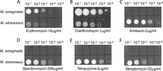
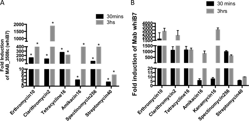
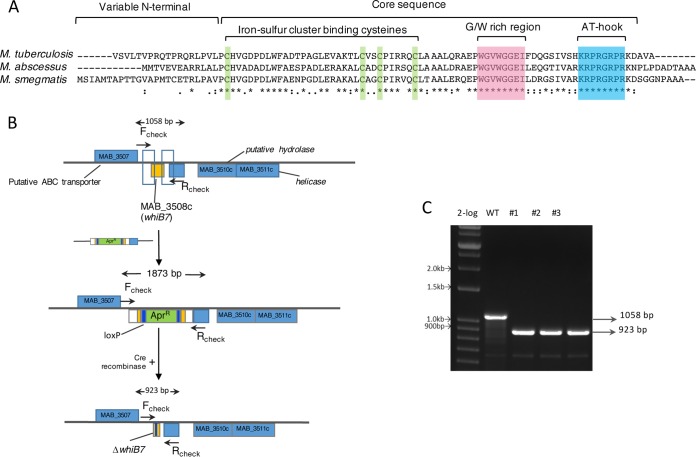
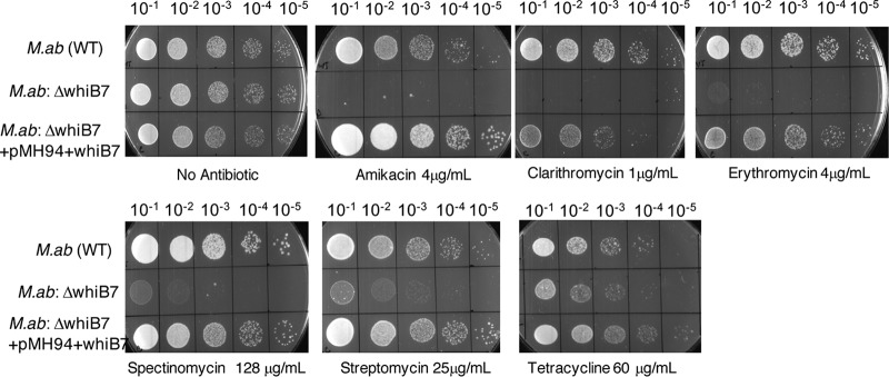
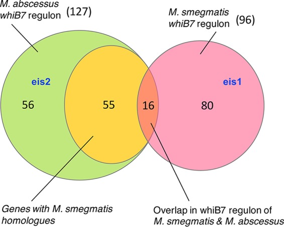
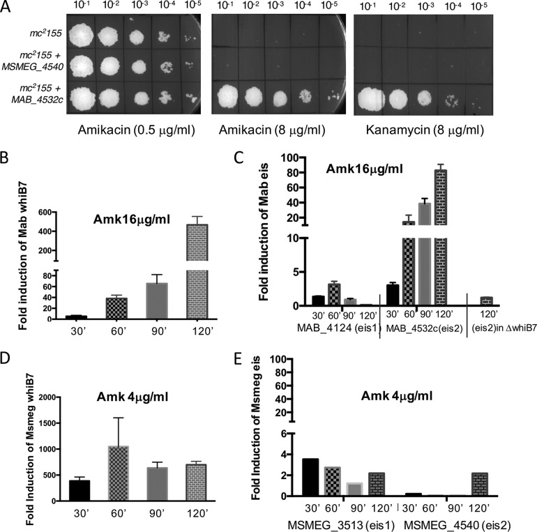
Mycobacterium abscessus causes acute and chronic bronchopulmonary infection in patients with chronic lung damage, of which cystic fibrosis (CF) patients are particularly vulnerable. The major threat posed by this organism is its high intrinsic antibiotic resistance. A typical treatment regimen involves a 6- to 12-month-long combination therapy of clarithromycin and amikacin, with cure rates below 50% and multiple side effects, especially due to amikacin. In the present work, we show that M. abscessuswhiB7, a homologue of Mycobacterium tuberculosis and Mycobacterium smegmatis whiB7 with previously demonstrated effects on intrinsic antibiotic resistance, is strongly induced when exposed to clinically relevant antibiotics that target the ribosome: erythromycin, clarithromycin, amikacin, tetracycline, and spectinomycin. The deletion of M. abscessuswhiB7 results in sensitivity to all of the above-mentioned antibiotics. Further, we have defined and compared the whiB7 regulon of M. abscessus with the closely related nontuberculous mycobacterium (NTM) M. smegmatis to demonstrate the induction of a species-specific repertoire of genes. Finally, we show that one such gene, eis2, is specifically induced in M. abscessus by whiB7 and contributes to its higher levels of intrinsic amikacin resistance. This species-specific pattern of gene induction might account for the differences in drug susceptibilities to other antibiotics and between different mycobacterial species.

Keywords: Mycobacterium; abscessus; antibiotic resistance; intrinsic; whiB7.


Figures

FIG 1
FIG 1
Differences in antibiotic sensitivities between M. abscessus ATCC 19977 and M. smegmatis mc2155 (A to F). Ten-fold serial dilutions of M. abscessus ATCC 19977 and M. smegmatis mc2155 grown to A600 of 0.7 and spotted on Middlebrook 7H10 containing indicated concentrations of antibiotics. M. abscessus is more resistant to amikacin, tetracycline, streptomycin, and spectinomycin than M. smegmatis but the sensitivities to clarithromycin and erythromycin are comparable.
FIG 2
FIG 2
Time course of induction of whiB7 upon antibiotic exposure. (A) The fold induction of transcript of MAB_3508c in wild-type M. abscessus ATCC 19977 exposed to amikacin (16 μg/ml), clarithromycin (2 μg/ml), erythromycin (10 μg/ml), tetracycline (16 μg/ml), streptomycin (40 μg/ml), and spectinomycin (256 μg/ml) over unexposed samples was determined in previous RNA-Seq studies. The asterisks (*) indicate q values of <0.001 (B) The results were verified by quantitative PCR (qPCR) and expressed as a fold overexpression over unexposed samples. Data represent the mean ± standard deviation (SD), n = 3. sigA was used as an endogenous control.
FIG 3
FIG 3
Deletion of M. abscessus whiB7. (A) Multiple-sequence alignment of WhiB7 from M. abscessus, M. smegmatis, and M. tuberculosis showing identical residues (*) and conserved motifs. (B) Schematic representation of creating a deletion of MAB_3508c (whiB7) using phage recombineering and unmarking using the Cre-lox system. (C) Three clones were selected (no. 1 to 3), and the whiB7 gene was amplified using the Fcheck and Rcheck primers, followed by confirmation by Sanger sequencing.
FIG 4
FIG 4
Deletion of MAB_3508c (whiB7) renders M. abscessus hypersensitive to multiple antibiotics. Ten-fold dilutions of the ΔwhiB7 mutant cells were spotted on Middlebrook 7H10/OADC containing indicated concentrations of antibiotics. The mutant is hypersensitive to clarithromycin, erythromycin, amikacin, streptomycin, and spectinomycin but marginally more sensitive to tetracycline than the wild-type parent strain. A complementing strain containing an integrated copy of whiB7 expressed from a constitutive promoter Phsp60 restores antibiotic resistance. M.ab, M. abscessus.
FIG 5
FIG 5
Species-specific regulons of WhiB7 in M. smegmatis and M. abscessus. M. smegmatis and M. abscessus whiB7 were expressed from constitutive promoters in respective ΔwhiB7 mutant strains. RNA-Seq analysis was performed to determine the regulon of WhiB7 in each species, and the overlap in the regulon is represented. eis2 is WhiB7 regulated exclusively in M. abscessus, whereas eis1 is exclusively induced in M. smegmatis by WhiB7. Numbers refer to the number of genes in each category.
FIG 6
FIG 6
MAB_4532c is induced exclusively in M. abscessus as part of the whiB7 regulon and confers amikacin (Amk) resistance. (A) Heterologous expression of MAB_4532c (eis2) from a constitutive promoter integrated at the L5 attB site increases amikacin and kanamycin resistance of M. smegmatis from 0.4 μg/ml to 8 μg/ml. Similar overexpression of MSMEG_4540 (eis2) is insufficient to enhance amikacin/kanamycin resistance in M. smegmatis. (B to E) Wild-type M. smegmatis and M. abscessus and the ΔMabwhiB7 mutant were grown to an A600 of 0.7, exposed to sublethal concentrations of amikacin, as indicated, and the amount of whiB7, eis1, and eis2 transcripts were determined by qPCR and plotted as fold induction over an unexposed control. Data represent the mean ± SD, n = 3. Although whiB7 is induced in both bacteria, eis2 is induced only in M. abscessus in a whiB7-dependent manner and confers species-specific amikacin sensitivity.

References

    1. Leung JM, Olivier KN. 2013. Nontuberculous mycobacteria: the changing epidemiology and treatment challenges in cystic fibrosis. Curr Opin Pulm Med 19:662–669. doi: 10.1097/MCP.0b013e328365ab33. - DOI - PMC - PubMed
    1. Prevots DR, Shaw PA, Strickland D, Jackson LA, Raebel MA, Blosky MA, Montes de Oca R, Shea YR, Seitz AE, Holland SM, Olivier KN. 2010. Nontuberculous mycobacterial lung disease prevalence at four integrated health care delivery systems. Am J Respir Crit Care Med 182:970–976. doi: 10.1164/rccm.201002-0310OC. - DOI - PMC - PubMed
    1. Qvist T, Gilljam M, Jonsson B, Taylor-Robinson D, Jensen-Fangel S, Wang M, Svahn A, Kotz K, Hansson L, Hollsing A, Hansen CR, Finstad PL, Pressler T, Hoiby N, Katzenstein TL, Scandinavian Cystic Fibrosis Study Consortium (SCFSC). 2015. Epidemiology of nontuberculous mycobacteria among patients with cystic fibrosis in Scandinavia. J Cyst Fibros 14:46–52. doi: 10.1016/j.jcf.2014.08.002. - DOI - PMC - PubMed
    1. Olivier KN, Weber DJ, Wallace RJ Jr, Faiz AR, Lee JH, Zhang Y, Brown-Elliot BA, Handler A, Wilson RW, Schechter MS, Edwards LJ, Chakraborti S, Knowles MR, Nontuberculous Mycobacteria in Cystic Fibrosis Study Group. 2003. Nontuberculous mycobacteria. I: multicenter prevalence study in cystic fibrosis. Am J Respir Crit Care Med 167:828–834. doi: 10.1164/rccm.200207-678OC. - DOI - PubMed
    1. Brown-Elliott BA, Wallace RJ Jr. 2002. Clinical and taxonomic status of pathogenic nonpigmented or late-pigmenting rapidly growing mycobacteria. Clin Microbiol Rev 15:716–746. doi: 10.1128/CMR.15.4.716-746.2002. - DOI - PMC - PubMed

MeSH terms

LinkOut - more resources