

Regulation of AMH by oocyte-specific growth factors in human primary cumulus cells
- PMID: 28874516
- PMCID: PMC5665699
- DOI: 10.1530/REP-17-0421
Regulation of AMH by oocyte-specific growth factors in human primary cumulus cells
Abstract
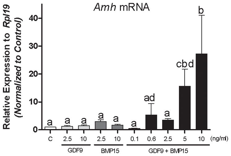
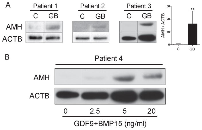
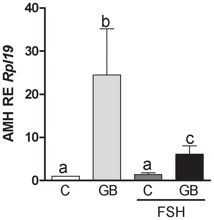
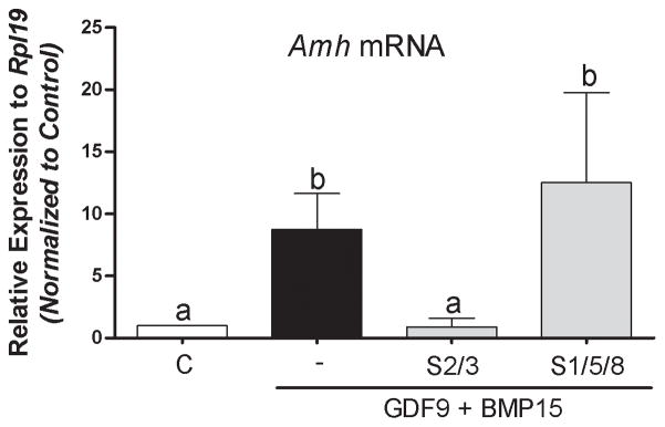
The regulation of AMH production by follicular cells is poorly understood. The purpose of this study was to determine the role of the oocyte-secreted factors, growth differentiation factor 9 (GDF9) and bone morphogenetic protein 15 (BMP15), on AMH production in primary human cumulus cells. Cumulus cells from IVF patients were cultured with a combination of GDF9, BMP15, recombinant FSH and specific signaling inhibitors. Stimulation with GDF9 or BMP15 separately had no significant effect on AMH mRNA levels. In contrast, simultaneous stimulation with GDF9 and BMP15 (G + B) resulted in a significant increase in AMH mRNA expression. Increasing concentration of G + B (0.6, 2.5, 5 and 10 ng/mL) stimulated AMH in a dose-dependent manner, showing a maximal effect at 5 ng/mL. Western blot analyses revealed an average 16-fold increase in AMH protein levels in cells treated with G + B when compared to controls. FSH co-treatment decreased the stimulation of AMH expression by G + B. The stimulatory effect of G + B on the expression of AMH was significantly decreased by inhibitors of the SMAD2/3 signaling pathway. These findings show for the first time that AMH production is regulated by oocyte-secreted factors in primary human cumulus cells. Moreover, our novel findings establish that the combination of GDF9 + BMP15 potently stimulates AMH expression.
© 2017 Society for Reproduction and Fertility.
Conflict of interest statement
The authors declare no conflict of interest that could be perceived as prejudicing the impartiality of the research reported.
Figures
References
-
- Al-Musawi SL, Walton KL, Heath D, Simpson CM, Harrison CA. Species differences in the expression and activity of bone morphogenetic protein 15. Endocrinology. 2013;154:888–899. - PubMed
-
- Andersen CY, Byskov AG. Estradiol and regulation of anti-Mullerian hormone, inhibin-A, and inhibin-B secretion: analysis of small antral and preovulatory human follicles’ fluid. J Clin Endocrinol Metab. 2006;91:4064–4069. - PubMed
-
- Andersen CY, Schmidt KT, Kristensen SG, Rosendahl M, Byskov AG, Ernst E. Concentrations of AMH and inhibin-B in relation to follicular diameter in normal human small antral follicles. Hum Reprod. 2010;25:1282–1287. - PubMed
-
- Baarends WM, Uilenbroek JT, Kramer P, Hoogerbrugge JW, van Leeuwen EC, Themmen AP, Grootegoed JA. Anti-mullerian hormone and anti-mullerian hormone type II receptor messenger ribonucleic acid expression in rat ovaries during postnatal development, the estrous cycle, and gonadotropin-induced follicle growth. Endocrinology. 1995;136:4951–4962. - PubMed
Publication types
MeSH terms
Substances
Grants and funding
LinkOut - more resources
Full Text Sources
Other Literature Sources
