. 2017 Oct 27;37(22):e00218-17.
doi: 10.1128/MCB.00218-17. Print 2017 Nov 15.

Internal Associations of the Acidic Region of Upstream Binding Factor Control Its Nucleolar Localization

Affiliations

Internal Associations of the Acidic Region of Upstream Binding Factor Control Its Nucleolar Localization

Shuhei Ueshima et al. Mol Cell Biol. .

Abstract

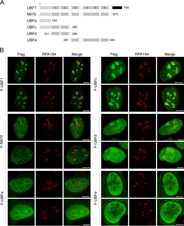
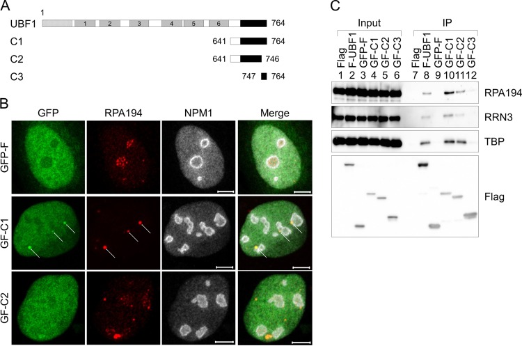
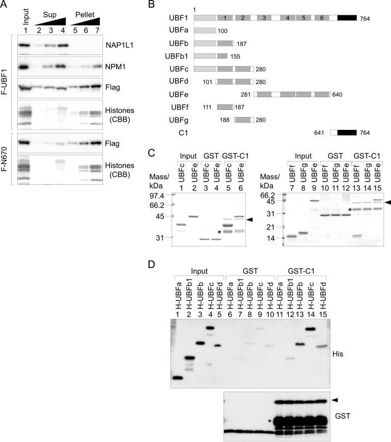
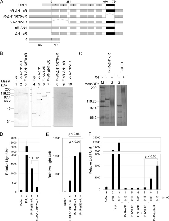
Upstream binding factor (UBF) is a member of the high-mobility group (HMG) box protein family, characterized by multiple HMG boxes and a C-terminal acidic region (AR). UBF is an essential transcription factor for rRNA genes and mediates the formation of transcriptionally active chromatin in the nucleolus. However, it remains unknown how UBF is specifically localized to the nucleolus. Here, we examined the molecular mechanisms that localize UBF to the nucleolus. We found that the first HMG box (HMG box 1), the linker region (LR), and the AR cooperatively regulate the nucleolar localization of UBF1. We demonstrated that the AR intramolecularly associates with and attenuates the DNA binding activity of HMG boxes and confers the structured DNA preference to HMG box 1. In contrast, the LR was found to serve as a nuclear localization signal and compete with HMG boxes to bind the AR, permitting nucleolar localization of UBF1. The LR sequence binds DNA and assists the stable chromatin binding of UBF. We also showed that the phosphorylation status of the AR does not clearly affect the localization of UBF1. Our results strongly suggest that associations of the AR with HMG boxes and the LR regulate UBF nucleolar localization.

Keywords: HMG box; nucleolus; ribosome biogenesis; transcription factors.


Figures

FIG 1
FIG 1
Multiple regions are required for efficient nucleolar localization of UBF. (A) Schematic diagram of truncated UBF mutants. The dotted, gray, and black boxes represent the dimerization domain, HMG boxes (1 to 6), and acidic region (AR), respectively. The numbers shown represent the positions of amino acids. (B) Immunofluorescence analysis. U2OS cells transiently expressing F-UBF1, -N670, -UBFc, -UBFa, -UBFd, and -UBFe were subjected to immunofluorescence assays 16 h after transfection with anti-Flag and anti-RPA194 (red) antibodies as indicated at the top of the panels. Bars indicate 5 μm.
FIG 2
FIG 2
AR of UBF shows potential ability to localize to the nucleoli. (A) Schematic diagram of truncated UBF1 mutants. (B) Localization of GFP-Flag (GF)-tagged AR mutants C1 and C2. U2OS cells transiently expressing GF, GF-C1, or GF-C2 were subjected to immunofluorescence assays 16 h after transfection with anti-RPA194 (red) and anti-B23 (NPM1) (white) antibodies as indicated at the top. Arrows represent the accumulated foci. Bars indicate 5 μm. (C) Association of the AR with the Pol I machineries. Flag (F), F-UBF1, GF, GF-C1, GF-C2, or GF-C3 was transiently expressed in 293T cells, and immunoprecipitation (IP) assays were performed with anti-Flag M2 affinity gel. Input (lanes 1 to 6) and immunoprecipitated (lanes 7 to 12) proteins were separated by SDS-PAGE and subjected to Western blotting with anti-RPA194, -RRN3, -TBP, and -Flag antibodies.
FIG 3
FIG 3
AR affects the localization of UBF by its association with HMG boxes. (A) Subcellular fractionation assay. HeLa cells transiently expressing Flag-UBF1 or -N670 were subjected to subcellular fractionation assays as described in Materials and Methods. Input (lane 1; 3 × 10⁴ cells), supernatant (Sup), and pellet fractions (lanes 2 to 4 and 5 to 7; 0.6 × 10⁴, 1.5 × 10⁴, and 3 × 10⁴ cells) were separated by SDS-PAGE and subjected to Western blotting with anti-Flag, anti-NAP1L1, and anti-NPM1 antibodies. Core histones were visualized by CBB staining. Two independent experiments showed similar results. (B) Schematic diagram of truncated UBF1 mutants used in panels C and D. The numbers shown represent the positions of amino acids. (C and D) Association of AR with the UBF mutants. GST, GST-C1, and His-tagged UBF mutants were expressed in E. coli and purified, and GST pulldown assays were performed using the purified proteins. Input and pulldown proteins with GST or GST-C1, as indicated, were separated by SDS-PAGE and subjected to CBB staining (C) or Western blotting with anti-His and -GST antibodies (top and bottom, respectively) (D). Positions of the full-length GST-C1 are indicated by arrowheads, and the bands shown by asterisks likely are premature translation termination products.
FIG 4
FIG 4
Associations between AR and HMG boxes occur intramolecularly. (A) Schematic diagram of the fusion proteins of the split Renilla luciferase fragments and UBF mutants. The boxes with horizontal and vertical lines represent N- and C-terminal Renilla luciferase (nR and cR, amino acids 1 to 229 and 230 to 311, respectively). The numbers shown represent the positions of amino acids. (B) Purified proteins. The fusion proteins purified from 293T cells (50 ng) were separated by SDS-PAGE and visualized by CBB staining. Positions of molecular size markers are shown at the left. Asterisks indicate contaminated or truncated proteins. (C) Chemical cross-link assay. F-nR-ΔN1-cR and F-UBF1, left untreated or treated with 0.05% glutaraldehyde, were separated by 7.5% SDS-PAGE and visualized with silver staining. (D and E) Detection of the interaction between AR and HMG boxes by the split Renilla luciferase complementation assay. Buffer alone, F-R, F-nR-ΔN1-cR, F-nR-ΔN1N670-cR, or F-nR-ΔN2-cR (0.15 and 0.3 pmol for D and E, respectively), as indicated, was mixed with the substrate of Renilla luciferase and the luminescence intensity was measured. (F) Inter- or intramolecular interaction between the AR and HMG boxes. Buffer alone, F-R, F-nR-ΔN1, F-ΔN1-cR, or F-nR-ΔN1-cR (0.05 or 0.15 pmol, as indicated) was mixed with the substrate of Renilla luciferase and luminescence intensity was measured. Equal amounts of F-nR-ΔN1 and F-ΔN1-cR (0.05 and 0.15 pmol of each protein) were mixed and luminescence intensity was measured (lanes 6 and 7). Error bars in panels D to F indicate ± standard deviations (SD) (n = 3). Statistical analyses were performed by two-tailed Student's t test.
FIG 5
FIG 5
AR attenuates the DNA binding activity of HMG boxes and confers DNA structure preference to HMG box 1. (A) Schematic diagram of UBF1 mutants. (B) DNA binding activity of His-tagged UBFb and UBFe. The 154-bp DNA fragment (0.04 μM) mixed and incubated alone or with increasing amounts of His-tagged proteins (0, 0.05, 0.1, 0.2, 0.3, and 0.4 μM) was separated by native PAGE and visualized by GelRed staining. (C) DNA binding activities of UBFb and UBFe are attenuated by the AR. His-UBFb (lanes 2 to 5) or His-UBFe (lanes 7 to 10) (0.3 μM) incubated without or with GST-ac1 (0.3, 1, and 3 μM) was mixed with 154 bp DNA (0.04 μM) and separated by native PAGE, and DNA was visualized by GelRed staining. Positions of free DNA are indicated by arrowheads. (D) UBF1 binds linear DNA fragments without specificity. Increasing amounts of Flag-UBF1 purified from 293T cells were mixed with 0.05 μM DNA fragments (150, 129, 150, and 124 bp, for panels A to D, respectively) harboring the rRNA gene promoter region and separated by native PAGE. DNA was visualized by GelRed staining. Positions of the DNA fragments relative to the transcription start site of rRNA (+1) are schematically shown at the bottom. The positions of the DNA are also shown in Fig. S4A in the supplemental material. (E) UBF1 preferentially binds to structured DNA. Supercoiled or linear pBluescript SKII plasmid (100 ng, 0.05 pmol) mixed with increasing amounts of Flag-UBF1 (0, 3, 6, 12, and 18 pmol for lanes 1 to 5 and 6 to 10) was separated by 0.8% agarose gel and visualized with GelRed staining. (F) AR confers DNA structure preference on UBFb. Supercoiled (lanes 1 to 12) and linear (lanes 13 to 24) DNA was mixed with His-UBFb (0, 1, 2, 3, 4, and 5 pmol as indicated) alone or preincubated with GST-ac1 (40 pmol). The mixture was separated and DNA was visualized as described for panel E.
FIG 6
FIG 6
Functions of the linker region in nuclear localization and the DNA binding activity of UBF1. (A) Schematic diagram of cloned UBF proteins. (B) Localization of the UBF proteins. U2OS cells transiently expressing Flag-UBF1, -UBF2, -UBFm1, or -UBFm2 were subjected to immunofluorescence assays with anti-Flag (green) and anti-NPM1 (red) antibodies as indicated at the top. (C) Amino acid sequences lacking in human UBFm2 and corresponding sequences of mouse and Xenopus UBF (UniProtKB accession numbers P17480, P25976, and P25980). (D) Localization of Flag-tagged UBFm2 in the presence of leptomycin B (LMB). U2OS cells transiently expressing Flag-UBFm2 were incubated in the absence or presence of 100 nM LMB for 3 h, and then immunofluorescence assays were performed with anti-Flag (green) and anti-NF-κB p65 (red) antibodies as indicated. (E) Localization of GFP and GFP fused with the 24-amino-acid sequence deleted in UBFm2 (GFP-LR). In panels C to E, DNA was counterstained with TO-PRO-3 iodide (blue). Bars indicate 5 μm. (F) Purified Flag-UBF1 and -UBFm2. Each protein (300 ng) was separated by SDS-PAGE and visualized by CBB staining. Positions of molecular size markers are shown at the left. (G) DNA binding activity of UBF1 and UBFm2. The 154-bp DNA fragment (0.03 μM) incubated with Flag-UBF1 or -UBFm2 (0, 0.09, 0.18, 0.27, 0.36, and 0.45 μM) was separated by native PAGE and visualized by GelRed staining. The position of the free DNA fragment is shown at the left.
FIG 7
FIG 7
LR associates with both DNA and AR. (A) Schematic diagram of truncated UBF1 mutants. Hatched boxes represent the LR, the 24-amino-acid segment deleted in UBFm2. Numbers shown are the positions of amino acids. (B) DNA binding activity of LR. The 154-bp DNA fragment incubated with GST or GST-LR (0, 0.06, 0.12, 0.18, 0.24, and 0.3 μM) was separated by native PAGE and visualized by GelRed staining. The position of free DNA is indicated by an arrowhead. (C) The LR associates with AR. GST, GST-ac1, MBP, and MBP-LR were expressed and purified, and GST pulldown assays were performed using the purified proteins. Input (lanes 1 and 2) and pulldown proteins with GST and GST-ac1 (lanes 3 to 6) were separated by SDS-PAGE and visualized by CBB staining. Positions of molecular size markers and those of GST and MBP are indicated. (D) Purified proteins. GST-ac1, His-UBFb, and GST-LR (300 ng) were separated by SDS-PAGE and visualized by CBB staining. Positions of molecular size markers are shown at the left. (E) The AR-binding activity of HMG box 1 and LR. GST-ac1 (1 μM) incubated with His-UBFb or GST-LR (0, 2, 4, 6, 8, and 10 μM) was separated by native PAGE and visualized by CBB staining. (F) Localization of Flag-N670 and -N640. U2OS cells transiently expressing Flag-N670 or -N640 were subjected to immunofluorescence assays with anti-Flag (green) and anti-NCL (red) antibodies. DNA was counterstained with TO-PRO-3 iodide (blue). Bars indicate 5 μm.
FIG 8
FIG 8
Long acidic tail interacts with multiple DNA binding domains of UBF. (A) Schematic diagram of truncated UBF1 mutants. (B) Subcellular fractionation assay. HeLa cells transiently expressing Flag-UBF1, -N670, -N706, or -N746 were subjected to subcellular fractionation assays. Input (lane 1; 3 × 10⁴ cells), supernatant (Sup), and pellet fractions (lanes 2 to 4 and 5 to 7; 0.6 × 10⁴, 1.5 × 10⁴, and 3 × 10⁴ cells) were separated by SDS-PAGE and subjected to Western blotting (WB) with anti-Flag antibody. Two independent experiments showed similar results. (C) AR associates simultaneously with two DNA binding domains. His-tagged UBFb (3, 10, and 30 pmol for lanes 3 to 5 and 7 to 9) or UBFe (3, 10, and 30 pmol for lanes 13 to 15 and 17 to 19) was added to GST-ac1 (10 pmol) and left unmixed (lanes 7 to 9 and 17 to 19) or mixed with LR (40 pmol) (lanes 2 to 5 and 12 to 15), and the complexes were separated by native PAGE and visualized with CBB staining. His-tagged proteins were detected by Western blotting with anti-His antibody. GST-ac1 alone (10 pmol, lanes 1 and 6), His-UBFb alone (30 pmol, lane 10), and His-UBFe alone (30 pmol, lane 20) were also separated as controls. Positions of free ac1, ac1-LR complex, ac1-LR-His-UBFb complex, or ac1-LR-His-UBFe complex are indicated.
FIG 9
FIG 9
HMG boxes and LR contribute to stable chromatin binding of UBF. (A) Schematic diagram of UBF mutants. (B and C) FRAP analyses of the UBF proteins. U2OS cells transiently expressing GFP-UBF1, -N670, -N640, or -UBFc were grown on glass-based dishes and subjected to FRAP assays. Cells showing similar GFP intensity were picked up and examined. Elliptical regions in the nucleus shown on the left (before bleach) were bleached with a 488-nm laser line, and the GFP intensity before and after photobleach was measured. (C) The recovery curves for GFP-UBF1, GFP-N670, GFP-N640, and GFP-UBFc were plotted as a function of time. Error bars of recovery curves indicate ±SD (n = 10). Statistical analyses were performed by two-tailed Student's t test for the data 5, 10, 20, 30, and 60 s after photobleach, and the P values are shown at the bottom of the graph. Typical images for GFP-UBF1, -N670, -N640, and -UBFc before and after photobleach are shown in panel B. The bar (B) indicates 5 μm.
FIG 10
FIG 10
Phosphorylation of AR does not clearly affect the nucleolar localization of UBF. (A) Schematic diagram of UBF mutants and the sequence of AR. Serine residues replaced by alanines are underlined at the bottom. (B) Phosphorylation of Flag-tagged UBF mutants. Flag-N670, -N706, -N728, -N748, and full-length UBF1, as indicated at the top, were expressed and purified from 293T cells, left untreated or treated with lambda phosphatase (PP), phosphorylated with asynchronous (As) or mitotic (M) cell extracts in the presence of [γ-32P]ATP, and separated on 7.5% SDS-PAGE, followed by CBB staining and autoradiography (top and bottom, respectively). Positions of molecular mass markers are shown at the top left. (C) Phosphorylation of wild-type and mutant UBF1 in vitro. Flag-UBF1-SA contains substitution mutations at all serine residues between 671 and 728 as shown in panel A. Flag-UBF1 and Flag-UBF1-SA were expressed in 293T cells and purified with anti-Flag affinity gel. The purified proteins (100, 200, and 400 ng) were phosphorylated with asynchronous cell extracts and analyzed as described for panel B. (D) Localization of wild-type and phosphorylation mutant UBF. Flag-UBF1 and -UBF1-SA were expressed in U2OS cells, and their localization was examined by immunofluorescence assay with anti-Flag and anti-RPA194 antibodies. Bars indicate 10 μm.

Similar articles

Cited by

References

    1. Jantzen HM, Admon A, Bell SP, Tjian R. 1990. Nucleolar transcription factor hUBF contains a DNA-binding motif with homology to HMG proteins. Nature 344:830–836. doi:10.1038/344830a0. - DOI - PubMed
    1. Henras AK, Soudet J, Gerus M, Lebaron S, Caizergues-Ferrer M, Mougin A, Henry Y. 2008. The post-transcriptional steps of eukaryotic ribosome biogenesis. Cell Mol Life Sci 65:2334–2359. doi:10.1007/s00018-008-8027-0. - DOI - PMC - PubMed
    1. Roussel P, Andre C, Masson C, Geraud G, Hernandez-Verdun D. 1993. Localization of the RNA polymerase I transcription factor hUBF during the cell cycle. J Cell Sci 104(Part 2):327–337. - PubMed
    1. Roussel P, Andre C, Comai L, Hernandez-Verdun D. 1996. The rDNA transcription machinery is assembled during mitosis in active NORs and absent in inactive NORs. J Cell Biol 133:235–246. doi:10.1083/jcb.133.2.235. - DOI - PMC - PubMed
    1. Jordan P, Mannervik M, Tora L, Carmo-Fonseca M. 1996. In vivo evidence that TATA-binding protein/SL1 colocalizes with UBF and RNA polymerase I when rRNA synthesis is either active or inactive. J Cell Biol 133:225–234. doi:10.1083/jcb.133.2.225. - DOI - PMC - PubMed

Substances

LinkOut - more resources