. 2017 Sep 19;114(38):E8072-E8080.
doi: 10.1073/pnas.1710566114. Epub 2017 Sep 5.

Schwann cells use TAM receptor-mediated phagocytosis in addition to autophagy to clear myelin in a mouse model of nerve injury

Affiliations

Schwann cells use TAM receptor-mediated phagocytosis in addition to autophagy to clear myelin in a mouse model of nerve injury

Amanda Brosius Lutz et al. Proc Natl Acad Sci U S A. .

Abstract

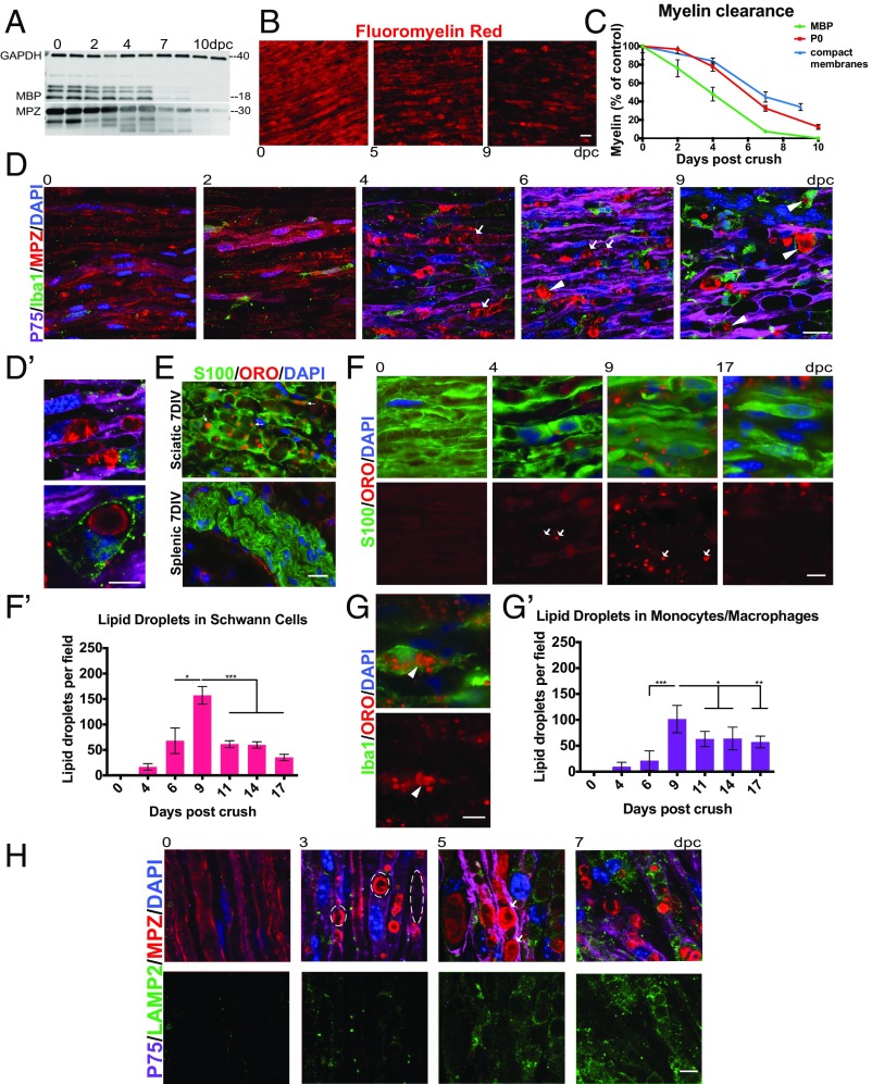
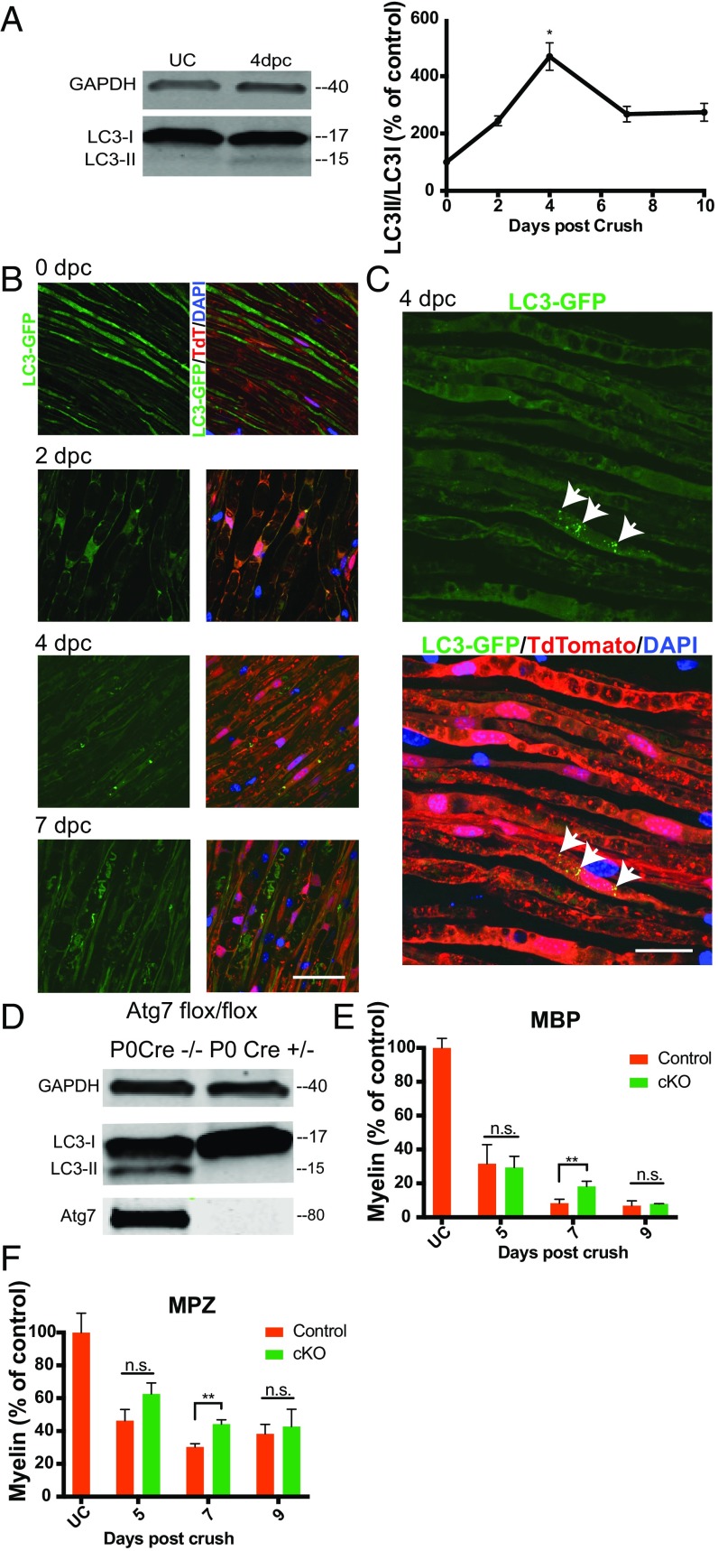
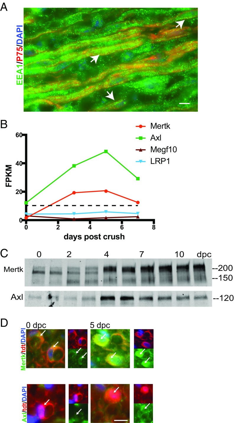
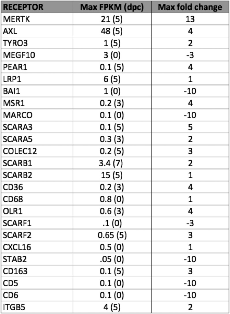
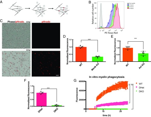
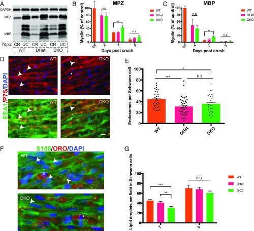
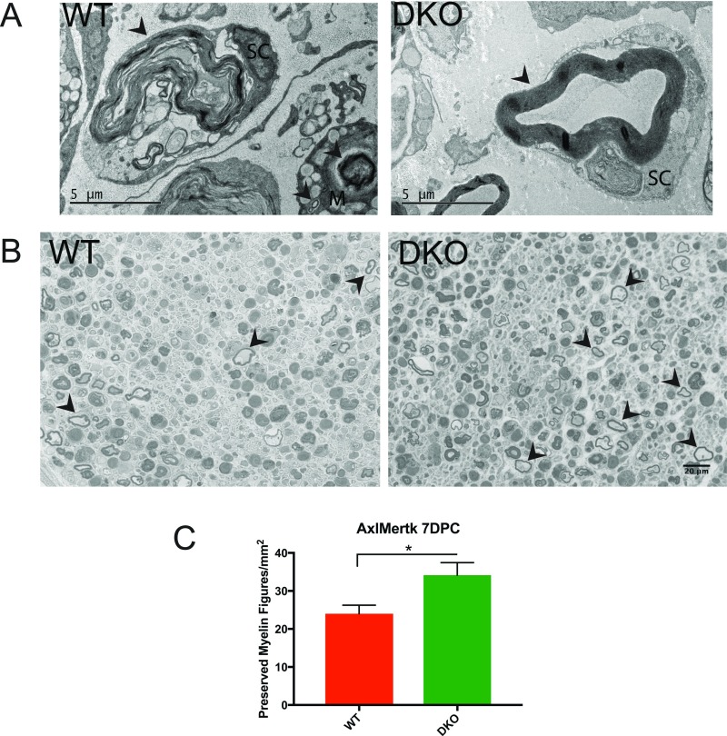
Ineffective myelin debris clearance is a major factor contributing to the poor regenerative ability of the central nervous system. In stark contrast, rapid clearance of myelin debris from the injured peripheral nervous system (PNS) is one of the keys to this system's remarkable regenerative capacity, but the molecular mechanisms driving PNS myelin clearance are incompletely understood. We set out to discover new pathways of PNS myelin clearance to identify novel strategies for activating myelin clearance in the injured central nervous system, where myelin debris is not cleared efficiently. Here we show that Schwann cells, the myelinating glia of the PNS, collaborate with hematogenous macrophages to clear myelin debris using TAM (Tyro3, Axl, Mer) receptor-mediated phagocytosis as well as autophagy. In a mouse model of PNS nerve crush injury, Schwann cells up-regulate TAM phagocytic receptors Axl and Mertk following PNS injury, and Schwann cells lacking both of these phagocytic receptors exhibit significantly impaired myelin phagocytosis both in vitro and in vivo. Autophagy-deficient Schwann cells also display reductions in myelin clearance after mouse nerve crush injury, as has been recently shown following nerve transection. These findings add a mechanism, Axl/Mertk-mediated myelin clearance, to the repertoire of cellular machinery used to clear myelin in the injured PNS. Given recent evidence that astrocytes express Axl and Mertk and have previously unrecognized phagocytic potential, this pathway may be a promising avenue for activating myelin clearance after CNS injury.

Keywords: Schwann cell; Wallerian degeneration; myelin; phagocytosis; regeneration.


Conflict of interest statement

The authors declare no conflict of interest.

Figures

Fig. 1.
Fig. 1.
Schwann cells and macrophages contribute to myelin clearance after nerve crush. (A) Western blot depicting myelin protein clearance from the crushed peripheral nerve. Each lane represents a separate sciatic nerve, and each well was loaded with the same amount of total nerve protein. (B) Representative images depicting myelin compact membrane clearance from the sciatic nerve visualized using FluoroMyelin dye (red). (Scale bar, 20 μm.) (C) Quantification of myelin protein and compact membrane clearance. n = 3 nerves for each FluoroMyelin time point. n = 5 nerves for each MBP and MPZ time point. All data are presented as mean ± SEM. (D) Confocal single-z-plane images of intact and degenerating whole-mount sciatic nerves at 0, 2, 4, 6, and 9 dpc stained with p75 (Schwann cells; purple), Iba1 (monocytes/macrophages; green), MPZ (myelin; red), and DAPI (nuclei; blue). Arrows indicate myelin that appears to be “inside” Schwann cells. Arrowheads indicate macrophages that have engulfed myelin debris. (Scale bar, 20 μm.) (D′) Higher-magnification image of a phagocytic macrophage at 9 dpc and Schwann cell association with myelin at 6 dpc. (Scale bar, 10 μm.) (E) Oil red O accumulation in sciatic and splenic nerves degenerated in vitro for 7 d. DIV, days in vitro. (Scale bar, 20 μm.) (F and G) Time course of oil red O lipid droplet accumulation in Schwann cells at 0, 4, 9, and 17 d after injury (F) and macrophages at 9 d after injury (G). Cryosections were stained with S100 (Schwann cells; green) and Iba1 (macrophages; green). Lipid droplets are red (oil red O). Nuclei are blue (DAPI). Arrows indicate lipid droplets in Schwann cells. Arrowheads indicate lipid droplets in macrophages. (Scale bars, 10 μm.) (F′ and G′) Quantification of lipid droplet accumulation in Schwann cells and macrophages from 0 to 17 dpc. n = 3 nerves and 6 fields of view for each time point. All data are presented as mean ± SEM. (H) Time course of lysosome accumulation in the sciatic nerve at 0, 3, 5, and 7 dpc. Whole-mount sciatic nerves are stained with p75 (Schwann cells; purple), MBP (myelin; red), LAMP2 (lysosomes; green), and DAPI (nuclei; blue). Dashed lines outline myelin ovoids. Arrows indicate myelin that appears to be inside Schwann cells. (Scale bar, 10 μm.) *P < 0.05, **P < 0.01, ***P < 0.001.
Fig. 2.
Fig. 2.
Schwann cell autophagy contributes to myelin clearance after nerve crush. (A) Western blot of whole-sciatic nerve protein lysate at 0 (UC) and 4 dpc and graph of LC3II/LC3I values for the first 10 d post injury. Each Western blot lane represents a separate sciatic nerve, and each well was loaded with the same amount of total nerve protein. All data are presented as mean ± SEM. n = 3 nerves for each time point. (B) Representative confocal single-z-plane images of whole-mount sciatic nerves from LC3-GFP+/−;P0 Cre+/−;loxSTOPlox tdtomato flox/+ mice at 0, 2, 4, and 7 dpc. Autophagosomes are green (LC3-GFP), Schwann cells are red (tdtomato), and nuclei are blue (DAPI). (Scale bar, 50 μm.) (C) Maximum-intensity projection from a stack of confocal images of tissue at 4 dpc. Arrows denote autophagosomes in a Schwann cell. (Scale bar, 20 μm.) (D) Western blot of immunopanned Schwann cells purified 5 d after crush from Atg7 flox/flox;P0 Cre+/− and Atg7 flox/flox;P0 Cre−/− mice. Each lysate was prepared from all of the Schwann cells purified from two sciatic nerves. (E and F) Quantification of Western blots of residual myelin proteins MBP and MPZ in sciatic nerves from Atg7 flox/flox P0 Cre+/− mice. Protein lysates were generated from single nerves, and each Western blot well was loaded with the same amount of total nerve protein. n = 3 or 4 nerves per time point per genotype. cKO, Atg7 flox/flox,P0 Cre+/−; control, Atg7 flox/flox;P0 Cre−/−. Data are presented as mean ± SEM. n.s., not significant; *P < 0.05, **P < 0.01.
Fig. 3.
Fig. 3.
Schwann cells up-regulate the phagocytic receptors Axl and Mertk after nerve crush. (A) Representative IHC image of a sciatic nerve 9 dpc stained for endosomes (EEA1; green), Schwann cells (p75; red), and nuclei (DAPI; blue). (Scale bar, 10 μm.) Arrows indicate regions of colocalization of EEA1 and p75 immunoreactivity. (B) Graphical illustration of the time course of expression of select phagocytic genes in acutely purified rat Schwann cells at 0, 3, 5, and 7 dpc. The dotted line represents an average FPKM of 11 for the dataset. FPKM values are averaged across two samples for each time point. (C) Western blot showing up-regulation of Mertk and Axl protein after mouse sciatic nerve crush. Protein lysates were generated from single nerves, and each Western blot well was loaded with the same amount of total nerve protein. (D) Cross-sections of loxSTOPlox tdtomato flox/+;P0 Cre+/− mouse sciatic nerve at 0 and 5 dpc stained by IHC with antibodies to Mertk and Axl (green) and DAPI. Arrows highlight Schwann cells in each field. (Scale bar, 10 μm.)
Fig. S1.
Fig. S1.
Table depicting rat Schwann cell expression of phagocytic receptors before and after sciatic nerve crush. Data obtained from RNAseq analysis of acutely purified P18 rat Schwann cells at 0, 3, 5, and 7 dpc. Schwann cells were purified according to ref. . Table indicates maximum FPKM across the time points sampled and timing of peak RNA abundance (in dpc) as well as fold change of FPKM relative to the uncrushed state. FPKM values represent average of two replicates for each time point.
Fig. 4.
Fig. 4.
Schwann cells use Axl and Mertk to clear myelin debris in vitro. (A) Schematic illustration of a Schwann cell in vitro myelin phagocytosis assay. (B) Representative FACS tracing of Schwann cells analyzed by flow cytometry after no exposure and 2-, 4-, and 8-h exposure to pHRODO-labeled myelin. (C) Representative live-cell images of Schwann cells before and 8 h following addition of pHRODO-labeled myelin to culture media. (Scale bar, 50 μm.) (DF) Results of an in vitro phagocytosis assay performed on Schwann cells purified from Axl−/−, Mertk−/−, and Axl/Mertk double-mutant Schwann cells and their littermate controls. Cells were purified from sciatic nerves 6 d after crush. Myelin phagocytosis was quantified using flow cytometry 2 to 3 h after addition of pHRODO-labeled PNS myelin debris. n = 4 for each genotype. Data are presented as mean ± SEM. (G) Quantification of integrated fluorescence per live-cell imaging field of Schwann cells coincubated with pHRODO-labeled myelin at 2 to 24 h after addition of myelin. n = 3 for each genotype: wild type, double heterozygote, and double knockout. Data are presented as mean ± SEM. **P < 0.01, ***P < 0.001.
Fig. 5.
Fig. 5.
Schwann cells use Axl and Mertk to clear myelin debris in vivo. (A) Representative Western blot showing myelin protein (MBP and MPZ) levels in wild-type, Axl/Mertk double-heterozygous, and Axl/Mertk double-knockout mouse sciatic nerves at 0 and 7 dpc. Protein lysates were generated from single nerves, and each Western blot well was loaded with the same amount of total nerve protein. CR, crushed; UC, uncrushed. (B and C) Quantification of MPZ- and MBP-stained Western blots of protein lysates from WT, Axl/Mertk double-heterozygous, and Axl/Mertk double-knockout mouse sciatic nerves at 0, 4, 7, and 9 dpc. n = 3 to 10 per genotype per time point. Data are presented as mean ± SEM. (D) Representative IHC images of cryosections of 9-dpc WT and Axl/Mertk DKO mouse sciatic nerves stained with antibodies to EEA1 (endosomes) and p75 (Schwann cells). Arrowheads indicate colocalization of EEA1 and p75, while asterisks indicate EEA1 immunoreactivity in a p75-negative cell, presumably a macrophage. (Scale bar, 10 μm.) (E) Quantification of IHC images of EEA1- and p75-labeled Schwann cells. Graph of average endosome abundance (EEA1 puncta) per Schwann cell at 9 dpc in Schwann cells from WT, Axl/Mertk double-heterozygous, and Axl/Mertk double-knockout mice. Eight cells were blindly selected and analyzed per animal. n = 4 or 5 animals per genotype. Data are presented as mean ± SEM. (F) Representative IHC images of 7-dpc cryosections of WT and Axl/Mertk DKO mouse sciatic nerves stained with ORO (lipid droplets) and antibodies to S100 (Schwann cells). Arrowheads indicate ORO within an S100-positive cell, while asterisks indicate ORO-positive droplets in S100-negative cells, presumably macrophages. (Scale bar, 10 μm.) (G) Quantification of ORO-positive lipid droplet abundance per field at 7 and 9 dpc in blindly selected and analyzed fields of IHC-stained tissue from WT, Axl/Mertk double-heterozygous, and Axl/Mertk double-knockout mice. n = 4 to 10 animals per genotype per time point. Four images were analyzed per animal. Data are presented as mean ± SEM. *P < 0.05, **P < 0.01, ***P < 0.001.
Fig. S2.
Fig. S2.
Loss of Axl/Mertk leads to retention of preserved myelin figures. (A) Electron microscopy images taken of wild-type (WT) and Axl/Mertk double-knockout (DKO) mouse sciatic nerves at 7 dpc. Arrowheads indicate myelin at various stages of degradation within or in close association with Schwann cells (SC) and macrophages (M). Note the basal lamina surrounding the Schwann cells. The DKO image features a preserved myelin figure, found to be significantly more abundant in DKO nerves than in WT nerves. (Scale bar, 5 μm.) (B) Representative images of toluidine blue-stained 1-μm sections of WT and DKO nerves at 7 dpc. Arrowheads indicate examples of preserved myelin figures (nonexhaustive labeling). (Scale bar, 20 μm.) (C) Quantification of preserved myelin figures in WT and DKO nerves at 7 dpc. n = 3 animals for each genotype. Two images were analyzed per animal. Data are presented as mean ± SEM. *P < 0.05.

References

    1. Arandjelovic S, Ravichandran KS. Phagocytosis of apoptotic cells in homeostasis. Nat Immunol. 2015;16:907–917. - PMC - PubMed
    1. Elliott MR, Ravichandran KS. Clearance of apoptotic cells: Implications in health and disease. J Cell Biol. 2010;189:1059–1070. - PMC - PubMed
    1. Burstyn-Cohen T, et al. Genetic dissection of TAM receptor-ligand interaction in retinal pigment epithelial cell phagocytosis. Neuron. 2012;76:1123–1132. - PMC - PubMed
    1. Hong S, et al. Complement and microglia mediate early synapse loss in Alzheimer mouse models. Science. 2016;352:712–716. - PMC - PubMed
    1. Gaudet AD, Popovich PG, Ramer MS. Wallerian degeneration: Gaining perspective on inflammatory events after peripheral nerve injury. J Neuroinflammation. 2011;8:110. - PMC - PubMed

Publication types

MeSH terms

Substances