. 2017 Oct 10;114(41):10954-10959.
doi: 10.1073/pnas.1710415114. Epub 2017 Sep 5.

Type 1 diabetes induction in humanized mice

Affiliations

Type 1 diabetes induction in humanized mice

Shulian Tan et al. Proc Natl Acad Sci U S A. .

Abstract

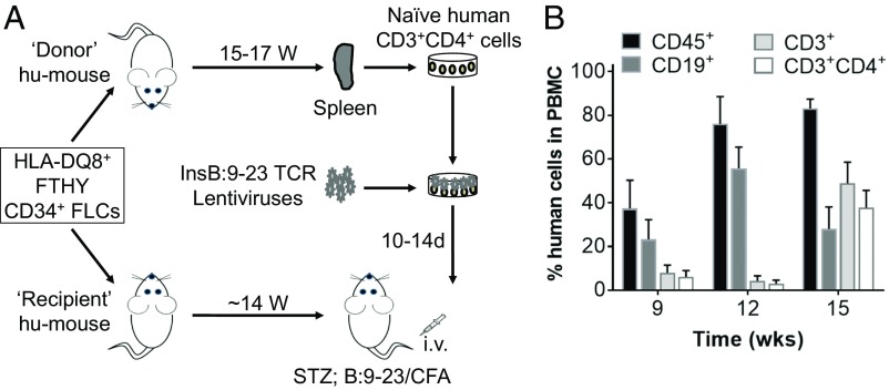
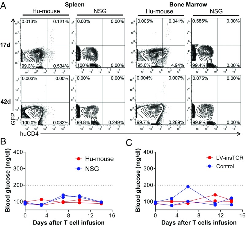
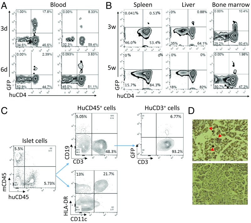
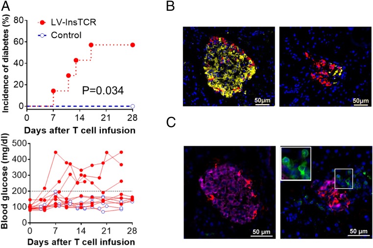
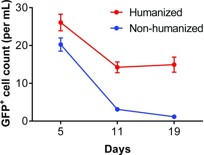
There is an urgent and unmet need for humanized in vivo models of type 1 diabetes to study immunopathogenesis and immunotherapy, and in particular antigen-specific therapy. Transfer of patient blood lymphocytes to immunodeficient mice is associated with xenogeneic graft-versus-host reactivity that complicates assessment of autoimmunity. Improved models could identify which human T cells initiate and participate in beta-cell destruction and help define critical target islet autoantigens. We used humanized mice (hu-mice) containing robust human immune repertoires lacking xenogeneic graft-versus-host reactivity to address this question. Hu-mice constructed by transplantation of HLA-DQ8+ human fetal thymus and CD34+ cells into HLA-DQ8-transgenic immunodeficient mice developed hyperglycemia and diabetes after transfer of autologous HLA-DQ8/insulin-B:9-23 (InsB:9-23)-specific T-cell receptor (TCR)-expressing human CD4+ T cells and immunization with InsB:9-23. Survival of the infused human T cells depended on the preexisting autologous human immune system, and pancreatic infiltration by human CD3+ T cells and insulitis were observed in the diabetic hu-mice, provided their islets were stressed by streptozotocin. This study fits Koch's postulate for pathogenicity, demonstrating a pathogenic role of islet autoreactive CD4+ T-cell responses in type 1 diabetes induction in humans, underscores the role of the target beta-cells in their immunological fate, and demonstrates the capacity to initiate disease with T cells, recognizing the InsB:9-23 epitope in the presence of islet inflammation. This preclinical model has the potential to be used in studies of the pathogenesis of type 1 diabetes and for testing of clinically relevant therapeutic interventions.

Keywords: humanized mice; insulin; type 1 diabetes.


Conflict of interest statement

The authors declare no conflict of interest.

Figures

Fig. S1.
Fig. S1.
Schematic of the LV-insTCR vector. Shown is the B:9–23-specific TCR-AcGFP fragment cloned into the pRRLSIN lentiviral vector.
Fig. S2.
Fig. S2.
Transgenic TCR expression in lentivirally transduced human TCR-deficient T-cell line cells. J.RT3-T3.5 cells were transduced with LV-insTCR lentiviruses (at a multiplicity of infection = 25). Shown are representative FCM profiles of human CD3 (A), TCRαβ (B), and TCRvβ11 (C) expression on nontransduced control and virally transduced (GFP+) cells.
Fig. 1.
Fig. 1.
Preparation of hu-mice for generating InsB:9–23-specific T cells and for induction of diabetes. (A) Schematic showing preparation of both donor hu-mice with HLA-DQ8+ human immune reconstitution, from which splenic human CD4+-naive T cells are prepared, transduced with LV-insTCR, expanded, and injected into recipient hu-mice for diabetes induction, and recipient HLA-DQ8–Tg hu-mice reconstituted with human immune cells autologous to the donor hu-mice, which are used as the recipients of LV-insTCR–transduced CD4+ T cells. (B). Levels (mean ± SEM) of human hematologic and immune cell chimerism in PBMCs of hu-mice from a representative experiment.
Fig. 2.
Fig. 2.
Purification and lentiviral transduction of hu-mouse–derived human CD3+CD4+CD45ROCD25-naive T cells. Spleen cells were prepared from donor hu-mice between 15 and 17 wk after humanization, as indicated in Fig. 1A, and human CD4+-naive T cells were prepared by MACS negative selection, using antibodies against mouse cells (anti-mCD45 and Ter119) and human cells expressing CD8, CD14, CD19, CD25, or CD45RO and transduced with LV-insTCR. (A) FCM profiles of human CD45+, CD3+, CD19+, CD3+CD4+, and CD4+CD8+ cells in the pooled spleen cells from donor hu-mice. (B) Purity of the MACS-selected hu-mouse spleen cells. Shown are the percentage of human CD3+CD4+ T cells and their expression of CD45RO, CD45RA, and CD25. (C) Expression of human CD3, CD4, and TCRvβ11 on the in vitro expanded LV-insTCR–transduced GFP+ cells.
Fig. S3.
Fig. S3.
Expansion of lentivirally transduced human CD4+ T cells in vitro. LV-insTCR–transduced hu-mouse–derived human CD4 T cells were expanded in vitro for 13 d. Data from two representative experiments are shown and presented as fold of expansion in cell numbers.
Fig. S4.
Fig. S4.
Survival of infused human T cells and levels of blood glucose in mice after infusion of human CD4 T cells. (A and B) LV-insTCR–transduced hu-mouse–derived human CD4 T cells were expanded for 13 d and injected into HLA-DQ8–Tg NSG mice (n = 2) or HLA-DQ8–Tg hu-mice grafted 14 wk earlier with human CD34+ FLCs (n = 2). (A) FCM analysis of surviving LV-insTCR–transduced GFP+CD4+ human T cells in spleen and bone marrow from hu-mouse (Left) and NSG mouse (Right) recipients at the indicated times after human T-cell transfer. (B) Blood glucose levels. (C) HLA-DQ8–Tg hu-mice grafted 14 wk earlier with human CD34+ FLCs and FTHY were treated by two injections of low-dose streptozotocin, followed 1 d later by infusion of expanded LV-insTCR–transduced (n = 2) or control (n = 2) hu-mouse–derived human CD4 T cells. Shown are blood glucose levels.
Fig. 3.
Fig. 3.
Survival of the infused LV-insTCR–transduced human CD4+ T cells in hu-mice. (A) Survival of GFP+CD4+ T cells in peripheral blood from hu-mice at 3 and 6 d after cell transfer. Data from two hu-mice at each point are shown. (B) Presence of GFP+CD4+ human T cells in spleen, liver, and bone marrow cells of two representative hu-mice analyzed at 3 and 5 wk, respectively, after cell transfer. (C) Pancreatic islets were prepared from hu-mice 11 d after infusion of LV-insTCR–transduced human CD4+ T cells, digested with trypsin, and stained with the indicated antibodies to detect human cell infiltration by flow cytometry. (D) Pancreatic tissue sections were prepared from hu-mice between 3 and 4 wk after injection of LV-InsTCR–transduced GFP+ human T cells (n = 3), and stained with anti-GFP antibodies. Shown are representative immunohistochemistry images of pancreas sections from hu-mice receiving LV-InsTCR–transduced GFP+ (Top) or control (Bottom) human T cells.
Fig. 4.
Fig. 4.
Induction of diabetes in HLA-DQ8–Tg hu-mice by adoptive transfer of autologous diabetogenic human CD4+ T cells. HLA-DQ8–Tg hu-mice were treated with low-dose streptozotocin and injected 1–2 d later with 5 × 10⁶ expanded LV-insTCR–transduced or control (i.e., the same hu-mouse–derived human CD4+ T cells that were similarly expanded in vitro as the LV-insTCR–transduced CD4+ T cells) human CD4+ T cells, followed 1 d later by immunization with InsB:9–23 peptides. (A) Cumulative incidence of diabetes (Top) and levels of blood glucose (Bottom) in hu-mice receiving LV-insTCR–transduced (solid symbol; n = 7) or control (opened symbol; n = 6) human T cells. Mice were defined as hyperglycemia if two consecutive blood glucose measurements >200 mg/dL (B and C) Immunofluorescent staining of pancreas samples prepared between 3 and 4 wk after injection of CD4+ T cells from hu-mice receiving control (Left) or LV-InsTCR–transduced (Right) human T cells (n = 3 per group). (B) Staining of mouse insulin (yellow) and glucagon (red). (C) Staining of human CD3+ cells (green), mouse insulin (pink) and glucagon (red). Nuclear is stained blue by DAPI.
Fig. S5.
Fig. S5.
Comparison of the survival of infused human T cells in humanized versus nonhumanized NSG mice. Ex vivo expanded hu-mouse–derived human T cells, which were transduced with insB:9–23-specific TCR/GFP, were injected i.v. to hu-mice with autologous human lymphohematopoietic cells or nonhumanized NSG mice. Blood was collected at days 5, 11, and 19 after adoptive transfer, and numbers of the transferred (GFP+) T cells were determined. Shown are GFP+ T-cell counts (mean ± SEM; n = 5 per group).

Comment in

  • Bridge between type 1 diabetes in mouse and man.
    Zeng D. Zeng D. Proc Natl Acad Sci U S A. 2017 Oct 10;114(41):10821-10823. doi: 10.1073/pnas.1715058114. Epub 2017 Oct 2. Proc Natl Acad Sci U S A. 2017. PMID: 28973949 Free PMC article. No abstract available.

References

    1. Bluestone JA, Herold K, Eisenbarth G. Genetics, pathogenesis and clinical interventions in type 1 diabetes. Nature. 2010;464:1293–1300. - PMC - PubMed
    1. Atkinson MA, Eisenbarth GS, Michels AW. Type 1 diabetes. Lancet. 2014;383:69–82. - PMC - PubMed
    1. Cryer PE. The barrier of hypoglycemia in diabetes. Diabetes. 2008;57:3169–3176. - PMC - PubMed
    1. Coppieters KT, et al. Demonstration of islet-autoreactive CD8 T cells in insulitic lesions from recent onset and long-term type 1 diabetes patients. J Exp Med. 2012;209:51–60. - PMC - PubMed
    1. Roep BO, Atkinson M, von Herrath M. Satisfaction (not) guaranteed: Re-evaluating the use of animal models of type 1 diabetes. Nat Rev Immunol. 2004;4:989–997. - PubMed

Publication types

MeSH terms