. 2017 Sep 19;114(38):10190-10195.
doi: 10.1073/pnas.1703100114. Epub 2017 Sep 5.

Treg-specific IL-27Rα deletion uncovers a key role for IL-27 in Treg function to control autoimmunity

Affiliations

Treg-specific IL-27Rα deletion uncovers a key role for IL-27 in Treg function to control autoimmunity

Jeongsu Do et al. Proc Natl Acad Sci U S A. .

Abstract

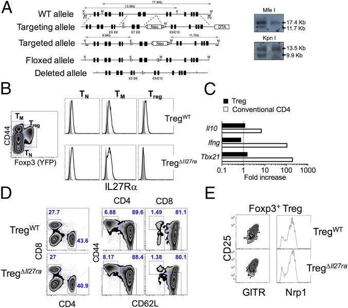
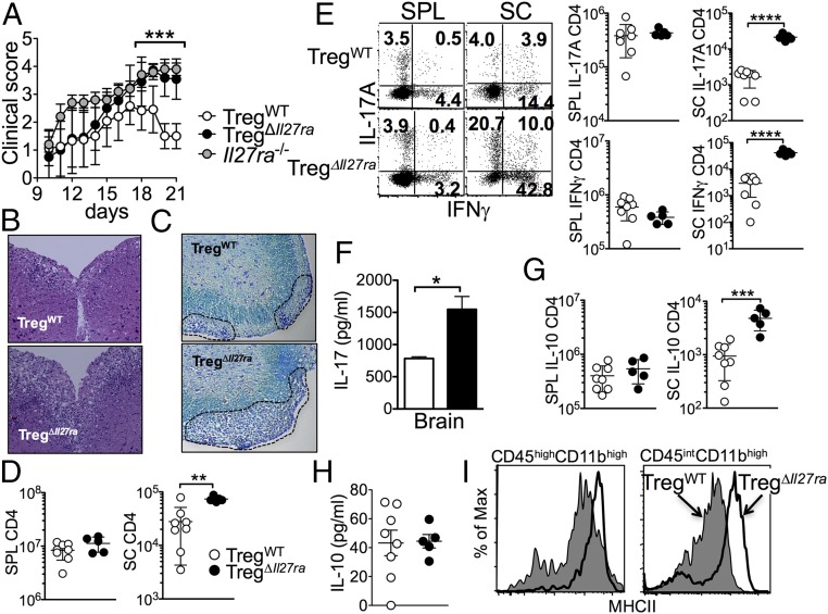
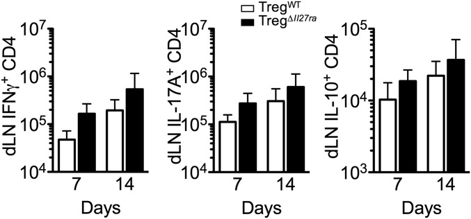
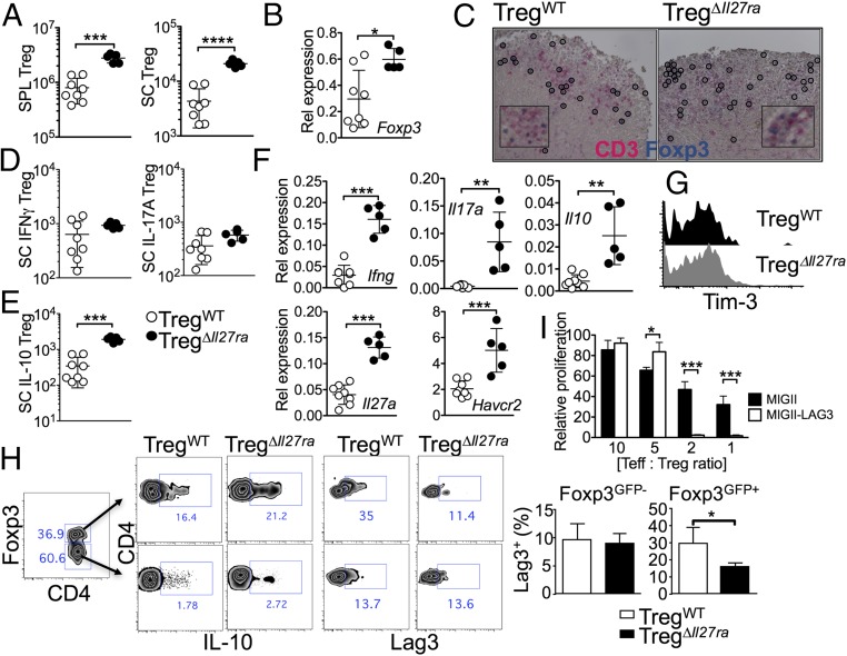
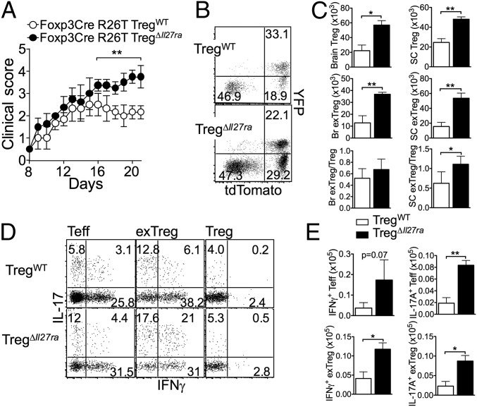
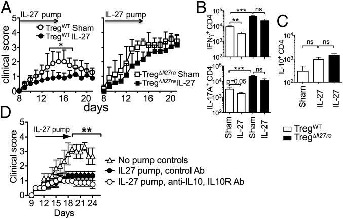
Dysregulated Foxp3+ Treg functions result in uncontrolled immune activation and autoimmunity. Therefore, identifying cellular factors modulating Treg functions is an area of great importance. Here, using Treg-specific Il27ra-/- mice, we report that IL-27 signaling in Foxp3+ Tregs is essential for Tregs to control autoimmune inflammation in the central nervous system (CNS). Following experimental autoimmune encephalomyelitis (EAE) induction, Treg-specific Il27ra-/- mice develop more severe EAE. Consistent with the severe disease, the numbers of IFNγ- and IL-17-producing CD4 T cells infiltrating the CNS tissues are greater in these mice. Treg accumulation in the inflamed CNS tissues is not affected by the lack of IL-27 signaling in Tregs, suggesting a functional defect of Il27ra-/- Tregs. IL-10 production by conventional CD4 T cells and their CNS accumulation are rather elevated in Treg-specific Il27ra-/- mice. Analysis with Treg fate-mapping reporter mice further demonstrates that IL-27 signaling in Tregs may control stability of Foxp3 expression. Finally, systemic administration of recombinant IL-27 in Treg-specific Il27ra-/- mice fails to ameliorate the disease even in the presence of IL-27-responsive conventional CD4 T cells. These findings uncover a previously unknown role of IL-27 in regulating Treg function to control autoimmune inflammation.

Keywords: Foxp3+ regulatory T cells; IL-27; Tr1 cells; autoimmunity.


Conflict of interest statement

The authors declare no conflict of interest.

Figures

Fig. 1.
Fig. 1.
Treg-specific Il27ra−/− mice. (A) Generation of neo-free Il27ra-floxed mice by the two-loxP and two-frt strategy. (B) IL-27Rα expression of naive (TN), memory phenotype (TM), and Foxp3+ Tregs (Treg) in Treg-specific Il27ra−/− (TregΔIl27ra) and littermate control (TregWT) mice. (C) Foxp3+ and conventional CD4 T cells were sorted from TregΔIl27ra mice and in vitro-stimulated with plate-coated anti-CD3/CD28 Abs in the presence of IL-27. Fold increases of the indicated genes were determined by qPCR analysis. (D and E) Lymph node cells were stained for CD4, CD8, CD44, and CD62L expression. Proportions of naive and effector/memory phenotype CD4 T cells (D) and surface phenotypes of Tregs (E) were examined. All of the experiments shown were repeated three times, and similar results were observed.
Fig. S1.
Fig. S1.
Treg-specific IL-27Rα deletion. Genomic DNA was isolated from FACS-sorted Foxp3 naive CD4, Foxp3+ Treg of TregΔIl27ra, and TregWT mice as well as naive CD4 T cells from C57BL/6 mice. PCR amplifying the Il27ra exons (3, 4, 7, 8) of the Il27ra gene was performed. Exons 7 and 8 represent genes targeted in TregΔIl27ra mice. Amplification of the indicated exon was normalized to the control exon 4. Each symbol represents an individually performed experiment.
Fig. S2.
Fig. S2.
Naive CD4 T cells from TregWT mice and TregΔIl27ra mice respond equally to IL-27 stimulation. Foxp3 CD44low naive CD4 T cells were FACS-sorted from TregWT mice and TregΔIl27ra mice and in vitro-stimulated in the presence of IL-27 for 48 h. The expression of the indicated genes was determined by qPCR analysis.
Fig. S3.
Fig. S3.
GFP expression of CD25+ CD4 T cells. Lymph node (LN) cells from TregWT mice and TregΔIl27ra mice were stained for CD25, CD4, and CD8 expression. CD25 and GFP expression of the indicated subsets was examined. The experiments were repeated three times, and similar results were observed.
Fig. 2.
Fig. 2.
Treg-specific Il27ra−/− mice develop severe EAE. (A) Groups of germline Il27ra−/− (n = 5), TregΔIl27ra (n = 10), and TregWT (n = 10) mice were induced for EAE. Clinical score was monitored daily. (B) Mice were killed 19 d post induction. H&E staining of the spinal cord. (Magnification: 10×.) (C) LFB staining of the spinal cord. (Magnification: 10×.) (D) Total CD4 T-cell numbers of the spleen and spinal cord at 19 d post induction. (E) Spleen and spinal cord cells were ex vivo-stimulated, and intracellular IFNγ/IL-17 expression was determined by FACS analysis. Absolute numbers of IFNγ/IL-17–producing CD4 T cells were enumerated. (F) CNS cells were isolated and ex vivo-stimulated with MOG peptide for 3 d. IL-17 secretion in the culture supernant was determined by ELISA. (G) Absolute numbers of IL-10–producing CD4 T cells. (H) IL-10 within inflamed brain tissues was determined by ELISA. (I) MHCII expression of microglia and infiltrating monocytes was measured. Each symbol represents an individually tested mouse. The results are representative of three independent experiments. *P < 0.05; **P < 0.01; ***P < 0.001; ****P < 0.0001.
Fig. S4.
Fig. S4.
CD4 T-cell cytokine production at different time points. Groups of TregΔIl27ra and TregWT mice were immunized with MOG peptide in CFA. Following 7 d and 14 d of immunization, draining inguinal LN cells were harvested, and IFNγ- and IL-17–producing CD4 T cells were enumerated by flow analysis. The data shown are representative of two independent experiments (n > 5).
Fig. 3.
Fig. 3.
Foxp3+ Treg responses in Treg-specific Il27ra−/− mice. (A) GFP+ Tregs were enumerated in the spleen and spinal cords of TregΔIl27ra and TregWT mice at 19 d post induction. (B) Foxp3 mRNA expression in the brain tissue was measured by qPCR. (C) Immunohistochemistry analysis for CD3 and Foxp3 expression. (Magnification: C, 20×; Inset, 40×.) (D) Spinal cord cells from TregΔIl27ra and TregWT mice were ex vivo-stimulated, and IFNγ- and IL-17–expressing GFP+ Tregs were enumerated by FACS analysis. (E) IL-10–producing Tregs in the spinal cord. (F) mRNA expression of the indicated genes in the brain tissue was measured by qPCR analysis. (G) Treg expression of TIM-3. (H) Lag3 and IL-10 expression of conventional CD4 and Tregs was measured by FACS analysis. (I) Foxp3+-inducible Tregs were transduced with retroviral vectors expressing Lag3. Empty vector-transduced Tregs were used as controls. The transduced Tregs were subsequently used in a suppression assay as described in Methods. Each symbol represents an individually tested mouse. Mean ± SD from two to three independent experiments is shown. *P < 0.05; **P < 0.01; ***P < 0.001; ****P < 0.0001.
Fig. S5.
Fig. S5.
Lag3 expression of brain-infiltrating T cells. TregΔIl27ra and TregWT mice were induced for EAE and killed at day 21 post induction. Mononuclear cells were isolated from the brain and stained for CD4 and Lag3. Zebra plots shown represent Lag3 expression of brain-infiltrating Foxp3+ Tregs and Foxp3 effector CD4 T cells. Similar results were observed in two independent experiments.
Fig. 4.
Fig. 4.
IL-27 signaling in Tregs controls Treg stability. (A) Groups of Rosa26CAG-tdTomato TregΔIl27ra and Rosa26CAG-tdTomato TregWT mice were induced for EAE. Clinical score was daily monitored. (B) Mice were killed 21 d post induction. YFP and tdTomato reporter expression of CD4 gated CNS cells is shown. (C) Absolute numbers of Tregs and exTregs in the CNS were calculated. (D) CNS cells were ex vivo-stimulated and intracellular IFNγ and IL-17 expression of Tconv, exTreg, and Tregs was determined by FACS analysis. (E) Absolute numbers of IFNγ- and IL-17–producing exTregs were calculated. The data shown are representative of two independent experiments (n = 6 ∼ 7). *P < 0.05; **P < 0.01.
Fig. 5.
Fig. 5.
IL-27 signaling in Tregs is essential to treat EAE. (A) TregWT and TregΔIl27ra mice were induced for EAE. Osmotic pump containing IL-27 was subcutaneously implanted (n = 9 ∼ 12) or sham surgery (n = 5 ∼ 6) was performed at 8 d post induction. The development of EAE was monitored. (B and C) Spinal cord cells from sham or IL-27–treated TregΔIl27ra and TregWT mice were ex vivo-stimulated, and intracellular IFNγ/IL-17 (B) and IL-10 (C) expression of CD4 T cells was determined by FACS analysis. The results shown are the mean ± SD of two to three independent experiments. (D) B6 mice (n = 5) were induced for EAE, and an osmotic pump containing IL-27 was implanted as described above. Anti–IL-10/anti–IL-10R mAbs (250 μg each) or control Ab was injected every 3 d. The development of EAE was monitored. *P < 0.05; **P < 0.01; ***P < 0.001.

References

    1. Long SA, Buckner JH. CD4+FOXP3+ T regulatory cells in human autoimmunity: More than a numbers game. J Immunol. 2011;187:2061–2066. - PMC - PubMed
    1. Pasare C, Medzhitov R. Toll pathway-dependent blockade of CD4+CD25+ T cell-mediated suppression by dendritic cells. Science. 2003;299:1033–1036. - PubMed
    1. Korn T, et al. Myelin-specific regulatory T cells accumulate in the CNS but fail to control autoimmune inflammation. Nat Med. 2007;13:423–431. - PMC - PubMed
    1. Hunter CA, Kastelein R. Interleukin-27: Balancing protective and pathological immunity. Immunity. 2012;37:960–969. - PMC - PubMed
    1. Yoshida H, Hunter CA. The immunobiology of interleukin-27. Annu Rev Immunol. 2015;33:417–443. - PubMed

Publication types

MeSH terms